Use Sophia to knock out your gen-ed requirements quickly and affordably. Learn more
×

Vertebral Column

Author: Sophia

what's covered

1. Regions of the Vertebral Column

The vertebral column is also known as the spinal column or spine. It consists of a sequence of vertebrae (singular, vertebra), each of which is separated and united by an intervertebral disc formed by connective tissue. Together, the vertebrae and intervertebral discs form the vertebral column. It is a flexible column that supports the head, neck, and body and allows for their movements. It also protects the spinal cord, which passes down the back through openings in the vertebrae.
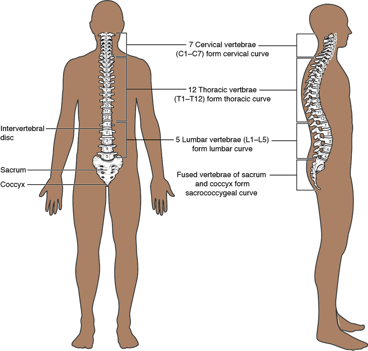

This image shows the structure of the vertebral column. The left panel shows the front view of the vertebral column and the right panel shows the side view of the vertebral column.
Vertebral Column - The adult vertebral column consists of 24 vertebrae, plus the sacrum and coccyx. The vertebrae are divided into three regions: cervical C1–C7 vertebrae, thoracic T1–T12 vertebrae, and lumbar L1–L5 vertebrae. The vertebral column is curved, with two primary curvatures (thoracic and sacrococcygeal curves) and two secondary curvatures (cervical and lumbar curves).

The vertebral column originally develops as a series of 33 vertebrae. However, as you developed, certain vertebrae fused together, leaving the adult human with 24 vertebrae, plus the sacrum and coccyx. The vertebral column is subdivided into five regions, with the vertebrae in each area named for that region and numbered in descending order.

  • In the cervical region (neck), there are 7 cervical vertebrae (C1–C7). Superiorly, the C1 vertebra articulates (forms a joint) with the occipital condyles of the skull. Inferiorly, C1 articulates with the C2 vertebra, and so on.
  • In the thoracic region (chest), there are the 12 thoracic vertebrae (T1–T12) that line the posterior portion of the thoracic cage.
  • In the lumbar region (lower back), there are 5 lumbar vertebrae (L1–L5).
  • In the sacral region, there is a single sacrum, which is also part of the pelvis. The sacrum is formed by the fusion of five sacral vertebrae.
  • At the inferior end of the vertebral column is the coccyx, or tailbone, which is formed from the fusion of four small coccygeal vertebrae. However, the sacral and coccygeal fusions do not start until age 20 and are not completed until middle age.
did you know
An interesting anatomical fact is that almost all mammals have seven cervical vertebrae, regardless of body size. This means that there are large variations in the size of cervical vertebrae, ranging from the very small cervical vertebrae of a shrew to the greatly elongated vertebrae in the neck of a giraffe. In a full-grown giraffe, each cervical vertebra is 11 inches tall.

learn more
To see additional bones and landmarks in this area, please visit the supplemental Axial Bones.pdf.

terms to know
Vertebral Column
A sequence of vertebrae that form the spine.
Intervertebral Disc
A connective tissue structure that separates and unites adjacent vertebrae.
Cervical Vertebra
A vertebral bone in the cervical region of the vertebral column.
Thoracic Vertebra
A vertebral bone in the thoracic region of the vertebral column.
Lumbar Vertebra
A vertebral bone in the lumbar region of the vertebral column.
Sacrum
A single fused bone in the sacral region, inferior to the lumbar vertebra.
Coccyx
A single fused bone at the most inferior end of the vertebral column; the tailbone.

2. Curvatures of the Vertebral Column

The vertebral column does not form a straight line, but instead curves. At birth, the entire vertebral column has a single anterior curvature. However, as an adult, there are four curves: two anterior and two posterior.

key concept
At birth, babies have a vertebral column that curves anteriorly in a C-shape, as if permanently slouching. This curvature of the spine is called primary curvature because it is the curvature that is present first in development. This is also one reason why babies are unable to support their heads or hold themselves upright when seated right away.

However, as a child learns to hold its head up, sit upright, stand, and walk, muscle tension will pull on the cervical and lumbar vertebrae and force them to curve posteriorly, forming what is called secondary curvature. The alternating curvatures in the spine cause the vertebral column to adopt an S-shape which increases the vertebral column’s strength, flexibility, and ability to absorb shock. When the load on the spine is increased, like by carrying a heavy backpack, the curvatures increase in depth (become more curved) to accommodate the extra weight. They then spring back when the weight is removed.
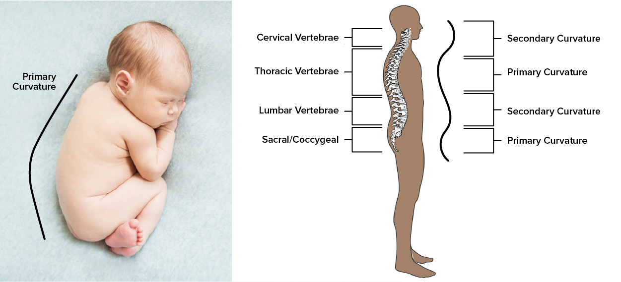

Image compares the c-shaped spine of a sleeping baby to the s-shaped curvature of an adult spine.
Vertebral Curvature - The adult vertebral column forms an S-shaped curve, alternating secondary and primary curvature.

Disorders associated with the curvature of the spine include kyphosis (an excessive posterior curvature of the thoracic region), lordosis (an excessive anterior curvature of the lumbar region), and scoliosis (an abnormal, lateral curvature, accompanied by twisting of the vertebral column).

Developmental anomalies, pathological changes, or obesity can enhance the normal vertebral column curves, resulting in the development of abnormal or excessive curvatures. Kyphosis, also referred to as humpback or hunchback, is an excessive posterior curvature of the thoracic region. This can develop when osteoporosis causes weakening and erosion of the anterior portions of the upper thoracic vertebrae, resulting in their gradual collapse. Lordosis, or swayback, is an excessive anterior curvature of the lumbar region and is most commonly associated with obesity or late pregnancy. The accumulation of body weight in the abdominal region results in an anterior shift in the line of gravity that carries the weight of the body. This causes an anterior tilt of the pelvis and a pronounced enhancement of the lumbar curve.

Scoliosis is an abnormal, lateral curvature accompanied by twisting of the vertebral column. Compensatory curves may also develop in other areas of the vertebral column to help maintain the head positioned over the feet. Scoliosis is the most common vertebral abnormality among girls. The cause is usually unknown, but it may result from weakness of the back muscles, defects such as differential growth rates in the right and left sides of the vertebral column, or differences in the length of the lower limbs. When present, scoliosis tends to get worse during adolescent growth spurts. Although most individuals do not require treatment, a back brace may be recommended for growing children. In extreme cases, surgery may be required.

Excessive vertebral curves can be identified while an individual stands in the anatomical position. Observe the vertebral profile from the side and then from behind to check for kyphosis or lordosis. Then have the person bend forward. If scoliosis is present, an individual will have difficulty bending directly forward, and the right and left sides of the back will not be level with each other in the bent position.
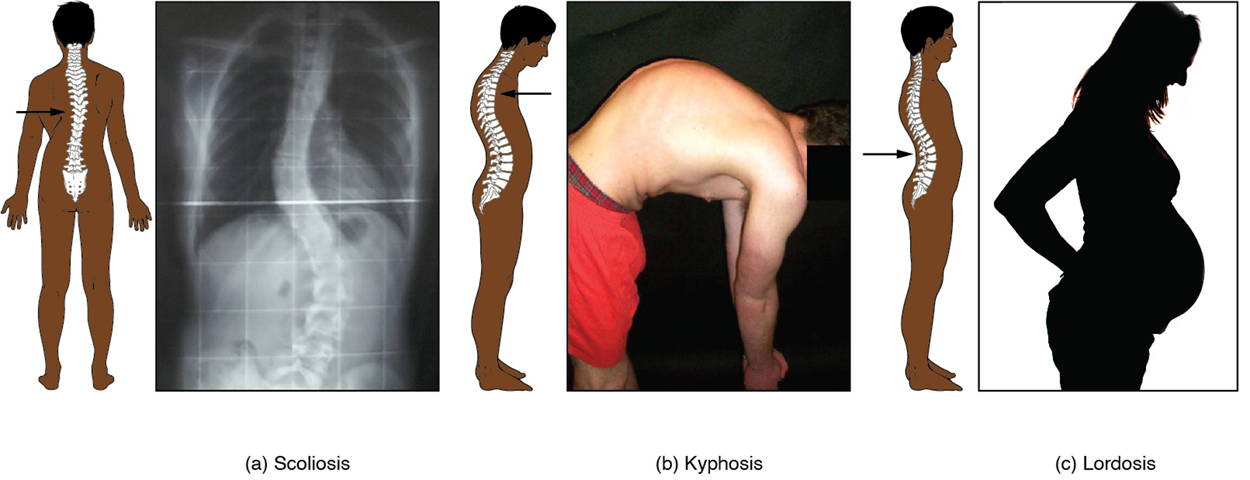
This image shows the changes to the abnormal curves of the vertebral columns in different diseases. The left panel shows the change in the curve of the vertebral column in scoliosis, the middle panel shows the change in the curve of the vertebral column in kyphosis, and the right panel shows the change in the curve of the vertebral column in lordosis.
Abnormal Curvatures of the Vertebral Column - (a) Scoliosis is an abnormal lateral bending of the vertebral column. (b) An excessive curvature of the upper thoracic vertebral column is called kyphosis. (c) Lordosis is an excessive curvature in the lumbar region of the vertebral column.

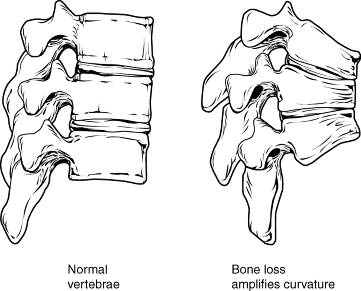
Osteoporosis - Osteoporosis is an age-related disorder that causes the gradual loss of bone density and strength. When the thoracic vertebrae are affected, there can be a gradual collapse of the vertebrae. This results in kyphosis, an excessive curvature of the thoracic region.lalt=This figure shows the changes to the spine in osteoporosis. The left panel shows the structure of normal vertebrae and the right panel shows the curved vertebrae in osteoporosis.

terms to know
Primary Curvature
The anterior curvature of the spine present at birth.
Secondary Curvature
The posterior curvature of the spine developed after birth.
Kyphosis
An excessive posterior curvature of the thoracic region of the vertebral column; humpback.
Lordosis
An excessive anterior curvature of the lumbar region; swayback.
Scoliosis
An abnormal, lateral curvature accompanied by twisting of the vertebral column.

3. General Structure of a Vertebra

Within the different regions of the vertebral column, vertebrae vary in size and shape, but they all follow a similar structural pattern. A typical vertebra will consist of a body, a vertebral arch, and seven processes.

The vertebral body is the anterior portion of each vertebra and is the part that supports the body weight. Because of this, the vertebral bodies progressively increase in size and thickness going down the vertebral column. The bodies of adjacent vertebrae are separated and strongly united by an intervertebral disc.

The vertebral arch forms the posterior portion of each vertebra. It consists of four parts, the right and left pedicles and the right and left laminae. Each pedicle forms one of the lateral sides of the vertebral arch. The pedicles are anchored to the posterior side of the vertebral body. Each lamina forms part of the posterior roof of the vertebral arch. The large opening between the vertebral arch and body is the vertebral foramen, which contains the spinal cord. In the intact vertebral column, the vertebral foramina of all of the vertebrae align to form the vertebral (spinal) canal, which serves as the bony protection and passageway for the spinal cord down the back. When the vertebrae are aligned together in the vertebral column, notches between the pedicles of adjacent vertebrae form an intervertebral foramen (intervertebral, between two vertebrae), the opening through which a spinal nerve exits from the vertebral column.

Seven processes arise from the vertebral arch. Each paired transverse process projects laterally and arises from the junction point between the pedicle and lamina. The single spinous process, also referred to as the vertebral spine, projects posteriorly at the midline of the back. The vertebral spines can easily be felt as a series of bumps just under the skin down the middle of the back. The transverse and spinous processes serve as important muscle attachment sites. A superior articular process extends or faces upward, and an inferior articular process faces or projects downward on each side of the vertebrae. The paired superior articular processes of one vertebra join with the corresponding paired inferior articular processes from the next higher vertebra or the skull in the case of the first vertebra (C1). These junctions form slightly moveable joints. The shape and orientation of the articular processes vary in different regions of the vertebral column and play a major role in determining the type and range of motion available in each region.
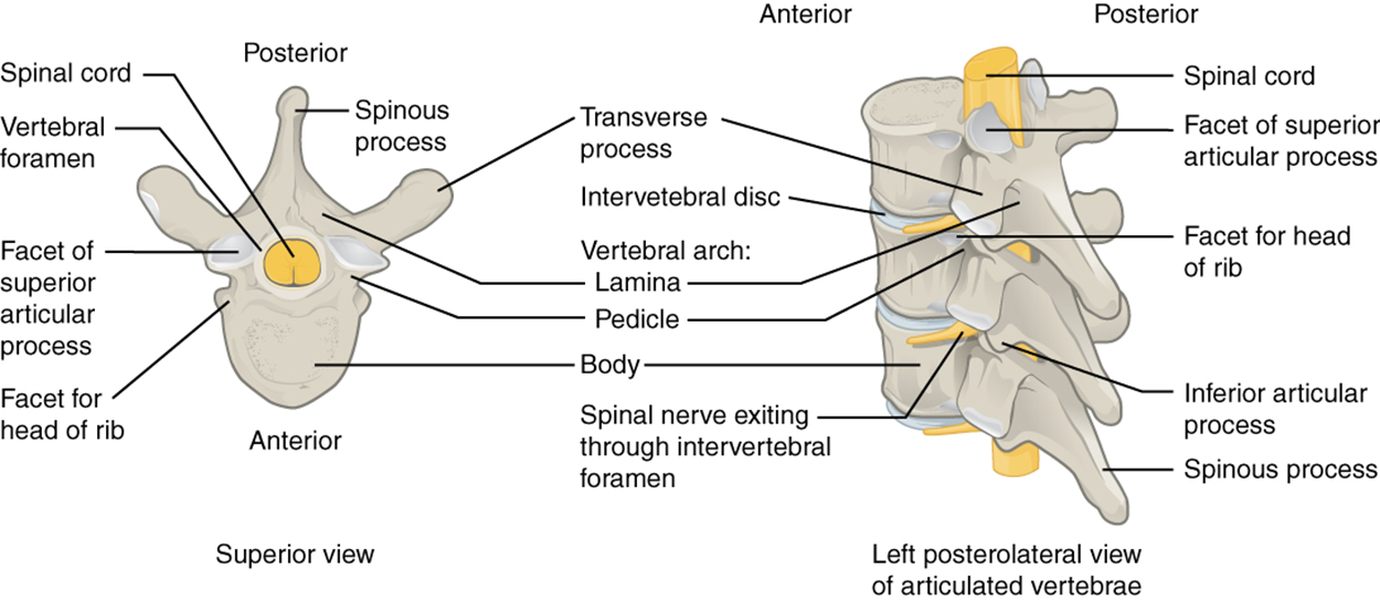

This image shows the detailed structure of each vertebra. The left panel shows the superior view of the vertebra and the right panel shows the left posterolateral view.
Parts of a Typical Vertebra - A typical vertebra consists of a body and a vertebral arch. The arch is formed by the paired pedicles and paired laminae. Arising from the vertebral arch are the transverse, spinous, superior articular, and inferior articular processes. The vertebral foramen provides passage to the spinal cord. Each spinal nerve exits through an intervertebral foramen, located between adjacent vertebrae. Intervertebral discs unite the bodies of adjacent vertebrae.

terms to know
Vertebral Body
The anterior portion of each vertebra and is the part that supports the body weight.
Vertebral Arch
The posterior portion of each vertebra that surrounds the vertebral foramen.
Pedicle
A lateral side of the vertebral arch.
Lamina
The posterior side of the vertebral arch
Vertebral Foramen
The large opening between the vertebral arch and body.
Vertebral Canal
The space formed by the stacked vertebral foramen which contains the spinal cord.
Intervertebral Foramen
The lateral opening between adjacent pedicles through which a spinal nerve exits the vertebral column.
Transverse Process
One of a pair of lateral bony projections from the pedicle-lamina junction.
Spinous Process
The posterior bony projection from the vertebral arch at the midline of the back.
Superior Articular Process
One of a pair of bony projections that extends upward from the vertebral arch to form a joint with the skull or inferior articular process of the vertebra above it.
Inferior Articular Process
One of a pair of bony projections that extends downward from the vertebral arch to form a joint with the superior articular process of the vertebra below it.

4. Regional Modifications of Vertebrae

In addition to the general characteristics of a typical vertebra described above, vertebrae also display characteristic size and structural features that vary between the different vertebral column regions. Thus, cervical vertebrae are smaller than lumbar vertebrae due to differences in the proportion of body weight that each supports. Thoracic vertebrae have sites for rib attachment, and the vertebrae that give rise to the sacrum and coccyx have fused together into single bones.

4a. Cervical Vertebrae

Cervical vertebrae show slight variations in their general structures that differentiate them from thoracic or lumbar vertebrae.
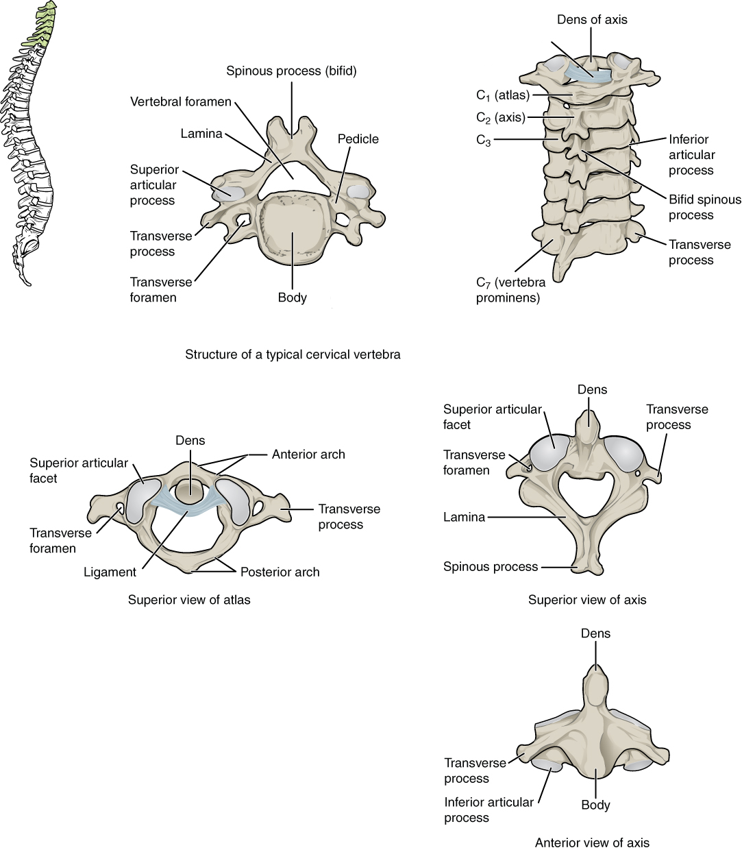
  • The vertebral bodies are small, reflecting the fact that they carry the least amount of body weight.
  • The spinous processes are bifid (Y-shaped).
  • The spinous processes of C3–C6 are short. However, the spinous process of C7 is longer. You can find this vertebra by running your finger down the midline of the posterior neck until you encounter the prominent projection at the base of the neck—this is the spinous process of C7.
  • The transverse processes are sharply curved (U-shaped) to allow for the passage of the cervical spinal nerves.
  • The articular processes are flattened and largely face upward or downward.
The cervical vertebrae also contain unique features that are not seen in other vertebrae. Each transverse process also has an opening called the transverse foramen. An important artery that supplies the brain ascends up the neck by passing through these openings.

The first and second cervical vertebrae are further modified, giving each a distinctive appearance. The first cervical (C1) vertebra is also called atlas, because this is the vertebra that supports the skull on top of the vertebral column (in Greek mythology, Atlas was the god who supported the Earth and heavens on his shoulders). The C1 vertebra does not have a body or spinous process. Instead, it is ring-shaped, consisting of an anterior arch and a posterior arch. The transverse processes of the atlas are longer and extend more laterally than do the transverse processes of any other cervical vertebrae. The superior articular processes face upward and are deeply curved for articulation with the occipital condyles on the base of the skull. The inferior articular processes are flat and face downward to join with the superior articular processes of the C2 vertebra.

The second cervical (C2) vertebra is called axis, because it serves as the axis for right and left rotation when turning the head. The axis resembles typical cervical vertebrae in most respects but is easily distinguished by the dens (odontoid process), a bony projection that extends upward from the vertebral body. The dens join with the inner aspect of the anterior arch of the atlas, where it is held in place by the transverse ligament.

This figure shows the structure of the cervical vertebrae. The left panel shows the location of the cervical vertebrae in green along the vertebral column. The middle panel shows the structure of a typical cervical vertebra and the right panel shows the superior and anterior view of the axis.
Cervical Vertebrae - A typical cervical vertebra has a small body, a bifid spinous process, transverse processes that have a transverse foramen and are curved for spinal nerve passage. The atlas (C1 vertebra) does not have a body or spinous process. It consists of an anterior and a posterior arch and elongated transverse processes. The axis (C2 vertebra) has upward projecting dens, which articulate with the anterior arch of the atlas.

4b. Thoracic Vertebrae

The thoracic vertebrae also have slight variations in their general structure that differentiate them from cervical or lumbar vertebrae.
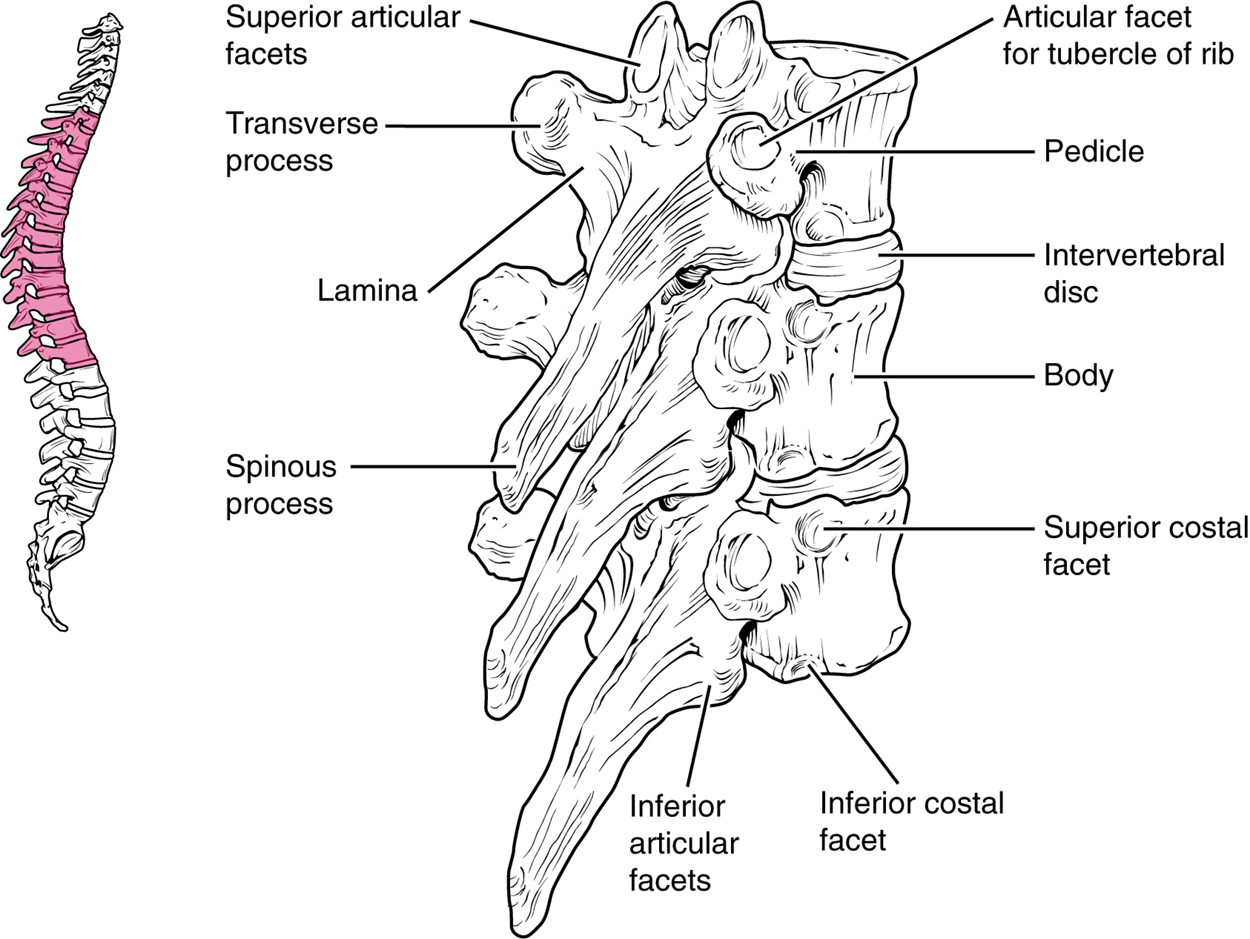
  • The bodies of the thoracic vertebrae are larger than those of cervical vertebrae and smaller than lumbar vertebrae.
  • The spinous processes are long and have a pronounced downward angle that causes them to overlap the next inferior vertebra.
  • The articular processes of thoracic vertebrae face anteriorly and posteriorly.
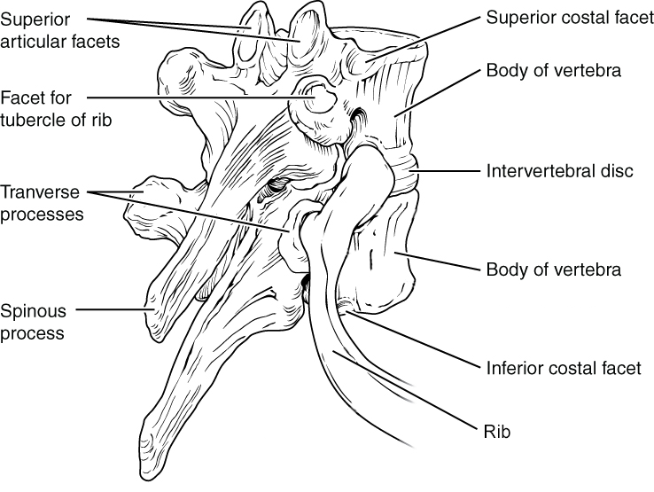
Thoracic vertebrae have several additional flat articulation sites, each of which is called a facet, where a rib is attached. Most thoracic vertebrae have two facets located on the lateral sides of the body, each of which is called a costal facet (costal, rib). These are for articulation with the head (end) of a rib. An additional facet is located on the transverse process for articulation with the tubercle of a rib.

This figure shows the structure of the thoracic vertebra. The left panel shows the vertebral column with the thoracic vertebrae highlighted in pink. The right panel shows the detailed structure of a single thoracic vertebra.
Thoracic Vertebrae - A typical thoracic vertebra is distinguished by the spinous process, which is long and projects downward to overlap the next inferior vertebra. It also has articulation sites (facets) on the vertebral body and a transverse process for rib attachment.

This diagram shows how the thoracic vertebra connects to the angle of the rib. The major parts of the vertebra and the processes connecting the vertebra to the rib are labeled.
Rib Articulation in Thoracic Vertebrae - Thoracic vertebrae have superior and inferior articular facets on the vertebral body for articulation with the head of a rib, and a transverse process facet for articulation with the rib tubercle.

4c. Lumbar Vertebrae

Lumbar vertebrae similarly have slight variations in their general structure.
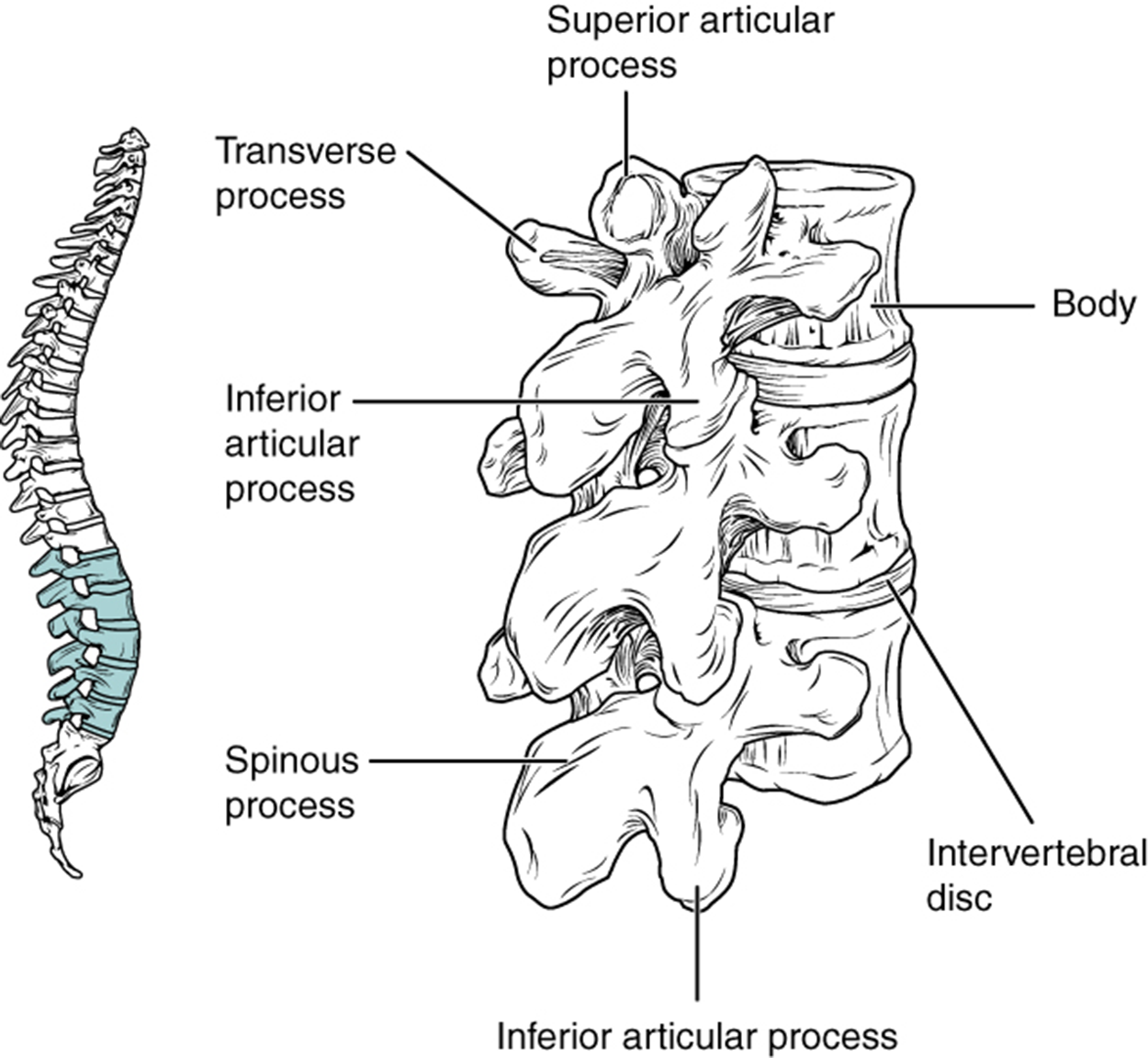
  • The vertebral bodies are large and thick, indicating they bear the largest amount of body weight and pressure.
  • The transverse processes are short.
  • The spinous processes are short and vertically flattened.
  • The articular processes are large, with the superior process facing medially and the inferior facing laterally.
This image shows the location and structure of the lumbar vertebrae. The left panel shows the location of the lumbar vertebrae (highlighted in green) along the vertebral column. The right panel shows the inferior articular process and the major parts are labeled.
Lumbar Vertebrae - Lumbar vertebrae are characterized by having a large, thick body and a short, rounded spinous process.

4d. Sacrum and Coccyx

The sacrum is a triangular-shaped bone that is thick and wide across its superior base where it is weight-bearing and then tapers down to an inferior, non-weight-bearing apex. It is formed by the fusion of five sacral vertebrae, a process that does not begin until after the age of 20. The coccyx, or tailbone, is derived from the fusion of four very small coccygeal vertebrae. It articulates with the inferior tip of the sacrum. It is not weight-bearing in the standing position but may receive some body weight when sitting.
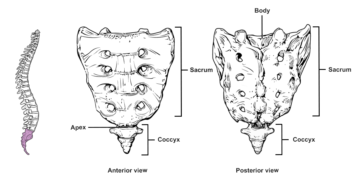
This figure shows the structure of the sacrum and coccyx. The left panel shows the vertebral column with the sacrum and coccyx highlighted in pink. The middle panel shows the anterior view and the right panel shows the posterior view of the sacrum and coccyx.
Sacrum and Coccyx - The sacrum is formed from the fusion of five sacral vertebrae. The coccyx is formed by the fusion of four small coccygeal vertebrae.

terms to know
Transverse Foramen
An opening in the transverse foramen of cervical vertebrae.
Atlas
The first cervical vertebra, C1.
Axis
The second cervical vertebra, C2.
Dens
A bony projection that extends upward from the vertebral body of the second cervical vertebra.
Facet
An articulation site.
Costal Facet
An articulation site with a rib bone.

5. Intervertebral Discs

The bodies of adjacent vertebrae are strongly anchored to each other by an intervertebral disc. This structure provides padding between the bones during weight bearing and, because it can change shape, also allows for movement between the vertebrae. Although the total amount of movement available between any two adjacent vertebrae is small, when these movements are summed together along the entire length of the vertebral column, large body movements can be produced. Ligaments that extend along the length of the vertebral column also contribute to its overall support and stability.

Recall that an intervertebral disc is a pad of connective tissue (fibrocartilage) that fills the gap between adjacent vertebral bodies. Each disc is anchored to the bodies of its adjacent vertebrae, thus strongly uniting them. The discs also provide padding between vertebrae during weight bearing. Because of this, intervertebral discs are thin in the cervical region and thickest in the lumbar region, which carries the most body weight. In total, the intervertebral discs account for approximately 25% of your body height between the top of the pelvis and the base of the skull. Intervertebral discs are also flexible and can change shape to allow for movements of the vertebral column.

Each intervertebral disc consists of two parts. The annulus fibrosus is the tough, fibrous outer layer of the disc. It forms a circle (annulus, ring) and is firmly anchored to the outer margins of the adjacent vertebral bodies. Inside is the nucleus pulposus, consisting of a softer, more gel-like material. It has a high water content that serves to resist compression and thus is important for weight bearing. With increasing age, the water content of the nucleus pulposus gradually declines. This causes the disc to become thinner, decreasing total body height somewhat, and reduces the flexibility and range of motion of the disc, making bending more difficult.
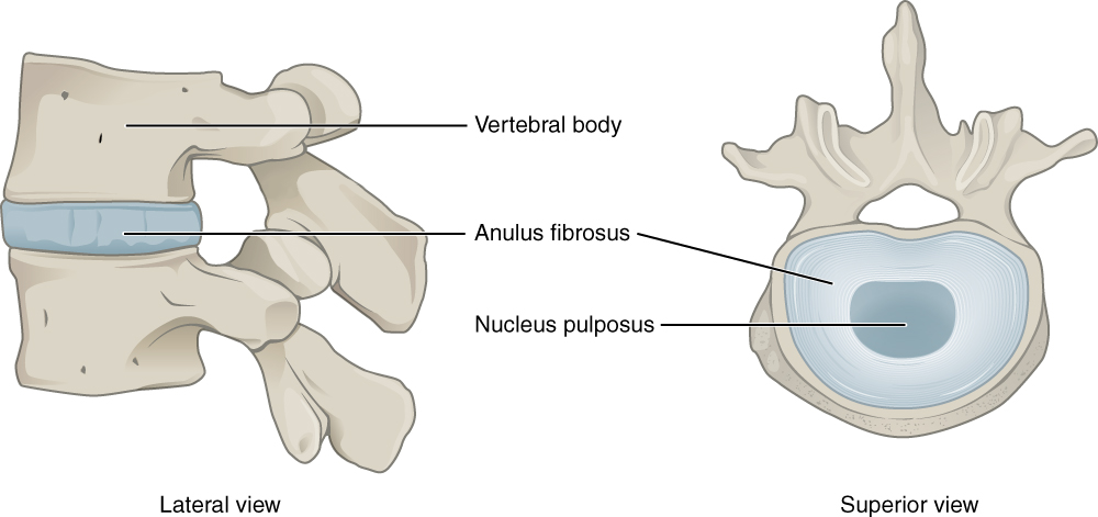
This image shows the structure of the intervertebral disk. The left panel shows the lateral view and the right panel shows the superior view.
Intervertebral Disc - The bodies of adjacent vertebrae are separated and united by an intervertebral disc, which provides padding and allows for movements between adjacent vertebrae. The disc consists of a fibrous outer layer called the annulus fibrosus and a gel-like center called the nucleus pulposus. The intervertebral foramen is the opening formed between adjacent vertebrae for the exit of a spinal nerve.

The gel-like nature of the nucleus pulposus also allows the intervertebral disc to change shape as one vertebra rocks side to side or forward and back in relation to its neighbors during movements of the vertebral column. Thus, bending forward causes compression of the anterior portion of the disc but expansion of the posterior disc. If the posterior annulus fibrosus is weakened due to injury or increasing age, the pressure on the disc when bending forward and lifting a heavy object can cause the annulus fibrosus to permanently stretch. If the material does not break, this is considered a bulging disc and can cause compression of a spinal nerve at the point where it exits through the intervertebral foramen, with resulting pain and/or muscle weakness in those body regions supplied by that nerve. If the pressure is greater and causes the nucleus pulposus to protrude (push) posteriorly through the annulus fibrosus, it will result in a herniated disc (“ruptured” or “slipped” disc). The most common sites for disc herniation are the L4/L5 or L5/S1 intervertebral discs, which can cause sciatica, a widespread pain that radiates from the lower back down the thigh and into the leg. Similar injuries of the C5/C6 or C6/C7 intervertebral discs, following forcible hyperflexion of the neck from a collision accident or football injury, can produce pain in the neck, shoulder, and upper limb.
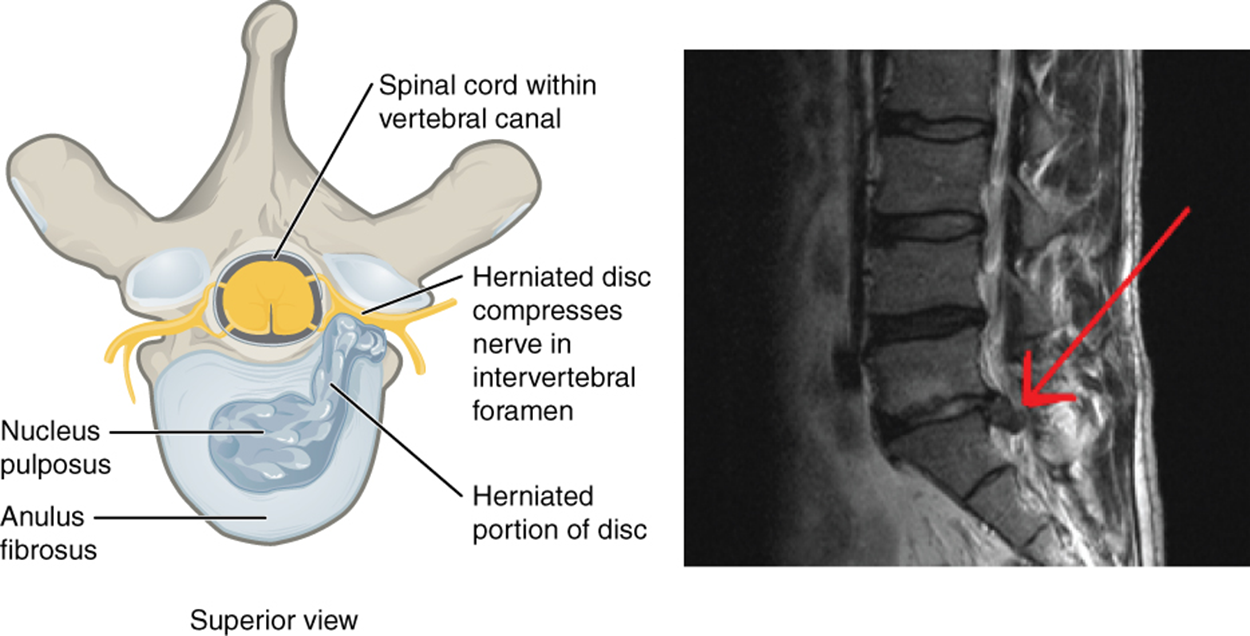
This figure shows a herniated disk. The left panel shows the superior view highlighting how the herniated disk compresses the nerve. The right panel shows a photograph of a herniated disk.
Herniated Intervertebral Disc - Weakening of the annulus fibrosus can result in herniation (protrusion) of the nucleus pulposus and compression of a spinal nerve, resulting in pain and/or muscle weakness in the body regions supplied by that nerve.

IN CONTEXT
Career Connection: Chiropractor
Chiropractors are health professionals who use nonsurgical techniques to help patients with musculoskeletal system problems that involve the bones, muscles, ligaments, tendons, or nervous system. They treat problems such as neck pain, back pain, joint pain, or headaches. Chiropractors focus on the patient’s overall health and can also provide counseling related to lifestyle issues, such as diet, exercise, or sleep problems. If needed, they will refer the patient to other medical specialists.

Chiropractors use a drug-free, hands-on approach for patient diagnosis and treatment. They will perform a physical exam, assess the patient’s posture and spine, and may perform additional diagnostic tests, including taking X-ray images. They primarily use manual techniques, such as spinal manipulation, to adjust the patient’s spine or other joints. They can recommend therapeutic or rehabilitative exercises, and some also include acupuncture, massage therapy, or ultrasound as part of the treatment program. In addition to those in general practice, some chiropractors specialize in sport injuries, neurology, orthopedics, pediatrics, nutrition, internal disorders, or diagnostic imaging.

To become a chiropractor, students must have 3–4 years of undergraduate education, attend an accredited, four-year Doctor of Chiropractic (D.C.) degree program, and pass a licensure examination to be licensed for practice in their state. With the aging of the baby-boom generation, employment for chiropractors is expected to increase.

terms to know
Annulus Fibrosus
The tough, fibrous outer layer of the intervertebral disc.
Nucleus Pulposus
The center gel-like material inside the intervertebral disc.
Bulging Disc
A condition in which the annulus fibrosus has expanded beyond its original boundary.
Herniated Disc
A condition in which the nucleus pulposus has protruded through the annulus fibrosus.
Sciatica
A widespread pain due to nerve compression that radiates from the lower back down the thigh and into the leg.

summary
In this lesson, you learned about the vertebral column. You learned to identify and differentiate the regions of the vertebral column by location and vertebral curvature. You learned the general structures of a vertebra as well as the regional modifications that exist in each vertebral region—cervical, thoracic, lumbar, sacrum, and coccyx. Lastly, you learned about the structure and function of the intervertebral discs.

Source: THIS TUTORIAL HAS BEEN ADAPTED FROM OPENSTAX "ANATOMY AND PHYSIOLOGY 2E." ACCESS FOR FREE AT openstax.org/books/anatomy-and-physiology-2e/pages/1-introduction. LICENSE: CC ATTRIBUTION 4.0 INTERNATIONAL. Accessed by October 2022.


Terms to Know
Annulus Fibrosus

The tough, fibrous outer layer of the intervertebral disc.

Atlas

The first cervical vertebra, C1.

Axis

The second cervical vertebra, C2.

Bulging Disc

A condition in which the annulus fibrosus has expanded beyond its original boundary.

Cervical Vertebra

A vertebral bone in the cervical region of the vertebral column.

Coccyx

A single fused bone at the most inferior end of the vertebral column; the tailbone.

Costal Facet

An articulation site with a rib bone.

Dens

A bony projection that extends upward from the vertebral body of the second cervical vertebra.

Facet

An articulation site.

Herniated Disc

A condition in which the nucleus pulposus has protruded through the annulus fibrosus.

Inferior Articular Process

One of a pair of bony projections that extends downward from the vertebral arch to form a joint with the superior articular process of the vertebra below it.

Intervertebral Disc

A connective tissue structure that separates and unites adjacent vertebrae.

Intervertebral Foramen

The lateral opening between adjacent pedicles through which a spinal nerve exits the vertebral column.

Kyphosis

An excessive posterior curvature of the thoracic region of the vertebral column; humpback.

Lamina

The posterior side of the vertebral arch.

Lordosis

An excessive anterior curvature of the lumbar region; swayback.

Lumbar Vertebra

A vertebral bone in the lumbar region of the vertebral column.

Nucleus Pulposus

The center gel-like material inside the intervertebral disc.

Pedicle

A lateral side of the vertebral arch.

Primary Curvature

The anterior curvature of the spine present at birth.

Sacrum

A single fused bone in the sacral region, inferior to the lumbar vertebra.

Sciatica

A widespread pain due to nerve compression that radiates from the lower back down the thigh and into the leg.

Scoliosis

An abnormal, lateral curvature accompanied by twisting of the vertebral column.

Secondary Curvature

The posterior curvature of the spine developed after birth.

Spinous Process

The posterior body projection from the vertebral arch at the midline of the back.

Superior Articular Process

One of a pair of bony projections that extends upward from the vertebral arch to form a joint with the skull or inferior articular process of the vertebra above it.

Thoracic Vertebra

A vertebral bone in the thoracic region of the vertebral column.

Transverse Foramen

An opening in the transverse foramen of cervical vertebrae.

Transverse Process

One of a pair of lateral bony projections from the pedicle-lamina junction.

Vertebral Arch

The posterior portion of each vertebra which surrounds the vertebral foramen.

Vertebral Body

The anterior portion of each vertebra and is the part that supports the body weight.

Vertebral Canal

The space formed by the stacked vertebral foramen which contains the spinal cord.

Vertebral Column

A sequence of vertebrae that form the spine.

Vertebral Foramen

The large opening between the vertebral arch and body.