Table of Contents |
Microbes differ in characteristics such as size, structure, habitat, metabolism, genetics, and more. Very broadly, microbes can be classified as acellular (without cells) or cellular. Both prokaryotes and eukaryotes have cells, but they differ in whether their cells have nuclei. However, acellular agents such as viruses do not have cells at all.
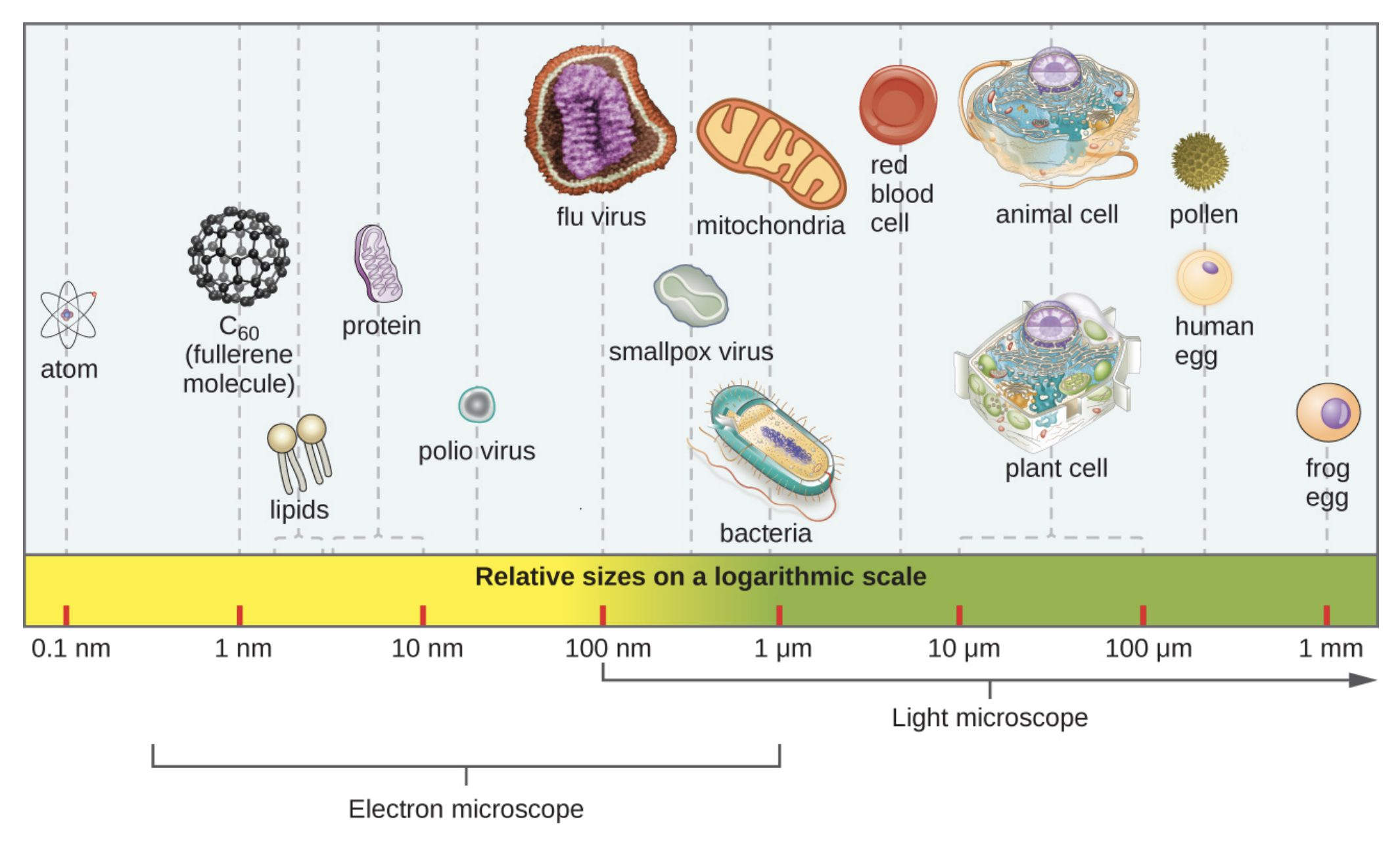
Microorganisms range in size considerably. Viruses are typically around 100 nanometers (nm) in length, although they can be larger or smaller. Bacteria are generally around 1 micrometer (μm) in length and 1 μm = 1 × 10³ nm. In general, plant and animal cells are approximately 10–100 μm in length. A microscope is required to view anything smaller than about 100 μm.
Although these are average sizes, there is considerable variation. Prokaryotic cells are generally smaller than eukaryotic cells, but cells of the bacterium Thiomargarita namibiensis have been documented to reach 750 μm in diameter. In 2022, a new bacterium was discovered that is so large it can be seen without a microscope. The average length of cells of this new species, Thiomargarita magnifica, are over 9,000 μm. Eggs, even those produced by ostriches, are single eukaryotic cells.
In the image below, notice the relative sizes of various microscopic and nonmicroscopic objects. Note that a typical virus measures about 100 nm, 10 times smaller than a typical bacterium (~1 µm), which is at least 10 times smaller than a typical plant or animal cell (~10–100 µm). An object must measure about 100 µm to be visible without a microscope.
Prokaryotic microorganisms are single-celled organisms that lack a true membrane-bound nucleus, in addition to many other differences from eukaryotic cells. You will learn more about the characteristics of prokaryotic and eukaryotic cells in other lessons.
The two major groups of prokaryotes are classified within the domains Bacteria and Archaea. Both of these groups are highly diverse and found in widely different habitats, including on and within humans.
The domain Bacteria includes prokaryotes that usually have a cell wall that contains peptidoglycan, which is a substance that provides rigidity to help protect the cell and maintain cell shape. Like all living cells, bacteria have a cell membrane. They also have a single circular chromosome of DNA.
One of the most obvious characteristics of bacteria viewed under a microscope is their shape, which can be useful in classification. The most common shapes are spherical (coccus), rod-shaped (bacillus), curved rods (vibrio), spherical rods (coccobacillus), rigid spirals (spirillum), and flexible spirals (spirochete).
The shape is often part of the name of the bacterium.
EXAMPLE
Bacillus subtilis is a well-studied, common rod-shaped bacterium that is common in soil. The medically important bacterium Staphylococcus aureus has clusters of spherical cells. S. aureus is common on the skin, but it can cause infections and has become a common cause of antibiotic-resistant infections.The images below show the common bacterial shapes. Note how coccobacillus is a combination of spherical (coccus) and rod-shaped (bacillus).

Bacteria have diverse lifestyles. Some, such as cyanobacteria, are photosynthetic and use energy from the sun to build organic molecules such as sugars from inorganic molecules such as carbon dioxide. Others, such as bacteria that live in hot water vents at the bottom of the ocean, are chemosynthetic and can use chemical reactions to do the same thing. Many bacteria cannot do this and must consume preformed organic molecules just as animals do. Most pathogens, or bacteria that cause disease, fall into this category and have to consume organic molecules.
EXAMPLE
The pathogens that cause tuberculosis (Mycobacterium tuberculosis) and strep throat (Streptococcus pyogenes) must consume organic molecules. You will learn more about these organisms and their lifestyles in other lessons.Although archaea may seem similar to bacteria at first glance, they are very different. These groups differ in their genetics, metabolic pathways, cell membrane composition, and cell wall composition. In some cases, archaea are more similar to eukaryotes than to prokaryotes.
One important difference between bacteria and archaea is that archaea do not have peptidoglycan cell walls. Some archaea have cell walls with pseudomurein, which is similar in function to peptidoglycan but has a different chemical structure. Some have other chemicals. S-layers are protein arrays found on the surface of most archaea and some bacteria. You will learn more about cell walls in other lessons.
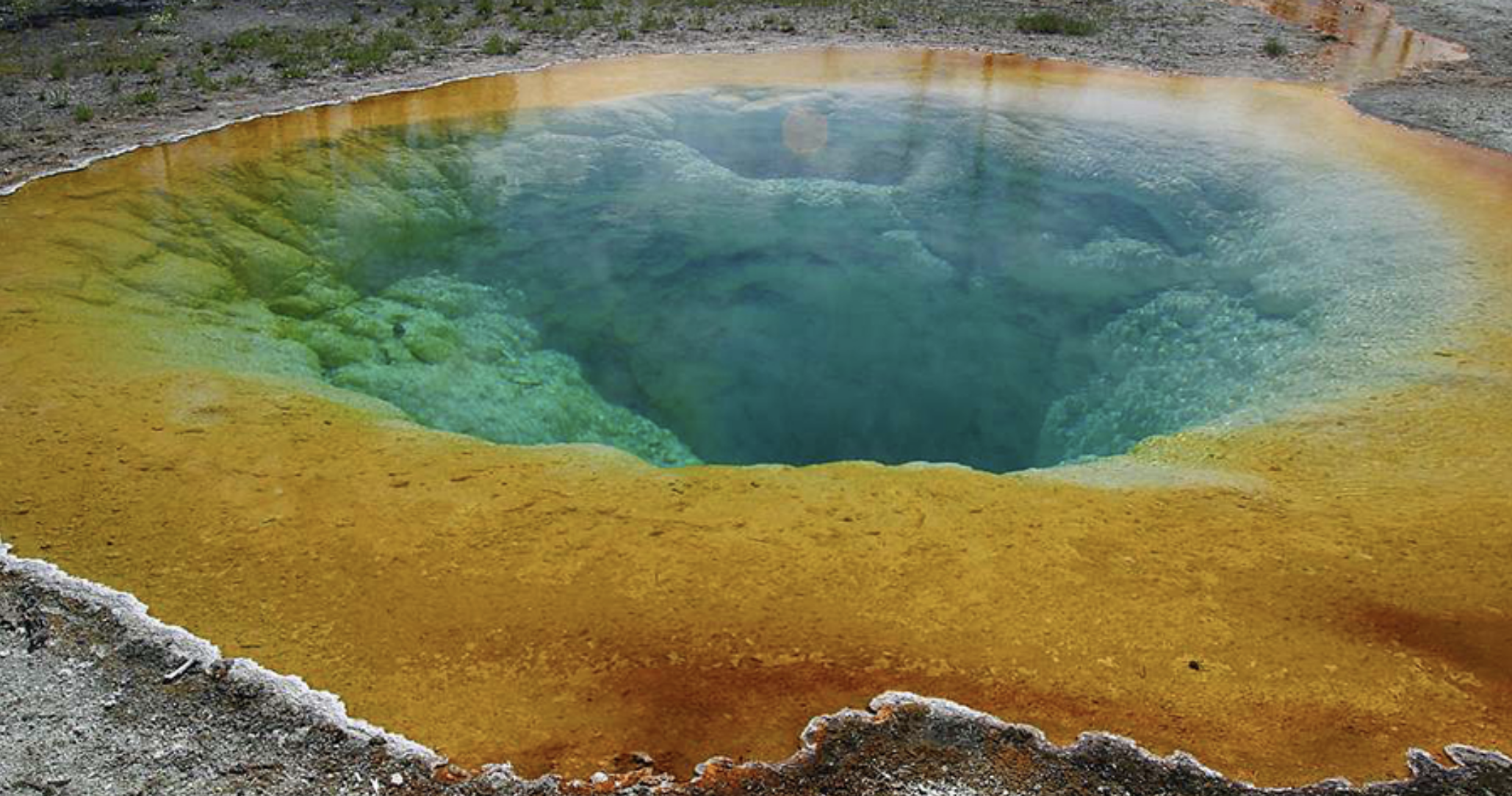
Archaea are found in many different places, including the human body and some environments that are very extreme in temperature or salt content, such as Halobacterium salinarum. Archaea are found in frozen ground in the Arctic, in ocean water, in very hot environments such as areas of geothermal activity like Yellowstone National Park, and in many other places.

Some archaea live in extreme environments, such as in the image below of Morning Glory Pool, a hot spring in Yellowstone National Park. The color differences in the pool result from the different communities of microbes that are able to thrive at various water temperatures.
Eukaryotic microorganisms all have cells that contain true membrane-bound nuclei. They include protists (such as algae) as well as fungi and helminths (which are worms). All of these groups include microscopic organisms (e.g., the larvae of helminths are microscopic) as well as some large organisms that are visible to the naked eye.
Although protists used to be grouped in a single kingdom, that is no longer the case as they are a very diverse group. Many are single celled, but some are multicellular.
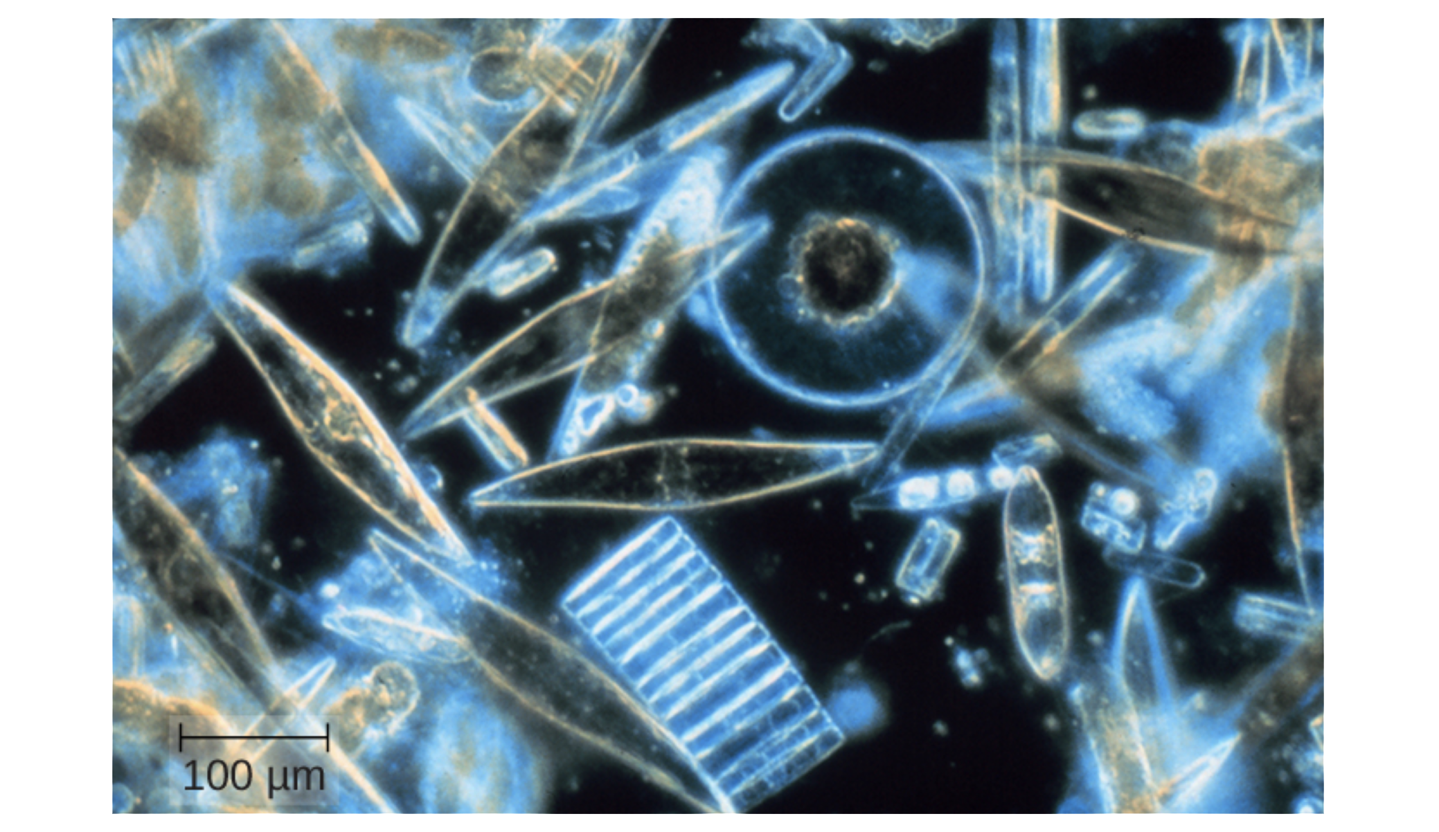
Photosynthetic protists are called algae (singular: alga). They are important at the base of food chains. Many organisms eat microscopic algae. Larger algae are often called seaweed and are another important food source. However, some species of algae can cause significant problems when something like fertilizer runoff causes their overgrowth. These organisms can become so abundant that they lead to large amounts of decomposition as they die, resulting in the depletion of the oxygen used by the organisms that consume them. Some algae produce toxins that harm or kill other organisms and become very problematic when they are overabundant.
The image below shows assorted diatoms, a kind of algae, that live in annual sea ice in McMurdo Sound, Antarctica. Diatoms range in size from 2 to 200 μm and are visualized here using light microscopy.
Protozoa are a diverse group of protists, many of which are nonphotosynthetic. Many protozoa can move using whip-like flagella, hair-like cilia, or protrusions of their cell membranes called pseudopodia.

Many protists are harmless, but some cause diseases.
EXAMPLE
Some diseases caused by protists include malaria, toxoplasmosis, and giardiasis (caused by G. lamblia). Additionally, Entamoeba histolytica causes a gastrointestinal illness, and Naegleria fowleri is the infamous “brain-eating” amoeba that causes an infection that is almost always fatal. You will learn more about these organisms and diseases in other lessons.Fungi (singular: fungus) are eukaryotes that can be single celled or multicellular and have cell walls containing chitin. Fungi are not photosynthetic but instead secrete digestive enzymes that are used to break down nutrients for absorption.
Single-celled fungi are called yeasts. Molds are an example of multicellular fungi. Mushrooms are fruiting bodies produced by some fungi.
Fungi can be beneficial in multiple ways. They are very important in nutrient cycling because of their role in decomposition.
EXAMPLE
Mushrooms and other fungi may be found growing on dead wood as they break it down. They can be useful in producing food as well. For example, yeast is used to produce bread. Some pharmaceuticals are produced by fungi, including penicillin.However, some types of fungi are harmful. Ringworm, athlete’s foot, and yeast infections are all caused by fungi. Mold often grows on bread or cheese. Fungi can sometimes cause serious systemic infections as well.
EXAMPLE
Inhaled fungal spores can cause a potentially dangerous lung infection called histoplasmosis. Chytrids have caused large-scale declines in the amphibian population around the world. Additionally, some fungi produce dangerous toxins. Poisonous mushrooms are one example. Aspergillus fungi produce two toxins, and infection is associated with a variety of symptoms and with the development of liver cancer.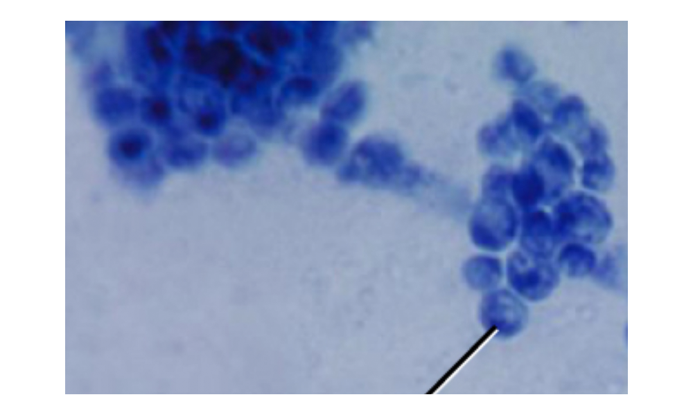
The image above shows Candida albicans, which is a unicellular fungus, or yeast. It causes vaginal yeast infections as well as oral thrush, a yeast infection of the mouth that commonly affects infants. C. albicans has a morphology similar to that of coccus bacteria; however, yeast is a eukaryotic organism (note the nuclei) and is much larger.

Helminths are multicellular parasitic worms. They are often included in the study of microbiology because they produce microscopic eggs and larvae.
Some helminths cause serious illnesses. People can become infected with guinea worms (Dracunculus medinensis) through contaminated water. Hookworms, which are picked up from the soil, cause gastrointestinal infections. Some flukes also cause human infections. Tapeworms are also helminths and infect humans when they are consumed in undercooked meat.
The roundworm Dirofilaria immitis causes heartworm disease in cats and dogs, among other animals. In dogs, this parasite enters the circulatory system and reproduces in the pulmonary arteries that carry blood from the heart to the lungs. Because the illness can be severe or even fatal, affecting blood flow and the function of the heart and lungs, many dogs are given treatment to prevent infection in areas where the parasite is common. If dogs become infected, treatment can be very difficult. Cats are not a normal host for these roundworms, which usually do not mature in cats. However, cats can experience respiratory illness or even die. The infection will often get better on its own, but treatment is very difficult as the medications that are effective in dogs are dangerous to cats. For this reason, preventive medication is often recommended for cats that are at risk of infection.
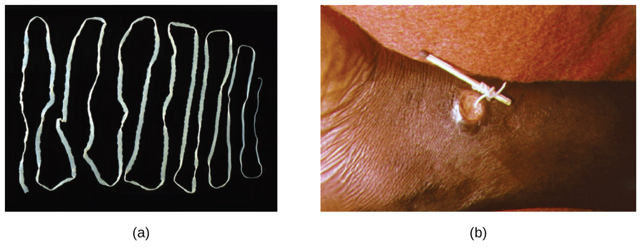
The image below shows (a) the beef tapeworm, Taenia saginata. It infects both cattle and humans. T. saginata eggs are microscopic (around 50 µm), but adult worms like the one shown here can reach 4–10 m, taking up residence in the digestive system. The image also shows an adult guinea worm (b), Dracunculus medinensis, being removed through a lesion in the patient’s skin by winding it around a matchstick.
Acellular agents are not technically alive because they are not made of cells, but they can replicate by infecting a host cell. In this way, they can potentially cause disease.
Viruses are the most well-known example of an acellular agent. They have genetic material (DNA or RNA) that can be single stranded or double stranded. In contrast, cells must have DNA and RNA. Viruses also have a structure made of protein (a capsid) that surrounds their genetic material.

Source: THIS TUTORIAL HAS BEEN ADAPTED FROM OPENSTAX "MICROBIOLOGY." ACCESS FOR FREE AT openstax.org/details/books/microbiology. LICENSE: CC ATTRIBUTION 4.0 INTERNATIONAL. Accessed by August 2022.
REFERENCES
Centers for Disease Control and Prevention. (2020). Mycoplasma pneumoniae infections. Retrieved July 24, 2022 from www.cdc.gov/pneumonia/atypical/mycoplasma/index.html
Centers for Disease Control and Prevention. (2020). Mycoplasma pneumoniae: Antibiotics and Resistance. Retrieved July 24, 2022 from www.cdc.gov/pneumonia/atypical/mycoplasma/hcp/antibiotic-treatment-resistance.html.
File: Halobacteria.jpg. (2022, June 28). Wikimedia Commons, the free media repository. Retrieved 16:55, July 27, 2022 from commons.wikimedia.org/w/index.php?title=File:Halobacteria.jpg&oldid=669376076.
Garrity, S., Lee-Fowler, T., & Reinero, C. (2019). Feline asthma and heartworm disease: Clinical features, diagnostics and therapeutics. Journal of feline medicine and surgery, 21(9), 825–834. doi.org/10.1177/1098612X18823348.
Klingl, A., Pickl, C., & Flechsler, J. (2019). Archaeal Cell Walls. Sub-cellular Biochemistry, 92, 471–493. doi.org/10.1007/978-3-030-18768-2_14.
Litster, A. L., & Atwell, R. B. (2008). Feline heartworm disease: a clinical review. Journal of feline medicine and surgery, 10(2), 137–144. doi.org/10.1016/j.jfms.2007.09.007.
Moissl-Eichinger, C., Probst, A.J., Birarda, G. et al. (2017). Human age and skin physiology shape diversity and abundance of Archaea on skin. Sci Rep, 7, 4039. doi.org/10.1038/s41598-017-04197-4.
Noack, S., Harrington, J., Carithers, D. S., Kaminsky, R., & Selzer, P. M. (2021). Heartworm disease - Overview, intervention, and industry perspective. International journal for parasitology. Drugs and drug resistance, 16, 65–89. doi.org/10.1016/j.ijpddr.2021.03.004
Schulz, H. N., & Jorgensen, B. B. (2001). Big bacteria. Annual review of microbiology, 55, 105–137. doi.org/10.1146/annurev.micro.55.1.105.
Volland, J. M., Gonzalez-Rizzo, S., Gros, O., Tyml, T., Ivanova, N., Schulz, F., Goudeau, D., Elisabeth, N. H., Nath, N., Udwary, D., Malmstrom, R. R., Guidi-Rontani, C., Bolte-Kluge, S., Davies, K. M., Jean, M. R., Mansot, J. L., Mouncey, N. J., Angert, E. R., Woyke, T., & Date, S. V. (2022). A centimeter-long bacterium with DNA contained in metabolically active, membrane-bound organelles. Science (New York, N.Y.), 376(6600), 1453–1458. doi.org/10.1126/science.abb3634