Table of Contents |
The term tissue is used to describe a group of cells found together in the body. The cells within a tissue share a common embryonic origin. Microscopic observation reveals that the cells in a tissue share morphological (form and organization) features and are arranged in an orderly pattern that achieves the tissue’s functions. From an evolutionary perspective, tissues appear in more complex organisms. For example, multicellular protists, which are ancient eukaryotes, do not have cells organized into tissues.
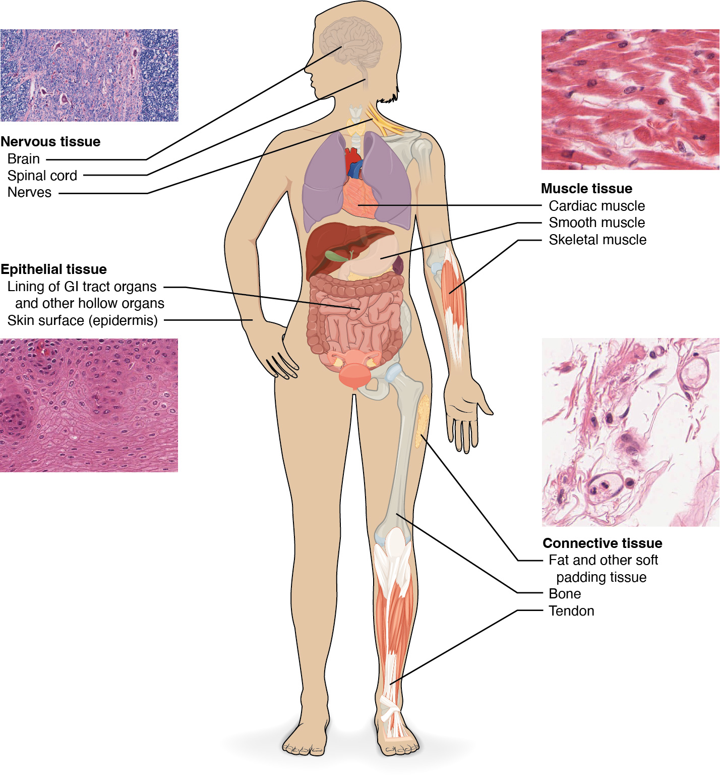
Although there are many types of cells in the human body, they are organized into four broad categories of tissues: epithelial, connective, muscle, and nervous. Each of these categories is characterized by specific functions that contribute to the overall health and maintenance of the body. A disruption of the structure is a sign of injury or disease. Such changes can be detected through histology, the microscopic study of tissue appearance, organization, and function.
Epithelial tissue, also referred to as epithelium, refers to the sheets of cells that cover exterior surfaces of the body, line internal cavities and passageways, and form certain glands. Connective tissue, as its name implies, binds the cells and organs of the body together and functions in the protection, support, and integration of all parts of the body. Muscle tissue is excitable, responding to stimulation and contracting to provide movement, and occurs as three major types: skeletal (voluntary) muscle, smooth muscle, and cardiac muscle in the heart. Nervous tissue is also excitable, allowing the propagation of electrochemical signals in the form of nerve impulses that communicate between different regions of the body.
The next level of organization is the organ, where several types of tissues come together to form a working unit. Just as knowing the structure and function of cells helps you in your study of tissues, knowledge of tissues will help you understand how organs function.
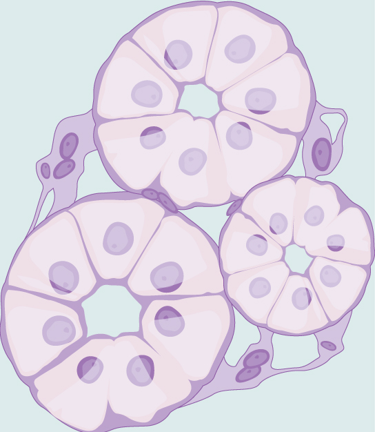
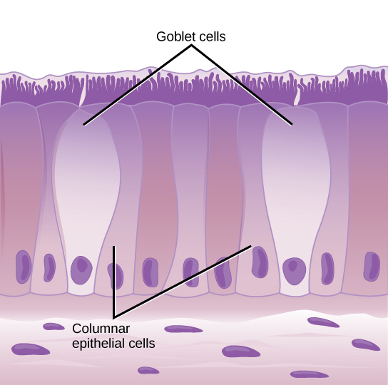
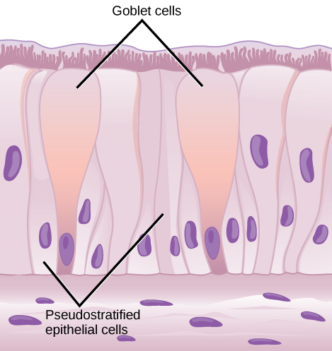
Epithelial tissues cover the outside of organs and structures in the body and line the lumens of organs in a single layer or multiple layers of cells. The types of epithelia are classified by the shapes of cells present and the number of layers of cells. Epithelia composed of a single layer of cells is called simple epithelia; epithelial tissue composed of multiple layers is called stratified epithelia. The table below summarizes the different types of epithelial tissues, and some examples are provided.
| Cell Shape | Description | Location |
|---|---|---|
| squamous | flat, irregular round shape | simple: lung alveoli, capillaries; stratified: skin, mouth, vagina |
| cuboidal | cube shaped, central nucleus | glands, renal tubules |
| columnar | tall, narrow, nucleus toward base; tall, narrow, nucleus along cell | simple: digestive tract; pseudostratified: respiratory tract |
| transitional | round, simple but appear stratified | urinary bladder |
All epithelia share some important structural and functional features. This tissue is highly cellular, with little or no extracellular material present between cells. Adjoining cells form a specialized intercellular connection between their cell membranes called a cell junction. The epithelial cells exhibit polarity with differences in structure and function between the exposed or apical-facing surface of the cell and the basal surface close to the underlying body structures. The basal lamina, a mixture of glycoproteins and collagen, provides an attachment site for the epithelium, separating it from underlying connective tissue. The basal lamina attaches to a reticular lamina, which is secreted by the underlying connective tissue, forming a basement membrane that helps hold it all together.

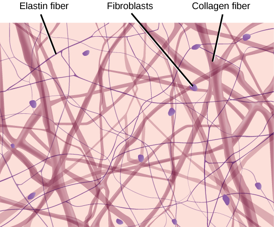
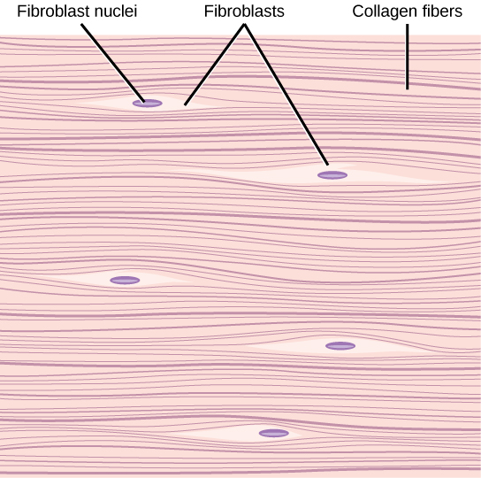
Connective tissues are made up of a matrix consisting of living cells and a nonliving substance called the ground substance. The ground substance is made of an organic substance (usually a protein) and an inorganic substance (usually a mineral or water). The principal cell of connective tissues is the fibroblast. This cell makes the fibers found in nearly all of the connective tissues. Fibroblasts are motile, able to carry out mitosis, and can synthesize whichever connective tissue is needed. Macrophages, lymphocytes, and, occasionally, leukocytes can be found in some of the tissues. Some tissues have specialized cells that are not found in the others. The matrix in connective tissues gives the tissue its density. When a connective tissue has a high concentration of cells or fibers, it has proportionally a less dense matrix.
The organic portion or protein fibers found in connective tissues are either collagen, elastic, or reticular fibers. Collagen fibers provide strength to the tissue, preventing it from being torn or separated from the surrounding tissues. Elastic fibers are made of the protein elastin; this fiber can stretch to one and one half of its length and return to its original size and shape. Elastic fibers provide flexibility to the tissues. Reticular fibers are the third type of protein fiber found in connective tissues. This fiber consists of thin strands of collagen that form a network of fibers to support the tissue and other organs to which it is connected.
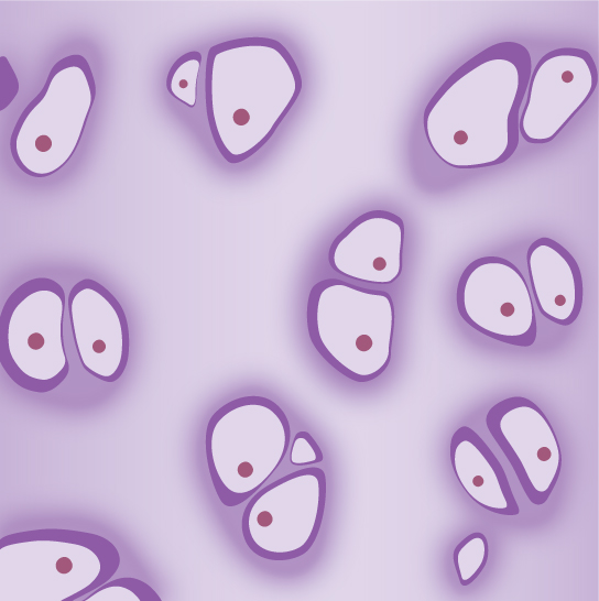
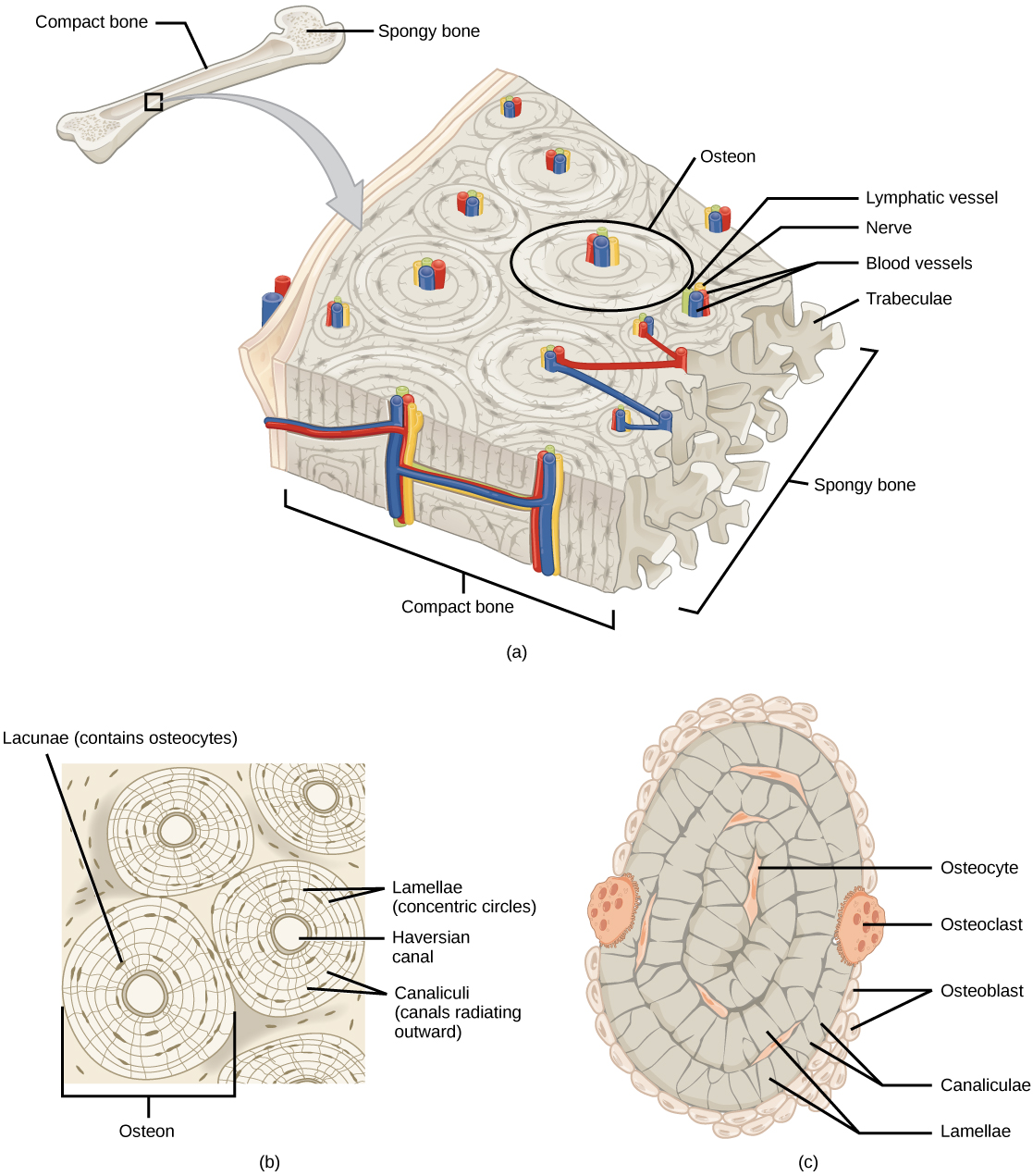
The various types of connective tissues, the types of cells and fibers they are made of, and sample locations of the tissues are summarized in the table below, and some examples are provided.
| Tissue | Cells | Fibers | Location |
|---|---|---|---|
| loose/areolar | fibroblasts, macrophages, some lymphocytes, some neutrophils | few: collagen, elastic, reticular | around blood vessels; anchors epithelia |
| dense, fibrous connective tissue | fibroblasts, macrophages | mostly collagen | irregular: skin; regular: tendons, ligaments |
| cartilage | chondrocytes, chondroblasts | hyaline: few: collagen fibrocartilage: large amount of collagen | shark skeleton, fetal bones, human ears, intervertebral discs |
| bone | osteoblasts, osteocytes, osteoclasts | some: collagen, elastic | vertebrate skeletons |
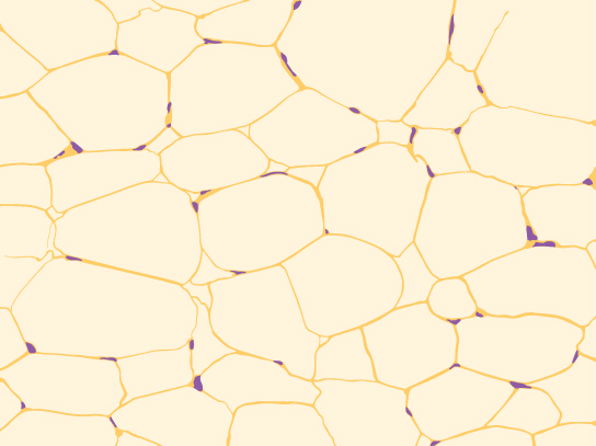
| adipose | adipocytes | few | adipose (fat) |
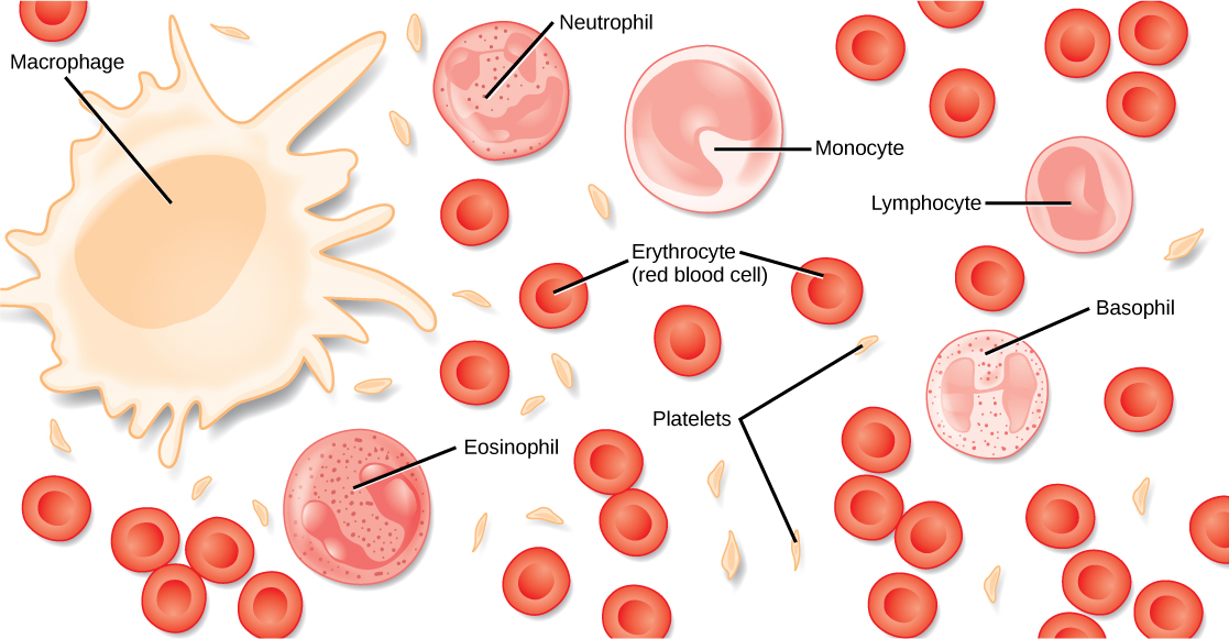
| blood | red blood cells, white blood cells | none | blood |
There are three types of muscle in animal bodies: smooth, skeletal, and cardiac. They differ by the presence or absence of striations (or bands), the number and location of nuclei, whether they are voluntarily or involuntarily controlled, and their location within the body. The table and figure below summarize these differences.
| Type of Muscle | Striations | Nuclei | Control | Location |
|---|---|---|---|---|
| smooth | no | single, in center | involuntary | visceral organs |
| skeletal | yes | many, at periphery | voluntary | skeletal muscles |
| cardiac | yes | usually single, in center (however, some develop two nuclei) | involuntary | heart |

Nervous tissues are made of cells specialized to receive and transmit electrical impulses from specific areas of the body and to send them to specific locations in the body. The main cell of the nervous system is the neuron, illustrated below.
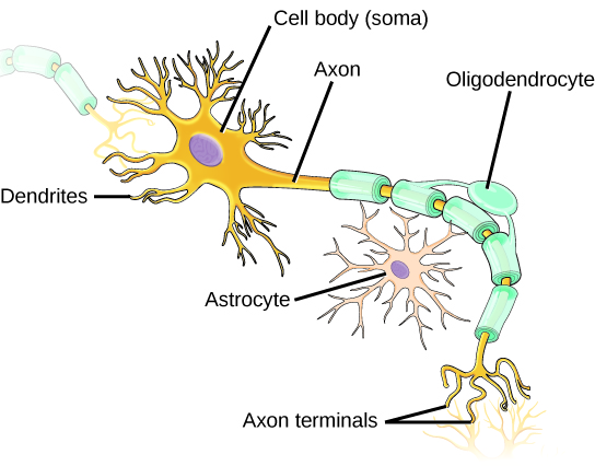
The large structure with a central nucleus is the cell body of the neuron. Projections from the cell body are either dendrites specialized in receiving input or a single axon specialized in transmitting impulses. Some glial cells are also shown. Astrocytes regulate the chemical environment of the nerve cell, and oligodendrocytes insulate the axon so the electrical nerve impulse is transferred more efficiently. Other glial cells that are not shown support the nutritional and waste requirements of the neuron. Some of the glial cells are phagocytic and remove debris or damaged cells from the tissue. A nerve consists of neurons and glial cells.
The zygote, or fertilized egg, is a single cell formed by the fusion of an egg and sperm. After fertilization, the zygote gives rise to rapid mitotic cycles, generating many cells to form the embryo. The first embryonic cells generated have the ability to differentiate into any type of cell in the body and, as such, are called totipotent, meaning each has the capacity to divide, differentiate, and develop into a new organism.
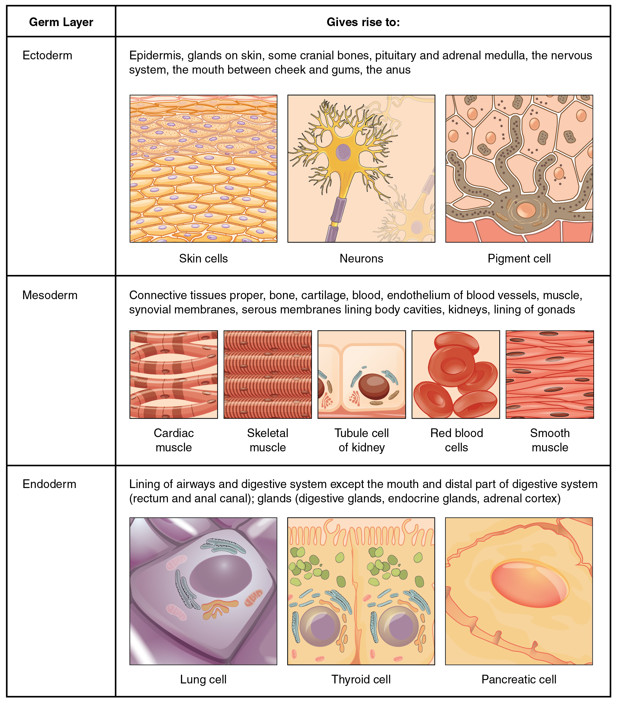
As cell proliferation progresses, three major cell lineages are established within the embryo. Each of these lineages of embryonic cells forms the distinct germ layers from which all the tissues and organs of the human body eventually form. Each germ layer is identified by its relative position: ectoderm (ecto- = “outer”), mesoderm (meso- = “middle”), and endoderm (endo- = “inner”).
The figure below shows the types of tissues and organs associated with each of the three germ layers. Note that epithelial tissue originates in all three layers, whereas nervous tissue derives primarily from the ectoderm and muscle tissue from mesoderm.
A tissue membrane is a thin layer or sheet of cells that covers the outside of the body (for example, skin), the organs (for example, pericardium), internal passageways that lead to the exterior of the body (for example, mucosa of stomach), and the lining of the moveable joint cavities. There are two basic types of tissue membranes: connective tissue and epithelial membranes.
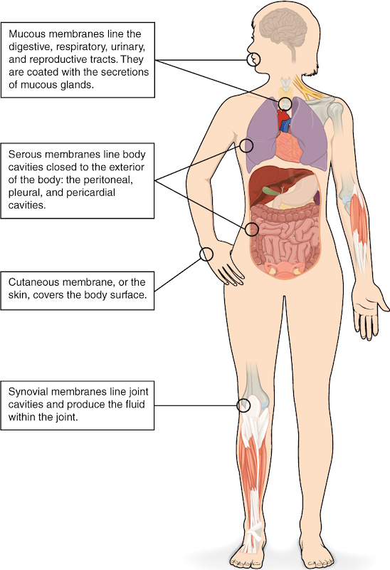
The connective tissue membrane is formed solely from connective tissue. These membranes encapsulate organs, such as the kidneys, and line our movable joints. A synovial membrane is a type of connective tissue membrane that lines the cavity of a freely movable joint. For example, synovial membranes surround the joints of the shoulder, elbow, and knee. Fibroblasts in the inner layer of the synovial membrane release a substance called hyaluronan into the joint cavity. The hyaluronan effectively traps available water to form the synovial fluid, a natural lubricant that enables the bones of a joint to move freely against one another without much friction. This synovial fluid readily exchanges water and nutrients with blood, as do all body fluids.
The epithelial membrane is composed of epithelium attached to a layer of connective tissue, for example, your skin. The mucous membrane is also a composite of connective and epithelial tissues. Sometimes called mucosae, these epithelial membranes line the body cavities and hollow passageways that open to the external environment, and include the digestive, respiratory, excretory, and reproductive tracts. Mucus, produced by the epithelial exocrine glands, covers the epithelial layer. The underlying connective tissue, called the lamina propria (literally “own layer”), helps support the fragile epithelial layer.
A serous membrane is an epithelial membrane composed of mesodermally derived epithelium called the mesothelium that is supported by connective tissue. These membranes line the coelomic cavities of the body (that is, those cavities that do not open to the outside), and they cover the organs located within those cavities. They are essentially membranous bags, with mesothelium lining the inside and connective tissue on the outside. Serous fluid secreted by the cells of the thin squamous mesothelium lubricates the membrane and reduces abrasion and friction between organs. Serous membranes are identified according to location. Three serous membranes line the thoracic cavity: the two pleura that cover the lungs and the pericardium that covers the heart. A fourth, the peritoneum, is the serous membrane in the abdominal cavity that covers abdominal organs and forms double sheets of mesenteries that suspend many of the digestive organs.
The skin is an epithelial membrane also called the cutaneous membrane. It is a stratified squamous epithelial membrane resting on top of connective tissue. The apical surface of this membrane is exposed to the external environment and is covered with dead, keratinized cells that help protect the body from desiccation and pathogens.
SOURCE: THIS TUTORIAL HAS BEEN ADAPTED FROM (1) OPENSTAX “BIOLOGY 2E”. ACCESS FOR FREE AT OPENSTAX.ORG/BOOKS/BIOLOGY-2E/PAGES/1-INTRODUCTION (2) OPENSTAX “ANATOMY AND PHYSIOLOGY 2E”. ACCESS FOR FREE AT OPENSTAX.ORG/BOOKS/ANATOMY-AND-PHYSIOLOGY-2E/PAGES/1-INTRODUCTION. LICENSING (1 & 2): CREATIVE COMMONS ATTRIBUTION 4.0 INTERNATIONAL.