Table of Contents |
The muscular system allows for movement, production of body heat, and the flow of blood and other substances.
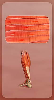
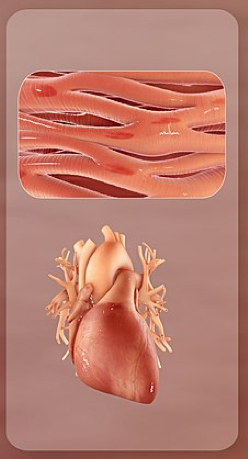
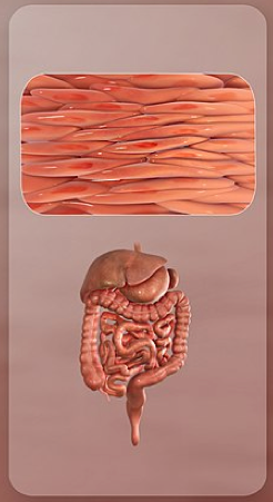
There are three main types of muscle found in the body:
As you previously learned, muscle is one of the four primary tissue types of the body. All three types of muscle tissues have some properties in common: they all exhibit a quality called excitability because their plasma membranes can change their electrical states (from polarized to depolarized) and send an electrical wave called an action potential along the entire length of the membrane. While the nervous system can influence the excitability of cardiac and smooth muscle to some degree, skeletal muscle completely depends on signaling from the nervous system to work properly. On the other hand, both cardiac muscle and smooth muscle can respond to other stimuli, such as hormones or local stimuli (such as cold making your hair stand on end).

The muscles all begin the actual process of movement by contracting (shortening) when a protein called actin is pulled by a protein called myosin. In skeletal and cardiac muscle, this occurs after specific binding sites on the actin have been exposed in response to the interaction between calcium ions (Ca⁺⁺) and proteins (troponin and tropomyosin) that “shield” the actin-binding sites. Ca⁺⁺ is also required for the contraction of smooth muscle, although its role is different: Here, Ca⁺⁺ activates enzymes, which in turn activate myosin heads. All muscles require adenosine triphosphate (ATP) to continue the process of contracting, and they all relax when the Ca⁺⁺ is removed and the actin-binding sites are re-shielded.
A muscle can return to its original length when relaxed due to a quality of muscle tissue called elasticity. It can recoil back to its original length due to elastic fibers. Energy stored in objects when they are under temporary strain or deformation, such as muscles being stretched, is called elastic energy. Muscle tissue also has the quality of extensibility; it can stretch or extend. Contractility allows muscle tissue to pull on its attachment points and shorten with force.
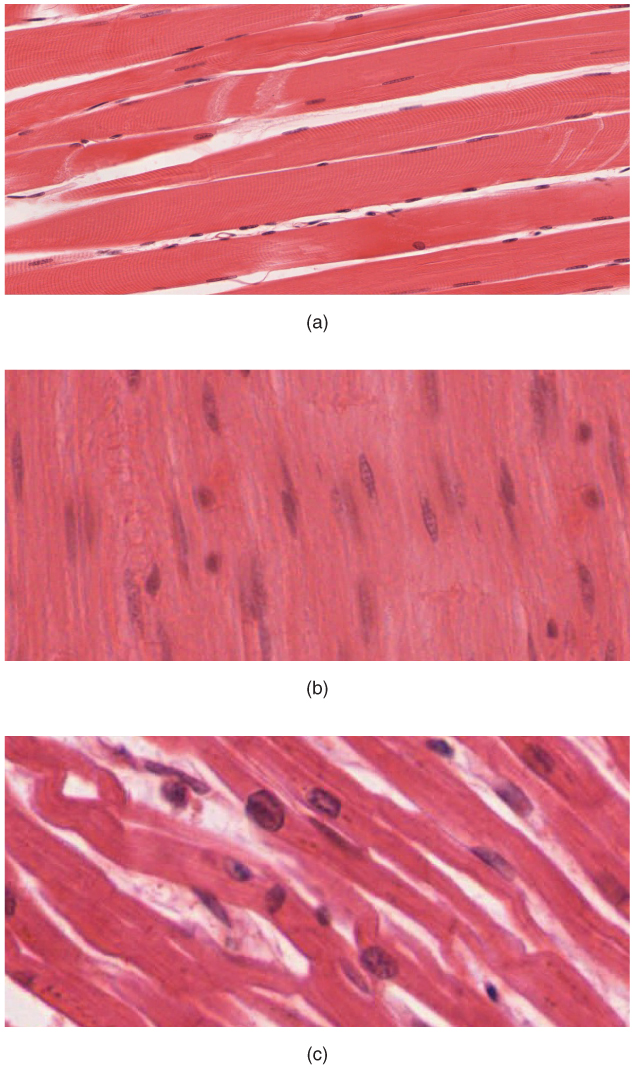
Differences among the three muscle types include the microscopic organization of their contractile proteins—actin and myosin. The actin and myosin proteins are arranged very regularly in the cytoplasm of individual muscle cells (referred to as fibers) in both skeletal muscle and cardiac muscle, which creates a pattern, or stripes, called striations. The striations are visible with a light microscope under high magnification (see the figure above).
Skeletal muscle fibers are multinucleated structures that compose the skeletal muscle, which is attached to bones or skin.

Cardiac muscle fibers each have one to two nuclei and are physically and electrically connected to each other so that the entire heart contracts as one unit (called a syncytium).

Because the actin and myosin are not arranged in such regular fashion in smooth muscle, the cytoplasm of a smooth muscle fiber (which has only a single nucleus) has a uniform, nonstriated appearance (resulting in the name smooth muscle). However, the less organized appearance of smooth muscle should not be interpreted as less efficient. Smooth muscle in the walls of arteries is a critical component that regulates blood pressure necessary to push blood through the circulatory system, and smooth muscle in the skin, visceral organs, and internal passageways is essential for moving all materials through the body.

Most muscle tissue of the body arises from embryonic mesoderm, which you learned about in a prior lesson. Paraxial (= next to the neural tube) mesodermal cells adjacent to the neural tube form blocks of cells called somites. Skeletal muscles excluding those of the head and limbs develop from mesodermal somites; skeletal muscle in the head and limbs develop from general mesoderm.
Somites give rise to myoblasts. A myoblast is a muscle-forming stem cell that migrates to different regions in the body and then fuses to form a myotube. As a myotube is formed from many different myoblast cells, it contains many nuclei but has a continuous cytoplasm.
Although the number of muscle cells is set during development, satellite cells help to repair skeletal muscle cells. A satellite cell is similar to a myoblast because it is a type of stem cell (an unspecialized cell that can divide without limit as needed); however, satellite cells are incorporated into muscle cells and facilitate the protein synthesis required for repair and growth. Satellite cells can regenerate muscle fibers to a very limited extent, but they primarily help to repair damage in living cells.
If a cell is damaged to a greater extent than can be repaired by satellite cells, the muscle fibers are replaced by scar tissue in a process called fibrosis. Because scar tissue cannot contract, muscle that has sustained significant damage loses strength and cannot produce the same amount of power or endurance as it could before being damaged.
Smooth muscle tissue can regenerate from a type of stem cell called a pericyte, which is found in some small blood vessels. Pericytes allow smooth muscle cells to regenerate and repair much more readily than skeletal and cardiac muscle tissue.
Similar to skeletal muscle tissue, cardiac muscle does not regenerate to a great extent. Dead cardiac muscle tissue is replaced by scar tissue, which cannot contract. As scar tissue accumulates, the heart can lose its ability to pump because of the loss of contractile power.
IN CONTEXT
Career Connection—Physical Therapist
As muscle cells die, they are not regenerated but instead are replaced by connective tissue and adipose tissue, which do not possess the contractile abilities of muscle tissue. Muscles atrophy, or waste away, when they are not used, and over time, if atrophy is prolonged, muscle cells die. It is therefore important that those who are susceptible to muscle atrophy exercise to maintain muscle function and prevent the complete loss of muscle tissue. In extreme cases, when movement is not possible, electrical stimulation can be introduced to a muscle from an external source. This acts as a substitute for endogenous neural stimulation, stimulating the muscle to contract and preventing the loss of proteins that occurs with a lack of use.
Physical therapists, also called physiotherapists, work with patients to maintain muscles. They are trained to target muscles susceptible to atrophy, and to prescribe and monitor exercises designed to stimulate those muscles. There are various causes of atrophy, including mechanical injury, disease, and age. After breaking a limb or undergoing surgery, muscle use is impaired and can lead to disuse atrophy. If the muscles are not exercised, this atrophy can lead to long-term muscle weakness.
A stroke, which occurs when brain tissue is damaged from lack of blood flow, can also cause muscle impairment by interrupting neural stimulation to certain muscles. Without neural inputs, these muscles do not contract and thus begin to lose structural proteins. Exercising these muscles can help to restore muscle function and minimize functional impairments. Age-related muscle loss is also a target of physical therapy, as exercise can reduce the effects of age-related atrophy and improve muscle function.
The goal of a physiotherapist is to improve physical functioning and reduce functional impairments; this is achieved by understanding the cause of muscle impairment and assessing the capabilities of a patient, after which a program to enhance these capabilities is designed. Some factors that are assessed include strength, balance, and endurance, which are continually monitored as exercises are introduced to track improvements in muscle function. Physiotherapists can also instruct patients on the proper use of equipment, such as crutches, and assess whether someone has sufficient strength to use the equipment and when they can function without it.
SOURCE: THIS TUTORIAL HAS BEEN ADAPTED FROM OPENSTAX “ANATOMY AND PHYSIOLOGY 2E”. ACCESS FOR FREE AT OPENSTAX.ORG/BOOKS/ANATOMY-AND-PHYSIOLOGY-2E/PAGES/1-INTRODUCTION. LICENSE: CREATIVE COMMONS ATTRIBUTION 4.0 INTERNATIONAL.