Table of Contents |
Imaging reports are critical to communicating the findings from imaging studies, such as CT scans and MRI scans. This allows specialists to share information when they consult on a patient. High quality, accurate communication helps specialists (such as radiologists who interpret imaging and physicians who use imaging to make clinical decisions) share information. It aids in interpreting diagnostic findings for proper treatment planning. It also reduces misinterpretation of imaging results in clinical practice.
Imaging reports typically contain certain information. This includes patient information, such as the patient’s name, age, and clinical history. The report identifies the type of study. For example, a report could specify that a patient underwent a CT chest with contrast, meaning that the patient had a CT of their chest with the addition of a contrast agent to make certain structures more visible. The findings present an objective description of the imaging results. The impression gives a summary and the radiologist’s interpretations of key findings. Finally, a comparison notes how the results compare with prior imaging studies if they are available. This can be helpful in documenting changes over time.
Common terms in radiology reports differ by the type of testing being done. Certain wording is common in imaging reports.
EXAMPLE
- “No acute findings” – No urgent abnormalities detected.
- “Findings consistent with…” – Imaging supports a possible diagnosis.
- “Cannot exclude…” – Imaging cannot confirm or rule out a condition.
- “Incidental finding” – An unexpected abnormality not related to the primary concern.
- “Follow-up recommended” – Additional imaging or testing is needed for further evaluation.
Imaging reports may also describe types of contrast and whether contrast was used. A contrast-enhanced study was performed with contrast dye to improve visualization. A non-contrast study was performed without a contrast agent.
Below, you will see examples of terms used for X-rays, CT scans, MRI images, and ultrasounds.
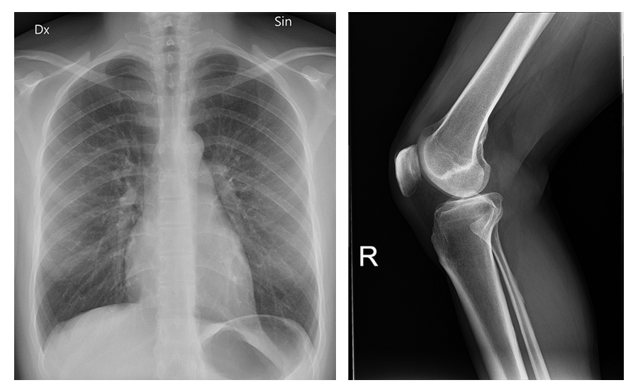
X-ray images are described based on how their opacity versus translucency to X-rays. Radiopaque materials such as bones and metallic implants block x-rays, while radiolucent materials such as air and gas allow X-rays to pass through.
The image below shows a chest X-ray on the left and a leg X-ray on the right. Note how the bones are more white (radiopaque) and the air in the lungs is dark (radiolucent).
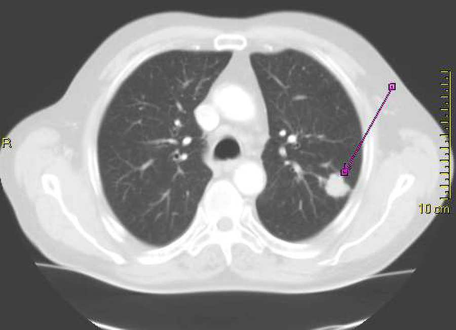
In CT reports, there is information on the density observed and on attenuation (meaning that radiation was less able to pass through, or attenuated). Hyperdense areas appear white on CT because they absorb more radiation. This is why areas of bone and/or hemorrhage appear white. Hypodense areas appear dark or black on CT because more radiation passes through. These include cysts, fluids, and infarcts (regions of tissue death). Isodense regions have a similar density to surrounding tissues.
CT scans can be performed with or without contrast. Contrast is administered orally or through a vein and makes certain structures brighter (e.g., tumors or regions of inflammation; Stanford Medicine, n.d.).
The image below is a chest CT that shows the lungs. Note that the image is a cross section, with the lungs to the left and right of the center. The bright white structure indicated by a purple line is a lung cancer. Note that most of the lungs are gray with some white spots and lines.
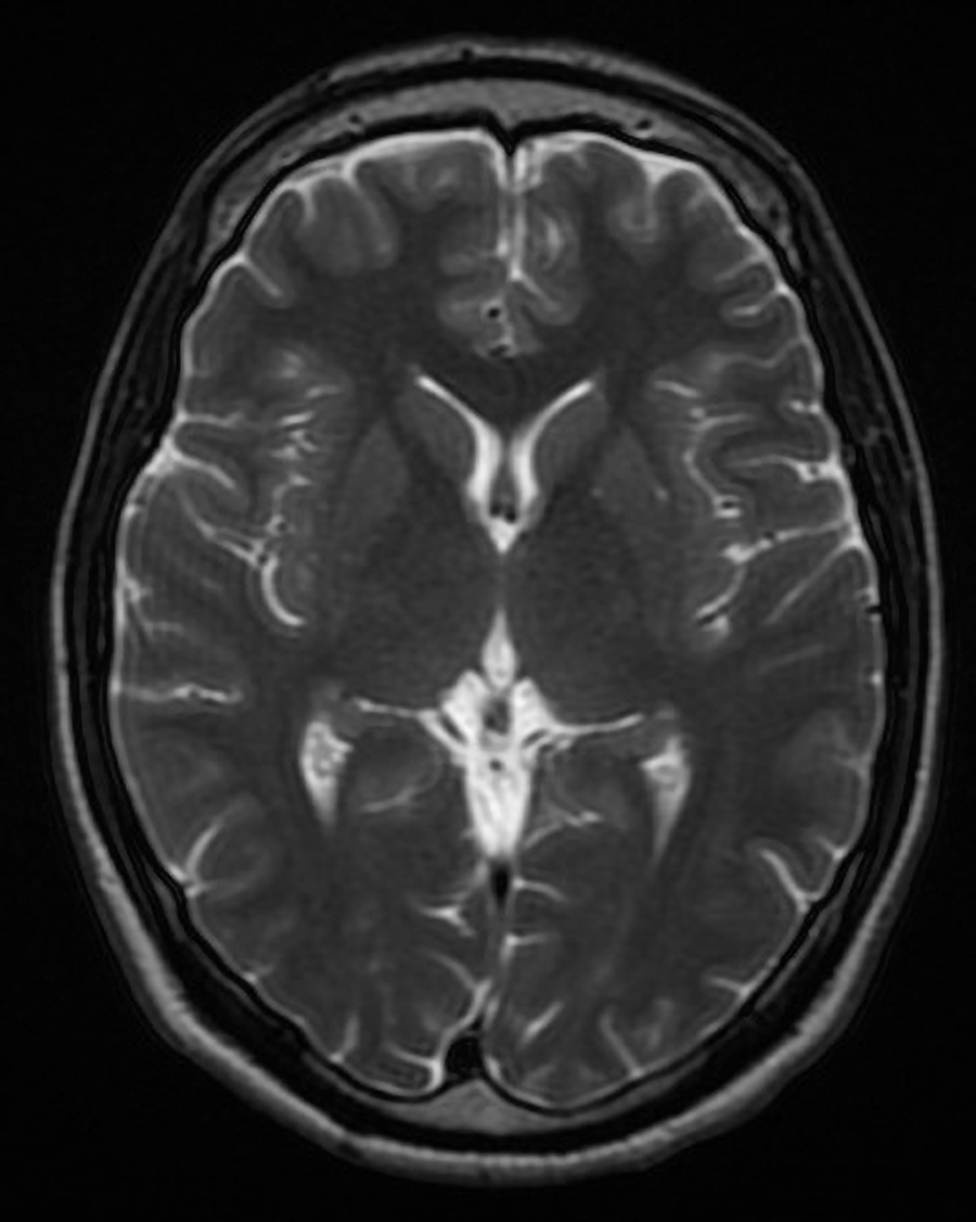
The images produced by MRI scans differ in signal intensity. Hyperintense regions are bright on MRI. Hypointense regions appear dark on MRI. As you learned previously, MRIs can produce T1-weighted images or T2-weighted images. T1 hyperintense regions can represent fat, hemorrhage, and similar structures. T2 hyperintense regions can represent edema, fluid, inflammation, and similar structures.
Contrast agents can be used for MRIs as well. Gadolinium-based contrast agents are typically used to highlight structures of interest. Although different agents act differently, these agents are commonly used to highlight structures such as tumors (Cluett, 2024).
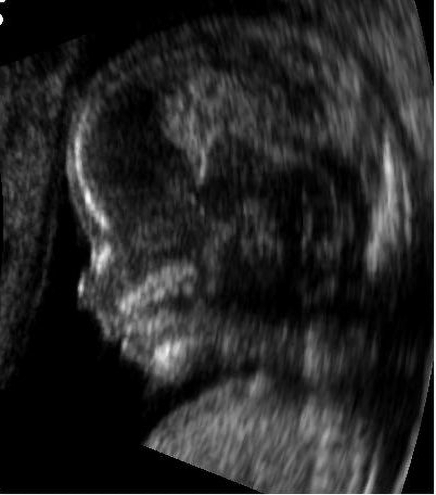
Echogenicity, meaning the way that structures respond to sound to produce an image, varies in ultrasounds. Hyperechoic (more echogenicity) means bright or white and represents structures such as bone, fat, and calcifications. Hypoechoic (less echogenicity) means darker than the surrounding tissues and represents structures such as tumors and cysts. Anechoic means completely black and represents structures such as fluid-filled cysts and blood vessels.
Acoustic shadowing occurs when there is a dark area behind a dense structure that is blocking sound waves.
The ultrasound below shows the head of a 14-week-old fetus.
Common pathological findings in imaging reports vary by body system. Examples by body system are summarized below.
Common imaging findings related to bone and the skeletal system include varied types of fractures and lesions. Lesions include osteolytic lesions (bone destruction seen in metastatic cancer) and osteoblastic lesions (bone-forming abnormalities, often associated with prostate cancer).
EXAMPLE
Cardiovascular findings may include aneurysms, aortic dissections, and coronary artery stenosis.An aneurysm is an abnormal dilation of a blood vessel. An aortic dissection is a tear in the inner layer of the aorta. Coronary artery stenosis is narrowing of the coronary arteries.
EXAMPLE
Pulmonary abnormalities may include consolidation (increased lung opacity due to infection), pleural effusion (fluid buildup in the pleural cavity), and pneumothorax (collapsed lung due to air in the pleural space).Abdominal and gastrointestinal abnormalities include hepatomegaly (an enlarged liver, which can occur when liver disease is present), cholelithiasis (gallstones, seen on ultrasound or CT), and bowel obstruction (a blockage in the intestines visible as dilated bowel loops seen on X-ray or CT; Cleveland Clinic, 2023). Dilated means that the bowel is wider than normal (constricted means narrower than normal; Merriam-Webster, n.d.a. and n.d.b.)
Neurological abnormalities include cerebral infarction (an ischemic stroke, seen as hypodense area on CT), hydrocephalus (fluid buildup in the brain’s ventricles), and a herniated disc (the protrusion of an intervertebral disc, which can cause nerve compression).
Findings associated with a brain tumor include a ring-enhancing lesion (a sign of brain abscess or metastases) and a midline shift (where the brain is displaced due to the effect of a newly forming mass).
Source: THIS TUTORIAL HAS BEEN ADAPTED FROM “CLINICAL NURSING SKILLS” BY Christy Bowen at OpenStax. ACCESS FOR FREE AT https://openstax.org/books/clinical-nursing-skills/pages/1-introduction. LICENSING: CREATIVE COMMONS ATTRIBUTION 4.0 INTERNATIONAL.
REFERENCES
Contrast Material for Computed Tomography Scan. (n.d.). Stanford Medicine Health Care. Contrast Material for Computed Tomography Scan | Stanford Health Care
Cluett, J. (2024, December 18). What is an MRI with Contrast? Verywell Health. MRI With Contrast: Uses, Procedure, and Side Effects
Bowel Obstruction. (2023, September 25). Cleveland Clinic. Bowel Obstruction: Signs & Symptoms, Causes, Treatment
Merriam-Webster. (n.d.a.). Dilated. In Merriam-Webster.com dictionary. Retrieved September 15, 2025, from www.merriam-webster.com/dictionary/dilated
Merriam-Webster. (n.d.b.). Constrict. In Merriam-Webster.com dictionary. Retrieved September 15, 2025, from www.merriam-webster.com/dictionary/constrict