Use Sophia to knock out your gen-ed requirements quickly and affordably. Learn more
×

Terminology for Burns and Wound Care

Author: Sophia

what's covered
In this lesson, you will learn about burns and wounds, both of which damage the skin and can increase infection risk. You will also learn about different types of burns and wounds, as well as some ways that they can be treated. Specifically, this lesson will cover:

Table of Contents

1. Introduction

In this lesson, you will learn about burns and wounds, including their treatment and care.

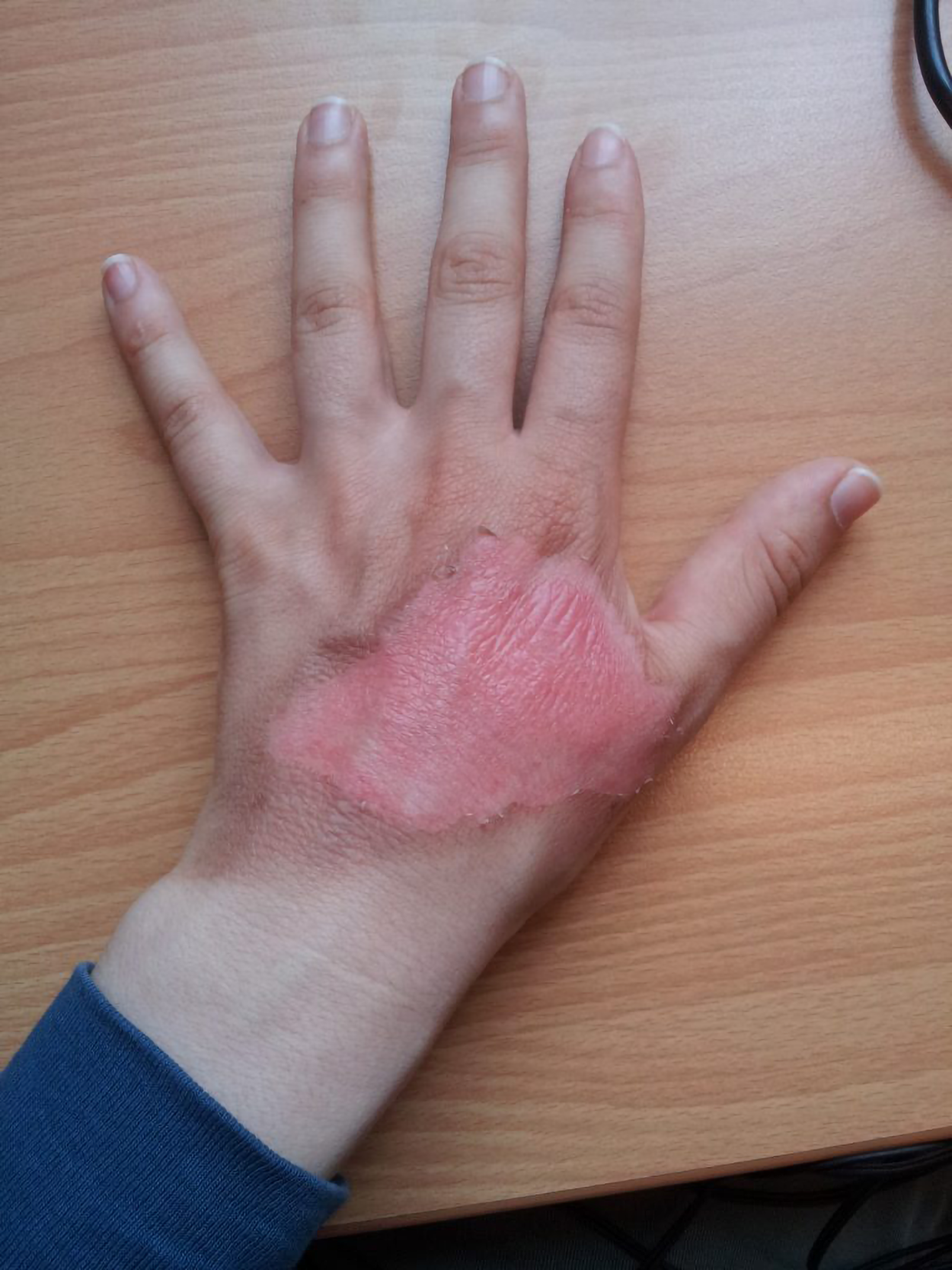
Burns are injuries (skin damage) caused by exposure to heat, flame, radiation, electricity, or chemicals. Burns cause damage to the skin layers, so deeper burns can seriously affect the integrity of the skin.

The term wound can be interpreted in several ways, but typically refers to an open wound caused when the skin is damaged due to some sort of external force, like an injury (Merriam-Webster, n.d.). Wounds can also reduce the effectiveness of skin as a protective barrier.

Burns and wounds are both included in this lesson because both involve some similar risks, such as infection.


2. Burns

Regardless of their cause, burns can lead to a massive fluid loss due to the loss of protection against dehydration by the skin. Burned skin is also extremely susceptible to infection due to the loss of protection against pathogens by the skin.

key concept
Burns were traditionally classified by their degree of severity as first-degree, second-degree, third-degree, and fourth-degree burns. This historic classification of burns has been replaced by a system reflecting the need for surgical intervention. They are referred to as superficial, superficial partial-thickness, deep partial-thickness, and full-thickness burns.

Burn Degree Updated Reference Description
First-degree burn Superficial burn Affects the epidermis. Although the skin may be painful and swollen, these burns typically heal on their own within a few days. Mild sunburn fits into the category of first-degree burns.
Second-degree burn Partial thickness burn Affects both the epidermis and a portion of the dermis. These burns result in swelling and painful blistering of the skin. It is important to keep the burn site clean to prevent infection. With good care, a second-degree burn will heal within several weeks.
Third-degree burn Full-thickness burn Extends fully into the epidermis and dermis, destroying the tissue and affecting the nerve endings and sensory function. These burns also affect subcutaneous tissue. These are serious burns that require immediate medical attention.
Fourth-degree burn Deep full-thickness burn More severe, affecting the underlying muscle and bone.

Full-thickness burns require debridement, surgical removal of dead tissue, followed by grafting skin from an unaffected part of the body or from skin grown in tissue culture. The general term for skin repair is dermatoplasty.

A cross-sectional of skin is depicted as a cube with three layers. The text ‘Epidermis (outer layer)’ points to the top layer of skin, depicted as pink and smooth. The text ‘Dermis (middle layer with vessels and glands)’ points to the middle layer of skin, depicted as a thicker wavy light pink line atop a slightly thinner wavy darker pink line. The text ‘Subcutaneous tissue (fat layer at the bottom)’ points to the third and bottom layer of skin, depicted as a thicker light yellow wavy line with small darker yellow circles throughout. There are two wavy horizontal lines that are superimposed over this layer, where one line is red and the other line is blue, and both lines have branches coming up vertically from them to depict the circulatory system’s place in the skin. The image of the cube of skin shifts over to the left and the text disappears except for the labels ‘Epidermis’, ‘Dermis’, and ‘Subcutaneous tissue’ still pointing out the respective areas of the skin. The word ‘Epidermis’ remains bold while the words ‘Dermis’ and ‘Subcutaneous tissue’ fade out slightly. A burn to the epidermis is shown, depicted as a darker pink irregular shape atop the pink and smooth layer of skin. A pink square with rounded corners shifts in from the right that contains the text ‘Superficial Burn (First Degree) – Affects the outermost skin layer. Heals without scarring’. This pink square and its containing text shifts out to the right. Next, the words ‘Epidermis’ and ‘Dermis’ remain bold while the words ‘Subcutaneous tissue’ fades out slightly, as a burn to the epidermis and dermis is shown, depicted as a darker pink irregular shape with smaller pink bubbling circles that is atop the pink and smooth layer of skin and extending down into the first wavy light pink line of the second layer of skin. At the same time, a darker pink square with round corners shifts in from the right that contains the text ‘Partial-Thickness Burn (Second Degree) – Involves both the epidermis and dermis. May scar’. This darker pink square and its containing text shifts out to the right. Next, all three words remain bold: ‘Epidermis’, ‘Dermis’ and ‘Subcutaneous tissue’, as a burn to all three layers of skin is shown, depicted as a maroon irregular shape with smaller, darker maroon irregular flat shapes inside, atop the pink and smooth layer of skin and extending down into the third layer of skin that was depicted as the thicker light yellow wavy line. At the same time, a dark pink square with round corners shifts in from the right that contains the text ‘Full-Thickness Burn (Third degree) – Eschar, necrosis’. All text fades out and the cube of skin, still with the depicted full-thickness burn, shifts to the left of the screen. An arrow accompanies the animation of the damaged maroon portion of skin lifting off the remaining healthy skin, while the following text on screen appears inside a light gray box: ‘Debridement – removal of dead tissue’.

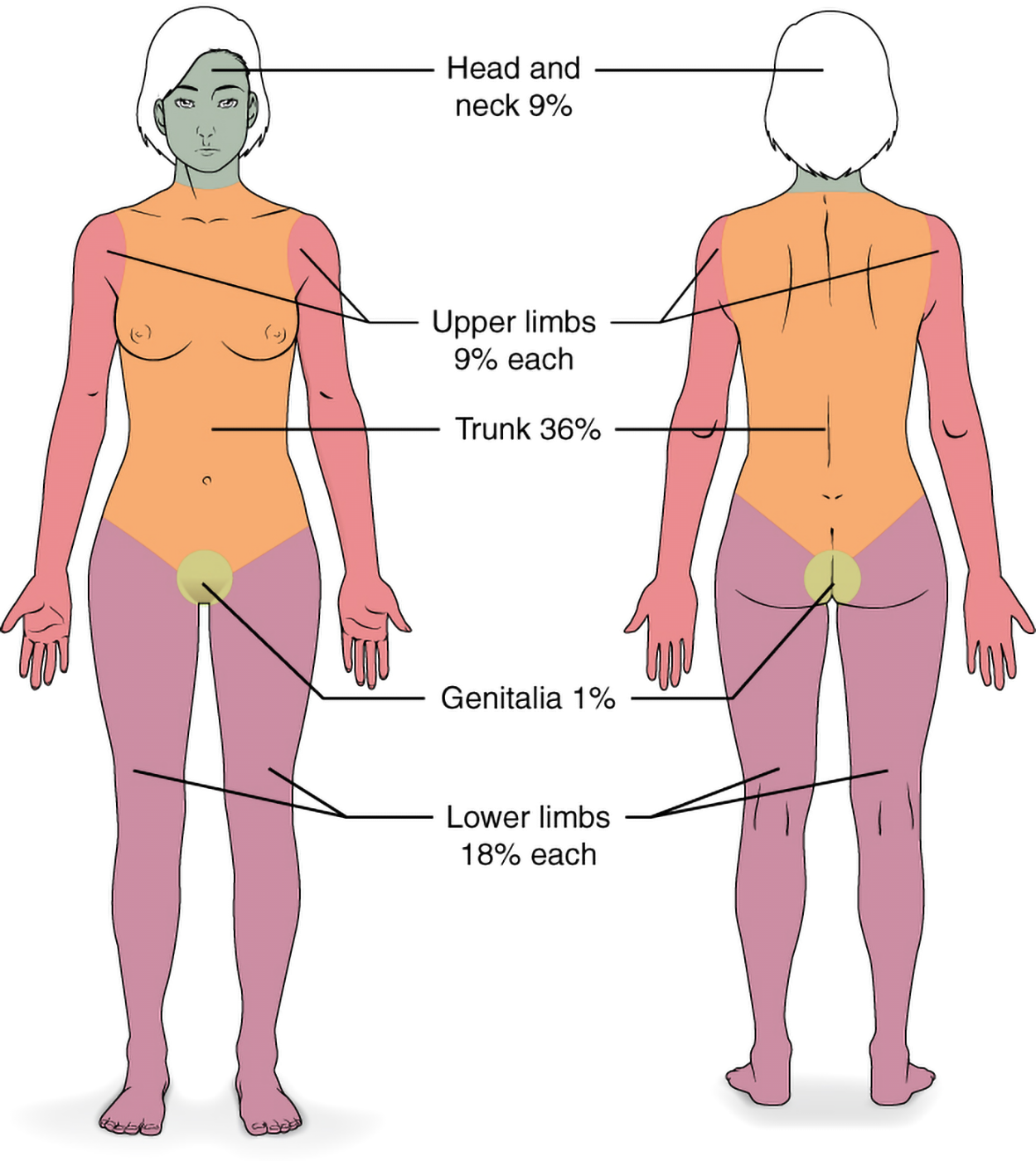
Severe burns are quickly measured in emergency departments using a tool called the “Rule of Nines,” which associates specific anatomical locations with a percentage of the body that is a factor of nine. This is used to calculate the total body surface area (TBSA) affected by burns. A rapid estimate of the burned surface area is used to estimate the amount of intravenous fluids needed to replace fluid loss. Using the Rule of Nines, the head is calculated as 9%, the upper limbs are 9% (4.5% on each side), the lower limbs are 18% (9% on each side), and the trunk is 36% (18% on each side), and the perineum/genitalia is 1%. The figure below illustrates the Rule of Nines. Note that there are specialized versions of this calculation for infants and children, as their body proportions are different.

Burns can occur in several ways:

  • Thermal burns are caused by fire, hot liquids, or steam.
  • Chemical burns are caused by acids and alkalis, including industrial chemicals.
  • Electrical burns are caused by contact with electrical currents.
  • Radiation burns are caused by radiation exposure, including UV exposure (sunburn), and radiation therapy.
  • Friction burns include road rash and carpet burn.
Burns can cause both local effects and systemic effects. Local effects only affect a limited amount of tissue; in this case, it's the tissue where the burn occurred. Systemic effects are whole-body effects.

The local effects of burns include tissue destruction, inflammation, and fluid loss. The systemic effects include shock, electrolyte imbalances, and infection risk. Shock, which is potentially life-threatening, occurs when there is insufficient blood supply to body tissues. For example, hypovolemic shock occurs when there is significant blood or fluid loss (e.g., through diarrhea), and anaphylactic shock occurs during anaphylaxis, a severe, potentially life-threatening allergic reaction. Burn shock specifically occurs when there is hypovolemia (low blood volume) due to fluid loss from skin damage.


3. Wounds

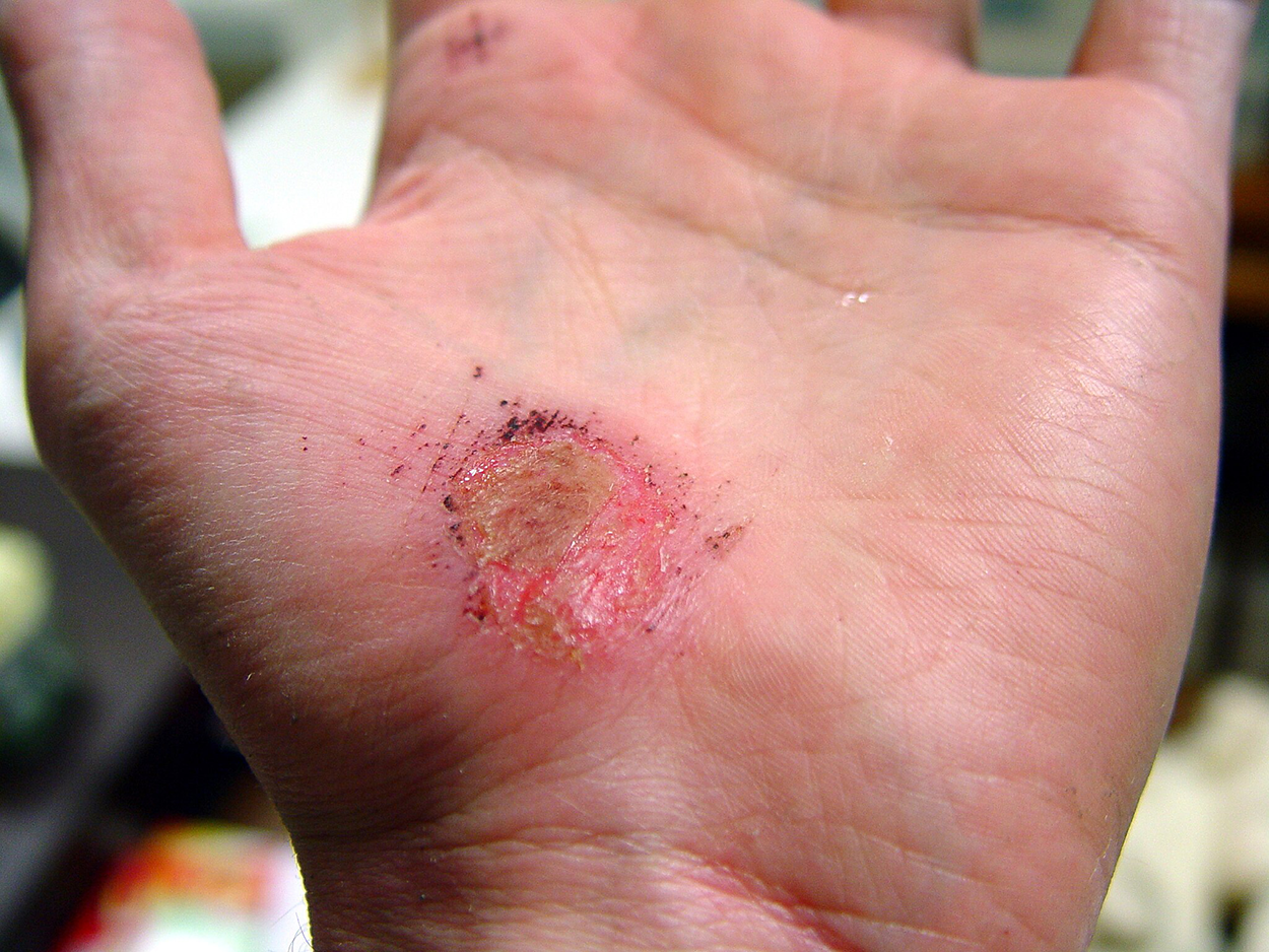
There are many types of wounds. Some are simple abrasions and lacerations that may heal easily even without stitches. Others may require considerable care and management.

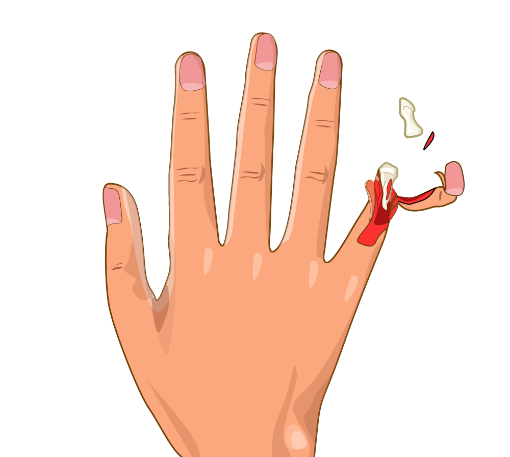
Acute wounds are wounds such as scratches and abrasions that occur quickly and then heal over a relatively short period of time. Examples of acute wounds include abrasions and lacerations, which you have already learned about. Puncture wounds are small but deep penetration wounds (e.g., the type of wound someone would get by stepping on a nail). Avulsion injuries can occur within the body, but skin avulsion injuries occur specifically when skin is torn away from its normal location. A skin avulsion injury can be relatively minor, or something more substantial like degloving, which occurs when skin and subcutaneous tissue are torn away, as shown in the figure below (which shows degloving and amputation).

Chronic wounds do not heal quickly. The exact amount of time required to classify a wound as chronic can vary, but these are wounds that can persist for very long times.

Examples of chronic wounds include:

  • Pressure injuries (formerly called pressure ulcers): Include decubitus ulcers and bedsores; caused by prolonged pressure on bony areas.
  • Diabetic ulcers: Neuropathy-related non-healing wounds in diabetes.
  • Venous ulcers: Leg ulcers that develop due to poor circulation.
  • Arterial ulcers: Ulcers that result from reduced blood flow to the extremities.

step by step
Wound healing follows a predictable series of phases:

  1. Inflammatory Phase (0–6 days): White blood cell response to clear bacteria and debris.
  2. Proliferation Phase (4–24 days): Granulation tissue (defined below) forms, new blood vessels grow.
  3. Maturation (Remodeling) Phase (21 days–1 year): Collagen remodeling, wound contracts.

An animated medical infographic illustrating the four stages of wound healing in a cross-sectional view of human skin. A header at the top says ‘Wound Healing’. In each stage of wound healing, the human skin is depicted as a cubical cross-section showing each layer of skin. Each stage is labeled and visually represented with cartoon-style graphics. The first phase is labeled as ‘Bleeding’. A nail is showing as piercing the skin. A blood vessel is labeled and depicted in the first cubical cross-section as a red, wavy horizontal line with smaller lines branching vertically up off it. Bacteria is labeled and depicted in the first cubical cross-section as small green ovals surrounding the pierced skin. Platelets are labeled and depicted in the first cubical cross-section as small yellow circles surrounding the pierced skin. The second phase is labeled as ‘Inflammatory’. A scab is labeled and showing as forming on the top layer of the skin. Fibroblasts are labeled and depicted in the second cubical cross-section as small tan irregular shapes with protrusions. Macrophages are labeled and depicted in the second cubical cross-section as small round light blue irregular shapes with protrusions. The third phase is labeled as ‘Proliferative’. The scabbed area from the previous phase is showing as closing up. The bottommost part of the third cubical cross-section is labeled ‘Subcutaneous fat’ and depicted as a thick yellow wavy line. Fibroblasts are labeled and depicted the same way as in the previous phase, but are now starting to surround the healing area. The fourth phase is labeled as ‘Remodeling’. No damage is evident on the surface of the skin any longer and this area is labeled ‘Freshly Healed Epidermis’. There is a slightly darker pink spot on the layer of skin underneath to designate where the injury had occurred, but no damage is evident, and this part is labeled ‘Freshly Healed Dermis’.

Wound healing can be affected by many factors, including age, diabetes, infection, and malnutrition.

As wounds heal, they produce exudate (drainage). Exudate can take several forms:

  • Serous: Clear, watery fluid.
  • Sanguineous: Bright red, fresh blood.
  • Serosanguineous: Pale pink, watery mix of blood and serous fluid.
  • Purulent: Pus-containing; yellow, green, thick pus (sign of infection).

Sometimes, wounds become infected. Signs that an infection has developed include:

  • Erythema: Redness around wound.
  • Edema: Swelling and fluid buildup.
  • Foul odor: Indicates bacterial overgrowth

Additional terms used in this context are suppuration (producing pus) and pyogenic (related to pus formation). A wound with purulent drainage produces pus-filled exudate.


4. Treatments, and Procedures

When a burn first occurs, it needs rapid treatment. For minor burns, it may be sufficient to run cool water on the burn. Do not use ice.

Fluid resuscitation can be used for severe burns. IV fluids may be given to prevent hypovolemic shock.

During burn and wound care, debridement can be necessary. This can be accomplished in several ways. There are several different types of debridement available. Mechanical debridement includes scrubbing and irrigation. Autolytic debridement uses dressings to allow the body to break down tissue naturally (“auto” means “self” and “-lysis” means breakdown or separation). As the name suggests, enzymatic debridement involves the application of special enzymes that break down necrotic tissue. In some cases, surgical debridement is needed. In surgical debridement, the dead tissue is surgically removed using a scalpel.

When monitoring burns and wounds, some descriptive terms can be helpful:

  • Eschar: Dead, blackened tissue after burns.
  • Slough: Yellow, moist dead tissue.
  • Granulation tissue: Red, bumpy new tissue forming during the healing process.
  • Maceration: Skin softening due to excess moisture.

Dressings are commonly used on wounds, including burns. Examples of dressing types include:

  • Occlusive dressings: Maintain moist environment (e.g., hydrocolloid).
  • Gauze dressings: Used for absorption and wound coverage.
  • Antimicrobial dressings: Silver-containing dressings to prevent infection.

Sometimes, the damage is so severe that skin grafting is needed. Skin grafting is used for severe burns and large wounds. The skin can be obtained from the patient who needs a graft (autograft), from another person (allograft), or from another species, such as a pig (xenograft).

A header reads ‘Skin Grafting Process’. On the left is a cross-section labeled ‘Burn Site’, showing a burn affecting the top two layers of skin. On the right is a healthy skin cross-section labeled ‘Healthy Donor Site’. Both images and the header fade upward. A human hand and arm appear, with a dotted rectangle near the wrist. A rectangle of skin lifts from this area, labeled ‘Skin removed from healthy donor’. The left half of the screen becomes transparent, and three rectangles slide in with text:

In some cases, hyperbaric oxygen therapy (HBOT) is used to increase the oxygenation of wounds to promote healing. HBOT is performed by having patients enter a pressurized chamber with a pure oxygen atmosphere. HBOT is especially helpful in treating chronic wounds, crush injuries, and burns that are not healing properly.

summary
In this lesson, you had an introduction to burns and wounds. First, you learned about types of burns and risks posed by burns. Then you learned about different types of wounds, including acute and chronic wounds. Finally, you learned about treatments and procedures related to burns and wounds. These types of wounds are commonly encountered, and chronic wounds are a major concern in some patient populations, so knowing the terminology will help you be well-prepared to help your patients.

Source: THIS TUTORIAL HAS BEEN ADAPTED FROM “OPEN RN | MEDICAL TERMINOLOGY – 2e” BY ERNSTMEYER & CHRISTMAN AT OPEN RESOURCES FOR NURSING (Open RN). ACCESS FOR FREE AT https://wtcs.pressbooks.pub/medterm/ LICENSING: CREATIVE COMMONS ATTRIBUTION 4.0 INTERNATIONAL.

REFERENCES

Merriam-Webster. (n.d.). Wound. In Merriam-Webster.com dictionary. Retrieved 6 April 2025, from www.merriam-webster.com/dictionary/wound

Attributions
Terms to Know
Acute Wounds

Wounds that occur quickly, such as scrapes and abrasions, and then heal during a relatively short time frame.

Anaphylactic Shock

Shock caused by an anaphylactic reaction.

Antimicrobial Dressing

Silver-containing dressings to prevent infection.

Arterial Ulcers

Ulcers that result from reduced blood flow to the extremities.

Burn Shock

Shock due to hypovolemia due to burns.

Chemical Burn

Burn caused by acids or alkalis, including industrial chemicals.

Chronic Wounds

Wounds that take a prolonged time to heal and persist long-term.

Deep Full Thickness Burn

A burn that affects the epidermis, dermis, and subcutaneous tissue and extends into muscle and bone.

Dermatoplasty

A procedure to repair skin.

Diabetic Ulcers

Neuropathy-related non-healing wounds in diabetes.

Edema

Swelling and fluid build-up.

Electrical Burn

Burn caused by contact with electrical currents.

Eschar

Dead, blackened tissue present after burns.

Exudate

Drainage from a wound.

First-Degree Burn

A burn that only affects the epidermis.

Fourth-Degree Burn

A burn that affects the epidermis, dermis, and subcutaneous tissue and extends into muscle and bone.

Friction Burn

Type of burn that includes road rash and carpet rash.

Full Thickness Burn

A burn that fully affects the epidermis and dermis and part of the subcutaneous tissue.

Gauze Dressing

Used for absorption and wound coverage.

Granulation Tissue

Red, bumpy new tissue forming.

Hyperbaric Oxygen Therapy

Treatment in which patients enter a pressured chamber with a pure oxygen environment.

Hypovolemic Shock

A type of shock that occurs due to excessive blood or fluid loss.

Local Effect

Affects a limited area.

Maceration

Skin softening due to excess moisture.

Occlusive Dressing

Maintain moist environment (e.g., hydrocolloid).

Partial Thickness Burn

A burn that affects the epidermis and part of the dermis.

Purulent

Pus-containing.

Purulent Drainage

Pus-filled drainage.

Pyogenic

Related to pus formation.

Radiation Burn

Burn caused by radiation exposure, including UV exposure (sunburn) and radiation therapy.

Rule of Nines

A system used to estimate the total body surface area affected by burns; specific anatomical locations with a percentage of the body that is a factor of nine.

Sanguineous

Bright red, fresh blood.

Second-Degree Burn

A burn that affects the epidermis and part of the dermis.

Serosanguineous

Pale pink, watery mix of blood and serous fluid.

Serous

Clear, watery fluid.

Shock

A potentially life-threatening condition that occur when there is insufficient blood supply to body tissues.

Slough

Yellow, moist dead tissue.

Superficial Burn

A burn that only affects the epidermis.

Suppuration

Producing pus.

Systemic Effect

Affects the entire body.

Thermal Burn

Burn caused by fire, hot liquids, or steam.

Third-Degree Burn

A burn that fully affects the epidermis and dermis and part of the subcutaneous tissue.

Venous Ulcers

Leg ulcers that develop due to poor circulation.

Wound

Typically refers to an open wound caused when the skin is damaged due to some sort of external force, like an injury.