Table of Contents |
If food is left sitting for a period of time, organisms such as maggots may start to grow on it. This type of common observation led people of many backgrounds to wonder how these organisms appeared. One very old explanation, dating back to the ancient Greeks, was the theory of spontaneous generation.
Spontaneous generation means that life can arise spontaneously from nonliving matter. When there was no way to observe microscopic organisms (such as larvae and eggs), it could have easily looked as though nonliving matter such as food was spontaneously producing new life. For example, tiny eggs and larvae could grow in meat without giving evidence of their existence to the naked eye. As they became larger and/or metamorphosed, organisms that were easily visible may have seemed to appear suddenly.
Because it seemed to match what people could see without microscopes and rigorous scientific studies, the theory of spontaneous generation persisted until scientists carefully studied it in the 17th century.
The Greek philosopher Aristotle (384–322 BC) was one of the earliest recorded scholars to describe the theory of spontaneous generation. He proposed that life arose from nonliving matter if the material contained pneuma (“spirit” or “breath”). As evidence, he noted several instances of the appearance of animals from environments previously devoid of such animals, such as the seemingly sudden appearance of fish in a new puddle of water.
People suggested other examples of spontaneous generation that can be explained easily using modern knowledge. In general, these were examples in which habitats became suitable and animal populations were attracted to them and then thrived.
EXAMPLE
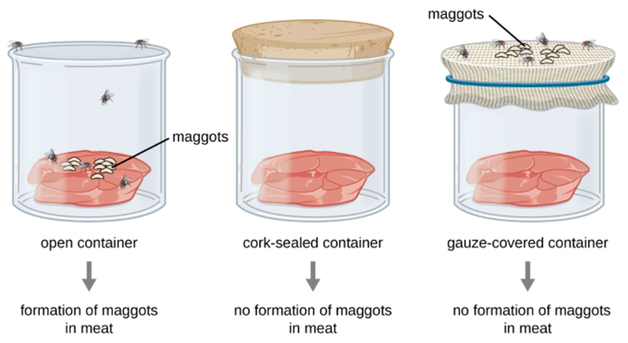
Some people argued that frogs seemed to simply appear along the banks of the Nile River in Egypt during flooding. Others noted that mice suddenly appeared among grain stored in barns with thatched roofs. Jan Baptista van Helmont, a 17th century Flemish scientist, proposed that mice could arise from rags and wheat kernels left in an open container for 3 weeks.One of the most important early experiments to test the theory of spontaneous generation was performed by Italian physician Francesco Redi (1626–1797) in 1668. To determine whether maggots could spontaneously appear on meat that was left out, he tested whether preventing adult flies from having direct access to the meat to lay eggs prevented maggots from appearing. Redi set up six containers and placed meat in each as shown in the figure below. Two of the containers were open to the air, two were covered with gauze, and two were tightly sealed with cork. His results supported his hypothesis that flies needed direct contact with meat for maggots to grow. Maggots grew on meat in the open containers and on the gauze above the meat in containers covered with gauze. No maggots appeared in the meat in the tightly sealed containers or gauze-covered containers.
In 1745, John Needham (1713–1781) published a report of his experiments investigating spontaneous generation. Needham briefly boiled broth infused with plant or animal matter. Boiling for a sufficient length of time kills vegetative (actively growing) microbes and you will learn more about its use in the lesson on physical control of microorganisms. Needham found that the broth became cloudy and that microbes were visible in drops of broth. He argued that this suggested that spontaneous generation had occurred. However, it is possible that the broth was not boiled long enough to kill all of the microbes present.
Lazzaro Spallanzani (1729–1799) conducted further experiments using heated broth in sealed and unsealed containers. He did not find growth in sealed containers unless they were later opened, allowing microbes to enter from the air. He believed that this evidence argued against spontaneous generation. However, Needham argued that the extended boiling used had destroyed the “life force” necessary for growth.
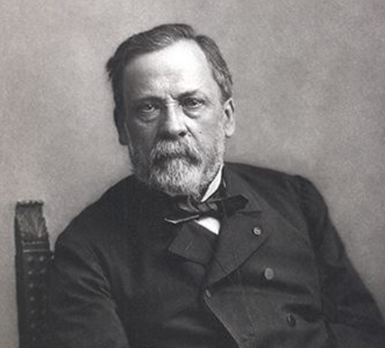
Although the work of Redi and Spallanzani suggested that spontaneous generation did not occur, the question was still debated extensively into the 19th century. Legendary microbiologist Louis Pasteur (1822–1895), introduced in the lesson on the history of microbiology, accepted a challenge posed by the Paris Academy of Science. Anyone who resolved the debate could win a prize.
In 1858, Pasteur began a series of important experiments by filtering air through a gun-cotton filter. When he examined the cotton afterward, he found that many microorganisms were present. This suggested that there were abundant microbes in air that could colonize exposed food sources.
Later, Pasteur made a series of flasks with long, twisted necks called “swan-neck” flasks. He boiled broth within the flasks. The twisted necks prevented airborne microbes from reaching the broth. The microbes became trapped on the bend of the neck as shown in the figure below. However, air could move in and out of the flask. This addressed the concern that a tightly sealed container was preventing entry of a “life force”. No microbes grew in the boiled broth within the flasks unless the necks of the flasks were broken, allowing microbes to enter from the air as shown in the image below.
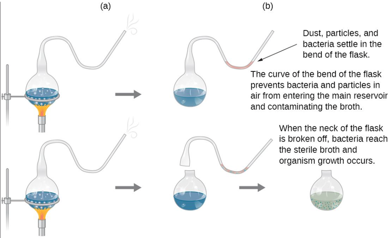
This set of experiments was considered definitive. Scientists moved away from considering spontaneous generation as a serious theory. Pasteur won the Alhumbert Prize from the Paris Academy of Sciences in 1862.
Pasteur summed up the results of the work himself. He said, “Omne vivum ex vivo” (“Life comes only from life”) and correctly noted that “…life is a germ and a germ is life. Never will the doctrine of spontaneous generation recover from the mortal blow of this simple experiment.”
Cell theory describes the origins of cells. We now know that cells do not arise spontaneously from nonliving matter as previously thought. Instead, modern cell theory describes the origin and role of cells in two basic tenets:
As mentioned in the lesson on observing microbes English scientist Robert Hooke (1635–1703) viewed cork cells through a microscope that he designed. He was the first person to describe these cells based on his microscopic observations, using the term “cells”. However, he did not realize that he was actually viewing dead cells that lacked internal structures or that cells were the foundational units of life.

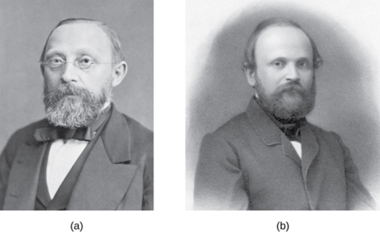
German botanist Matthias Schleiden made observations of plant tissues in 1838 and described them as being composed of cells. German physiologist Theodor Schwann (1810–1882) noted cells in animal tissue. When the two discussed their findings, they noted important similarities and this led to the foundation for realizing that cells are the building blocks of life.
As understanding of the role of cells in plant and animal tissues increased, so did understanding of the structures within cells. Nuclei, which are membrane-bound structures containing DNA, were first described in plant cells by Scottish botanist Robert Brown (1773–1858) in 1831. In the early 1880s, German botanist Andreas Schimper (1856–1901) was the first to describe the chloroplasts of plant cells, which are important in photosynthesis. Schimper noted that chloroplasts could divide independently of the host cell.
To understand endosymbiotic theory, it is important to understand how groups of cells differ. All cells can be divided into two major types: prokaryotic cells that lack a true membrane-bound nucleus and eukaryotic cells that have a true membrane-bound nucleus and other membrane-bound subcellular structures called organelles. Chloroplasts are an example of this type of organelle. Another important organelle is the mitochondrion (plural mitochondria), which generates energy. Prokaryotic and eukaryotic cells differ in a variety of other ways and you will learn more about these in the lessons on prokaryotic and eukaryotic cells.
American anatomist Ivan Wallin (1883–1969) followed up on this work by studying mitochondria, chloroplasts, and bacteria to compare them. Although Wallin found evidence for the endosymbiotic hypothesis, he argued that mitochondria could be cultured outside of their host cells; however, further work and understanding of the mitochondrial genome suggests that this is not the case.
In the 1960s, it was discovered that mitochondria and chloroplasts have their own DNA. This stimulated new interest in the endosymbiotic theory. In 1967, American geneticist Lynn Margulis (1938–2011) published her ideas regarding the endosymbiotic hypothesis of the origins of mitochondria and chloroplasts. She supported her ideas with microscopic, genetic, molecular, fossil, and geological data.
Some of the current evidence supporting the endosymbiotic theory includes genetic sequencing data showing the relatedness of mitochondrial and chloroplast DNA to bacterial DNA, that some genes from mitochondria and chloroplasts have moved to the nucleus, and that mitochondrial and chloroplast ribosomes resemble bacterial ribosomes. Additionally, bacteria, chloroplasts, and mitochondria use a similar type of binary fission to divide one cell into two.
EXAMPLE
There are examples of endosymbionts today. These include endosymbiotic bacteria that live in insect guts and photosynthetic bacteria-like organelles in protists.IN CONTEXT
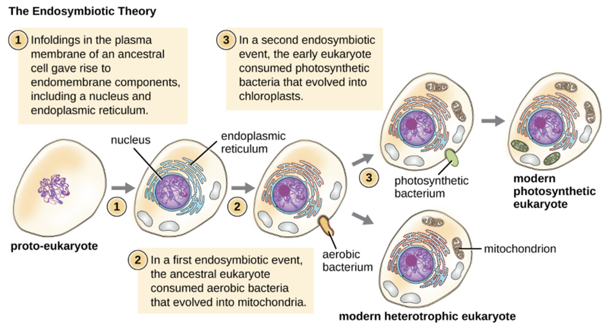
The basic steps of endosymbiosis are summarized in the figure below. Initially, there was a proto-eukaryote with loose DNA (i.e., the DNA was not enclosed in a membrane). In step 1, infoldings in the plasma membrane of the ancestral cell gave rise to endomembrane (internal membrane) components, including a nucleus and endoplasmic reticulum. You will learn more about these internal structures in the lesson on eukaryotic cells. In the first endosymbiotic event, the ancestral eukaryote took in an aerobic bacterium. The bacteria was an endosymbiont and provided an evolutionary advantage because it helped in energy production. The eukaryote and bacteria became increasingly interdependent and eventually the bacteria evolved into mitochondria. Some lineages developed into modern heterotrophic eukaryotes. Heterotrophs are organisms that need to consume organic (carbon-based) molecules such as proteins, fats, and carbohydrates (humans are heterotrophs!).
In other lineages, a second endosymbiotic event occurred. The early eukaryote, which already had a mitochondrion, took up a photosynthetic bacterium and an endosymbiotic relationship developed. This provided an advantage to the cell, which was now able to use sun energy to build sugars and other molecules from carbon dioxide. Over time, the photosynthetic cell evolved into a chloroplast and the relationship became obligatory so that neither the original cell nor the chloroplast could survive if they were separated. This gave rise to modern photosynthetic eukaryotes. Unlike heterotrophs, these photosynthetic organisms are autotrophs and can use inorganic molecules such as carbon dioxide to build complex organic molecules such as sugars.
Source: THIS TUTORIAL HAS BEEN ADAPTED FROM OPENSTAX “MICROBIOLOGY.” ACCESS FOR FREE AT openstax.org/details/books/microbiology. LICENSE: CC ATTRIBUTION 4.0 INTERNATIONAL.
RESOURCES
Parker, N., Schneegurt, M., Thi Tu, A.-H., Lister, P., & Forster, B. (2016). Microbiology. OpenStax. Access for free at openstax.org/books/microbiology/pages/1-introduction