Table of Contents |

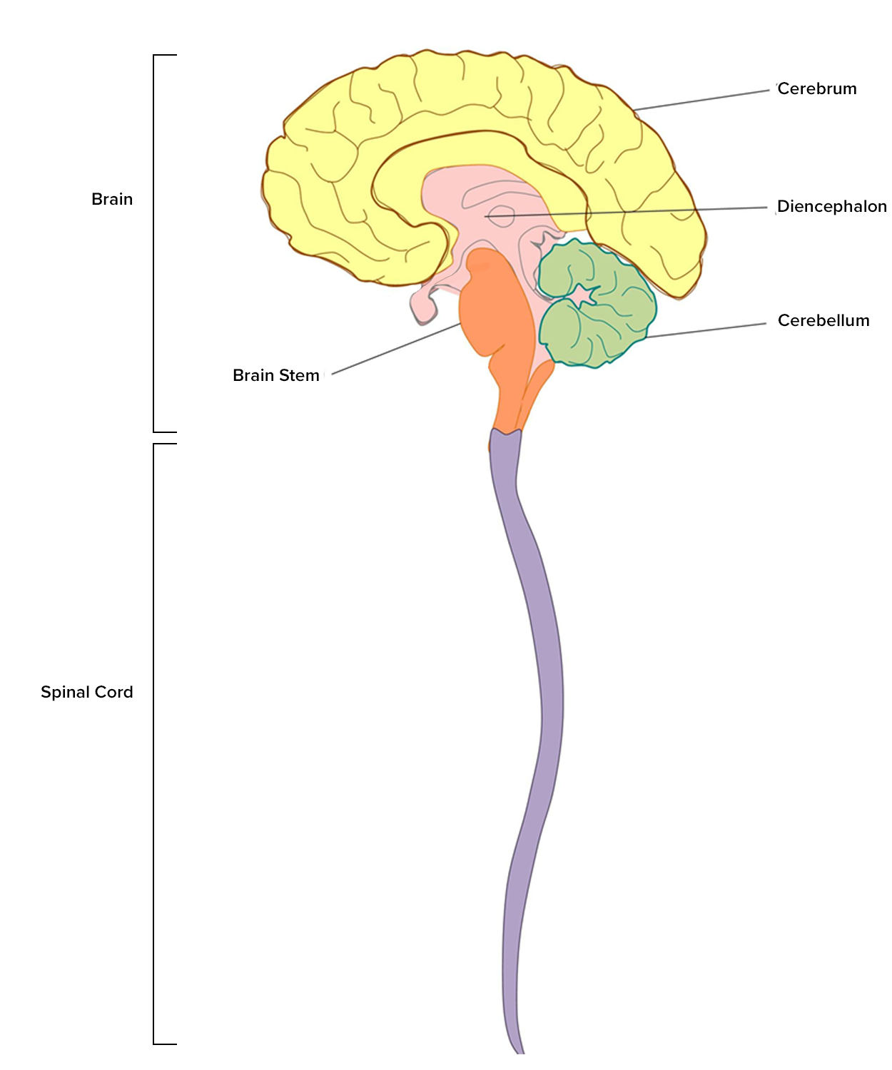
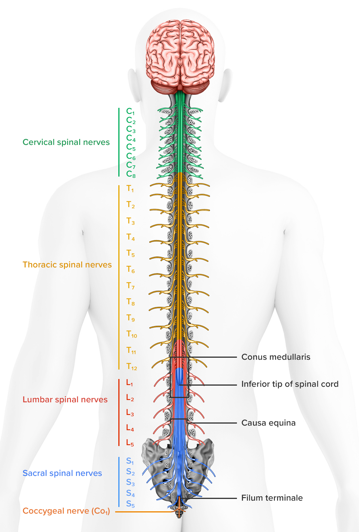
The spinal cord is an organ of the nervous system primarily made of nervous tissue and located in the vertebral canal formed by the vertebrae. At the superior end (toward the head), the spinal cord connects to the brain through the foramen magnum in the occipital bone. In the adult body, the inferior end of the spinal cord extends to approximately vertebrae L1 or L2 because the spinal cord stops growing before the vertebrae do. The inferior tip of the spinal cord is cone shaped and is called the conus medullaris. Beyond this, a strand of fibrous connective tissue called the filum terminale connects the conus medullaris to the sacrum (at the base of the lumbar vertebrae) to hold it in place.
Along the length of the spinal cord, 31 pairs of spinal nerves (collections of axons in the peripheral nervous system) exit the vertebral space between each vertebra and extend out to various points throughout the body. Each vertebra has its own set, which are named after the vertebral region where they originate—cervical, thoracic, lumbar, sacral, and coccygeal spinal nerves. At the inferior end of the spinal cord, beyond the conus medullaris, the lumbar and sacral spinal nerves extend into the vertebral cavity towards their exit. This collection of inferior spinal nerves resembles a horse's tail and is known as the cauda equina (cauda, tail; equine, horse).

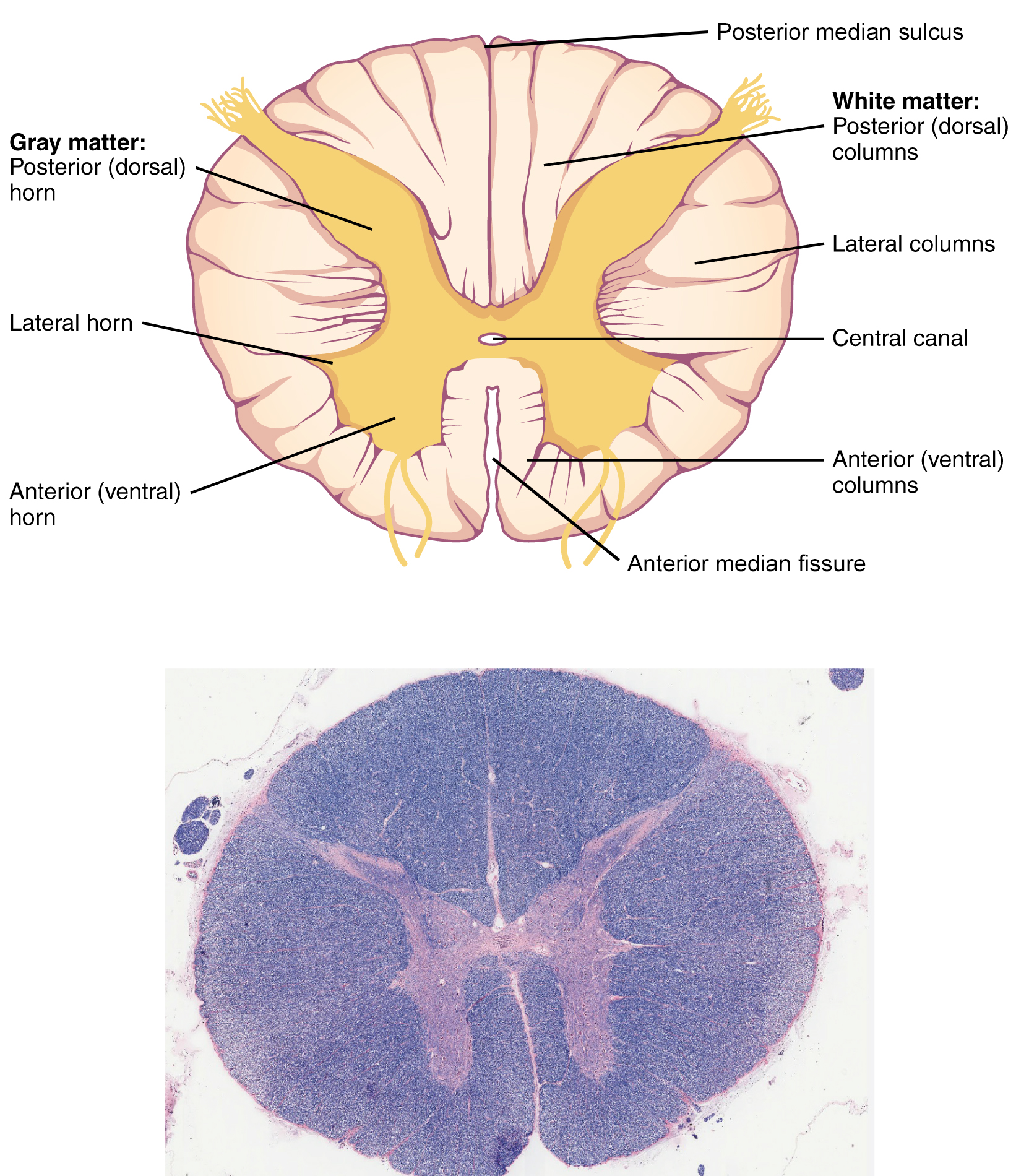
The anterior midline is marked by the deep anterior median fissure, and the posterior midline is marked by the shallow posterior median sulcus. These two grooves allow you to orient any view of the spinal cord to determine the anterior (front) and posterior (back) sides.
In the center of the spinal cord is a longitudinal hollow tube called the central canal, which circulates cerebrospinal fluid (CSF). CSF is produced in the brain and functions much like synovial fluid to circulate nutrients and remove wastes from a region that does not have direct access to blood. CSF also functions to protect the brain by acting as a shock absorber.

Recall that nervous tissue can be grouped into gray and white matter. Gray matter is primarily composed of cell bodies, dendrites, and nonmyelinated axons, whereas white matter is primarily composed of myelinated axons. The spinal cord is organized with the white matter being superficial and the gray matter being mostly deep. As you can see in the image above, the gray matter is organized in what some call a letter H, a butterfly, or an ink-blot test. The gray matter can be further subdivided into regions referred to as horns. The posterior horn is responsible for sensory processing. The anterior horn sends out motor signals to the skeletal muscles. The lateral horn, which is only found in the thoracic, upper lumbar, and sacral regions, contains cell bodies of motor neurons of the autonomic nervous system. There are no autonomic nervous system functions in the cervical region, so there are no lateral horns needed there.
Just as the gray matter is separated into horns, the white matter of the spinal cord is separated into columns. These columns contain tracts (groups of axons in the central nervous system) that transport action potentials up (ascending) or down (descending) the spinal cord. Ascending tracts of the spinal cord of nervous system fibers in these columns carry sensory information up to the brain, whereas descending tracts of the spinal cord carry motor commands from the brain.
Looking at the spinal cord longitudinally, the columns extend along its length as continuous bands of white matter. Between the two posterior horns of gray matter are the posterior columns. Between the two anterior horns and bounded by the axons of motor neurons emerging from that gray matter area are the anterior columns. The white matter on either side of the spinal cord, between the posterior horn and the axons of the anterior horn neurons, are the lateral columns. The posterior columns are composed of axons of ascending tracts. The anterior and lateral columns are composed of many different groups of axons of both ascending and descending tracts—the latter carrying motor commands down from the brain to the spinal cord to control output to the periphery.
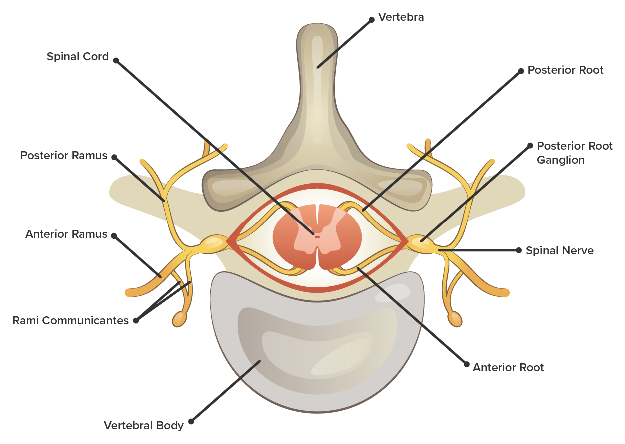
Attached to the spinal cord are the axons that connect the central nervous system to all peripheral structures such as the internal organs, muscles, skin, and more. Axons enter the posterior side through the posterior (dorsal) root. Axons emerge from the anterior side through the anterior (ventral) root. The directionality of information flow through the nerve roots is one-way traffic in which incoming sensory information travels through the posterior root to enter the spinal cord and outgoing motor information travels through the anterior root to travel to the effector. Along the posterior root is a ganglion (collection of neuron cell bodies in the peripheral nervous system) called the posterior root ganglion. This ganglion contains the cell bodies of the sensory neurons that make up the posterior root.
Where the posterior and anterior root meet, they combine together for a short time before separating again. This short segment is called the spinal nerve and contains both sensory and motor information. There are 31 pairs of spinal nerves named for the vertebral region where they are located: cervical, thoracic, lumbar, and sacral. Beyond the spinal nerve, the axons that make it up separate into three different pathways, each referred to as a ramus (ramus, branch). Each ramus (plural, rami) serves a different region of the body by transporting sensory signals from that region to the spinal cord and motor signals from the spinal cord to that region. The anterior rami serve the anterior and lateral parts of the body as well as the limbs. The posterior rami serve the posterior side of the body. The rami communicantes (communicating branches) transport signals to and from the internal organs and structures within the trunk.

SOURCE: THIS TUTORIAL HAS BEEN ADAPTED FROM OPENSTAX “ANATOMY AND PHYSIOLOGY 2E”. ACCESS FOR FREE AT OPENSTAX.ORG/BOOKS/ANATOMY-AND-PHYSIOLOGY-2E/PAGES/1-INTRODUCTION. LICENSE: CREATIVE COMMONS ATTRIBUTION 4.0 INTERNATIONAL.