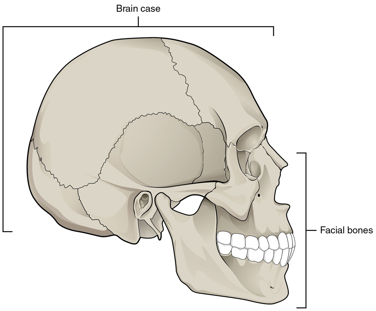
The cranium (skull) is the skeletal structure of the head that supports the face and protects the brain. The bones of the cranium are collectively referred to as the craniofacial bones. These can be subdivided into cranial and facial bones. Cranial bones form the brain casing, surrounding and protecting the brain. Facial bones underlie the facial structure, including the eye sockets, nose, upper and lower jaws, and more.
Note: You will learn the bones of the skull and their most prominent bony landmarks in this lesson.
learn more
To see additional bones and landmarks in this area, please visit the supplemental
Axial Bones.pdf.
Parts of the Skull - The skull consists of the rounded brain case that houses the brain and the facial bones that form the upper and lower jaws, nose, orbits, and other facial structures.
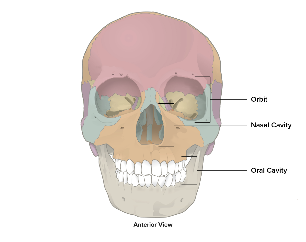
The craniofacial bones form multiple spaces within the cranium, some large and some small.
The orbit is also known as the eyesocket or the bony opening of the eye. Recall that the orbital region of anatomy relates to the eye.
The nasal cavity is the space enclosed by the nose which is split into right and left halves by the bony nasal septum.
The oral cavity is also known as the mouth.
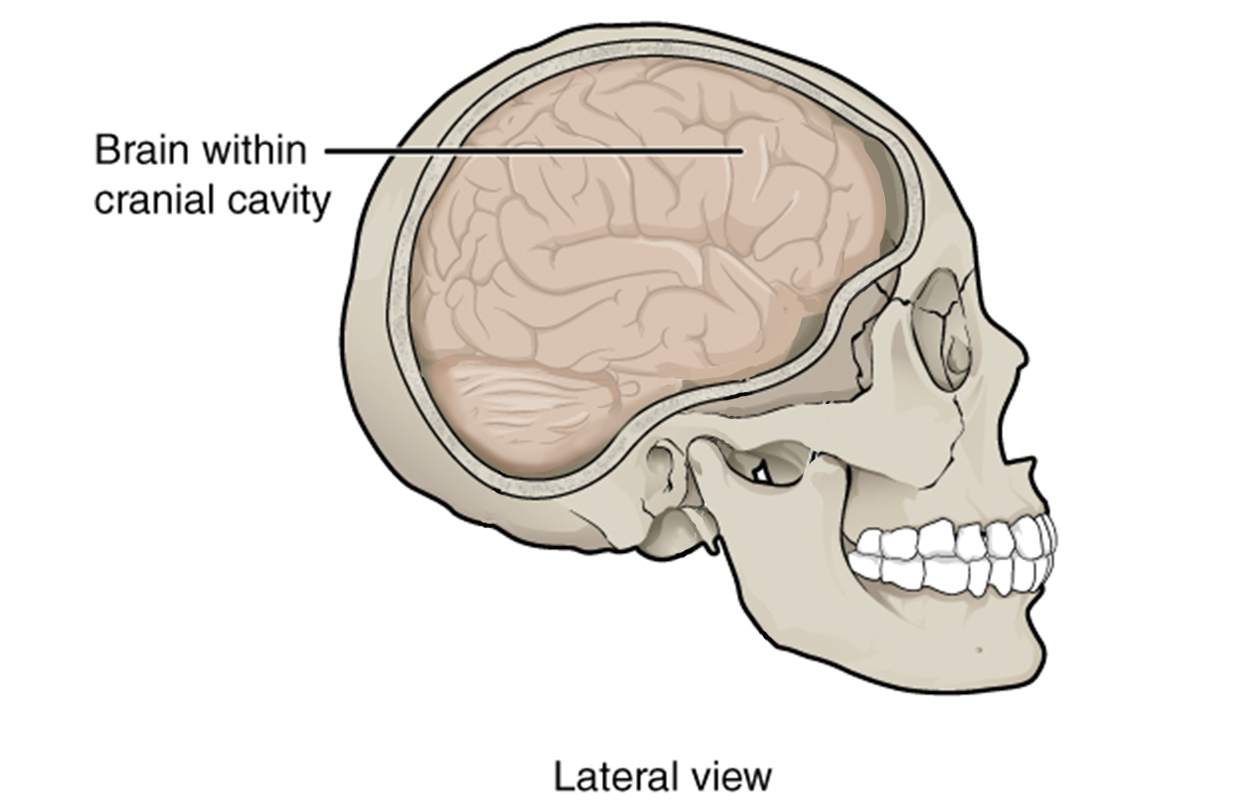
Recall that the cranial bones form the cranial cavity which protects the brain.
Multiple smaller sinuses are present in the anterior craniofacial bones. These will be discussed later in this lesson.
Skull Spaces - The craniofacial bones form multiple spaces within the skull including the cranial cavity, oral cavity, nasal cavity, and orbit.
terms to know
Cranium
The skeletal structure of the head that supports the face and protects the brain.
Craniofacial Bones
The bones of the skull.
Cranial Bones
The bones of the skull that form the brain casing.
Facial Bones
The bones of the skull that underlie facial structure.
Orbit
The bony opening of the eye.
Nasal Cavity
A large space in the skull behind the nose.
Nasal Septum
The bony separation of the right and left sides of the nasal cavity.
Oral Cavity
A large space in the skull behind the jaws, also known as the mouth.
2. Cranial Bones
The brain case contains and protects the brain. The interior space that is almost completely occupied by the brain is called the cranial cavity. This cavity space is defined by cranial bones on all sides. The floor of the brain case is referred to as the base of the skull. This is a complex area that varies in depth and has numerous openings for the passage of cranial nerves, blood vessels, and the spinal cord.
Cranial Cavity - The bones of the brain case surround and protect the brain, which occupies the cranial cavity.
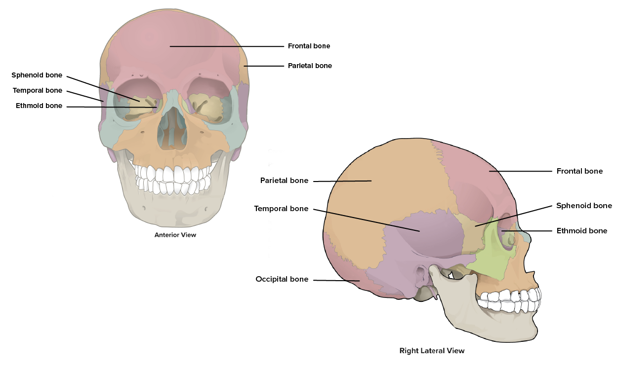
The brain case consists of eight bones. These include one frontal bone, a pair of parietal bones, one occipital bone, a pair of temporal bones, one ethmoid bone, and one sphenoid bone.
Cranial Bones - The cranial cavity is formed by eight bones.
2a. Frontal Bone
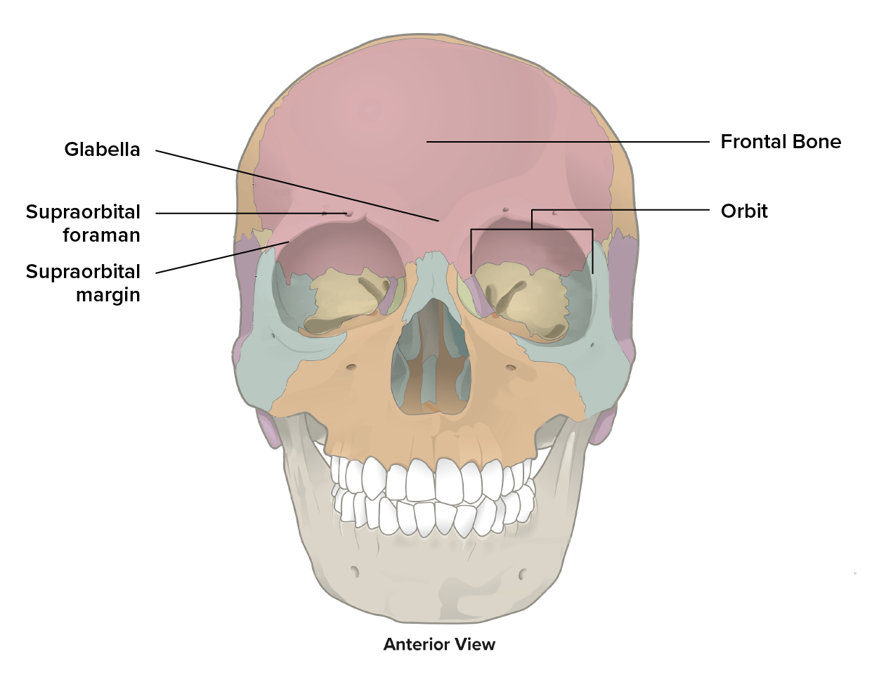
The frontal bone is the single flat bone that forms the anterior and part of the superior portion of the cranial cavity as well as the forehead. The frontal bone has a number of bony landmarks.
The glabella is a slight depression along the anterior midline, between the eyebrows. You can use your fingers to feel it.
The frontal bone forms the upper border of the orbit called the supraorbital margin (supra, superior; margin, border), literally the ‘border above the eye.’
The supraorbital foramen is a hole (foramen, hole) in the supraorbital margin that provides passage for a sensory nerve to the forehead.
The frontal bone is thickened just above each supraorbital margin, forming rounded brow ridges. These are located just behind your eyebrows and vary in size among individuals, although they are generally larger in males. Inside the cranial cavity, the frontal bone extends posteriorly. This flattened region forms both the roof of the orbit below and the floor of the anterior cranial cavity above.
Frontal Bone - This bone forms the forehead and a portion of the brain case.
2b. Parietal Bone
The parietal bones are the pair (right and left) of flat bones that form most of the superior and lateral side of the brain case. Each parietal bone is bounded anteriorly by the frontal bone, inferiorly by the temporal bone, and posteriorly by the occipital bone.
2c. Occipital Bone
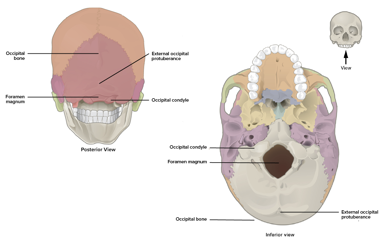
The occipital bone is the single flat bone that forms the posterior skull and posterior base of the cranial cavity. The occipital bone has a number of bony landmarks.
The external occipital protuberance (pro, in front of; tuberance, outgrowth) is a small projection (bump) at the posterior midline. This landmark serves as an attachment point for a ligament of the posterior neck.
The foramen magnum (magnum, large) is a large opening at the base of the skull which allows for passage of the spinal cord as it connects to the brain.
The occipital condyles are two oval-shaped projections located on either side of the foramen magnum and form joints with the first vertebra, supporting the skull on top of the vertebral (spinal) column.
Occipital Bone -This bone forms the posterior and part of the inferior border of the brain case, allows for muscle attachment, and supports the skull on top of the spinal column.
2d. Temporal Bone
The temporal bones are the pair (right and left) of flat bones that form the lower lateral side of the skull. Each temporal bone is bounded superiorly by the parietal bone, anteriorly by the sphenoid bone, and posteriorly by the occipital bone.
did you know
Common wisdom has it that the temporal bone (temporal, time) is so named because this area of the head (the temple) is where hair typically first turns gray, indicating the passage of time.
The temporal bone has a number of bony landmarks.
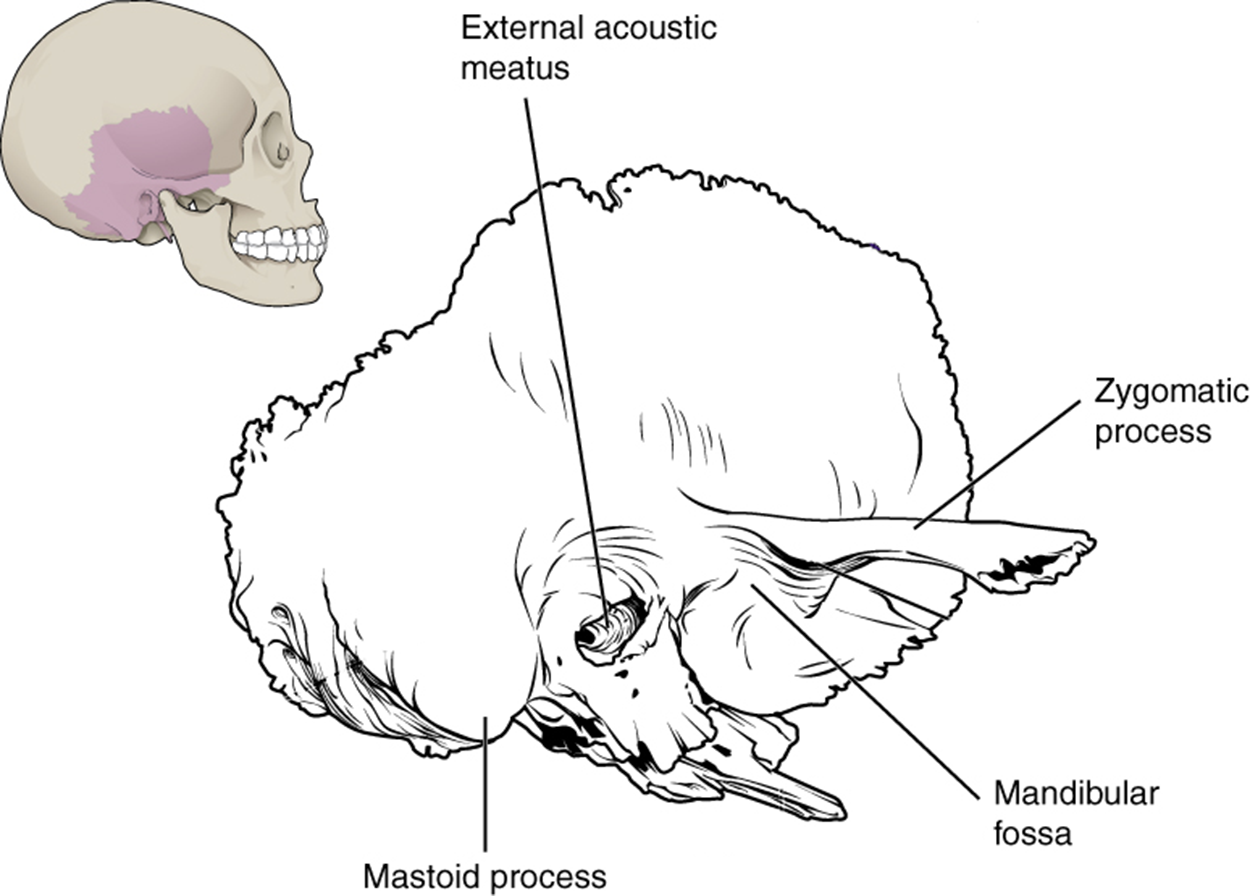
Temporal Bone - A lateral view of the isolated temporal bone shows the squamous, mastoid, and zygomatic portions of the temporal bone.
The external acoustic meatus (ear canal) is the large opening on the lateral side of the skull that is associated with the ear.
The mandibular fossa is the deep, oval-shaped depression located on the external base of the skull, just in front of the external acoustic meatus. The mandible (lower jaw) joins with the skull at this site as part of the temporomandibular joint (TMJ), which allows for movements of the mandible during opening and closing of the mouth.
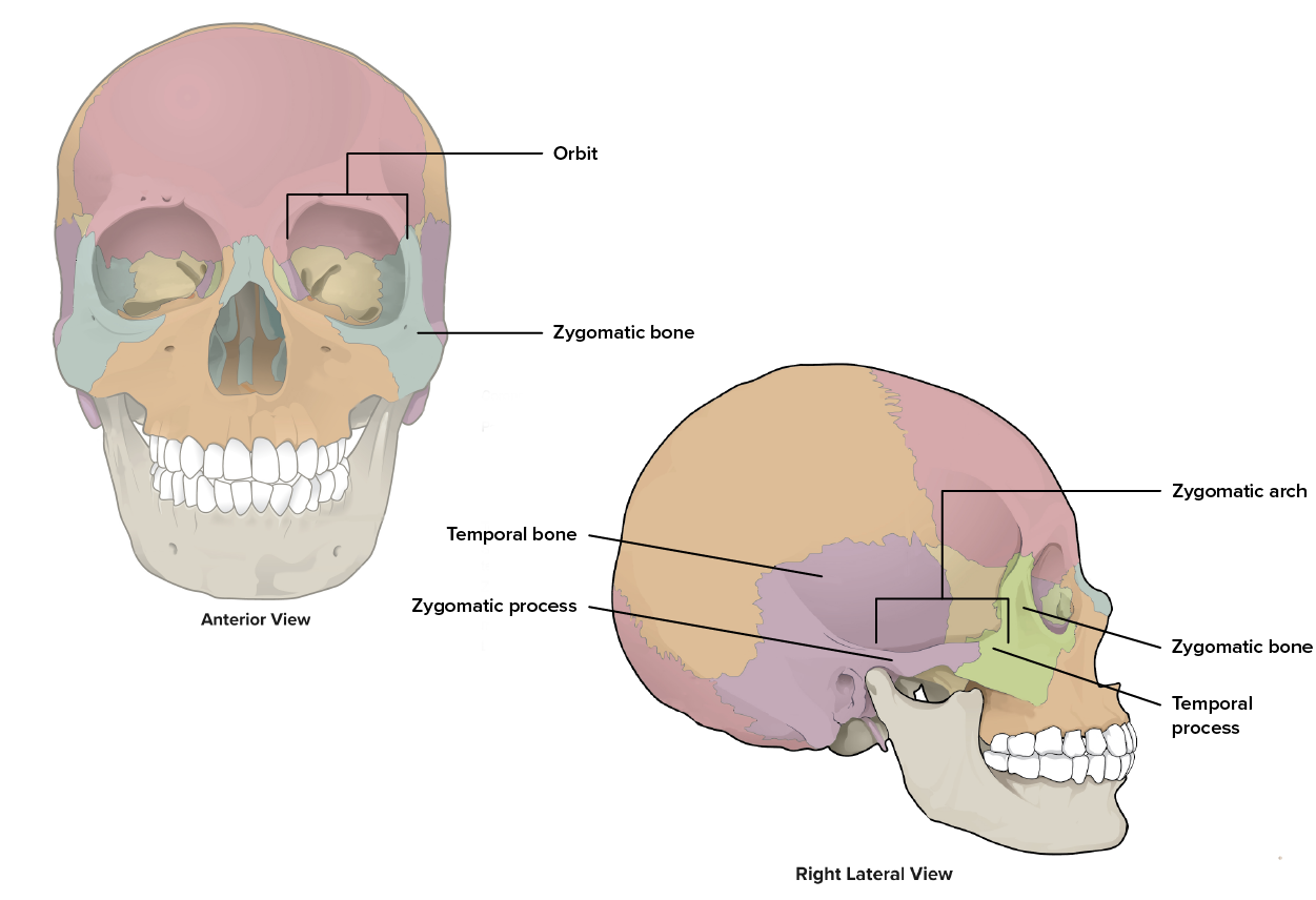
The zygomatic process of the temporal bone projects anteriorly towards the cheek (zygomatic) bone, forming the posterior portion of the zygomatic arch.
The mastoid process is a large prominence pointed inferiorly and located just posterior to the external acoustic meatus. It can easily be felt on the side of the head just behind your earlobe. This process serves as a site for muscle attachment.
2e. Sphenoid Bone
The sphenoid bone is a single, irregular bone of the skull which forms much of the base of the central skull and also extends laterally to contribute to the sides of the skull. Because of its position, the sphenoid bone makes contact with all of the other seven cranial bones of the skull, and is referred to as the 'cranial keystone.'
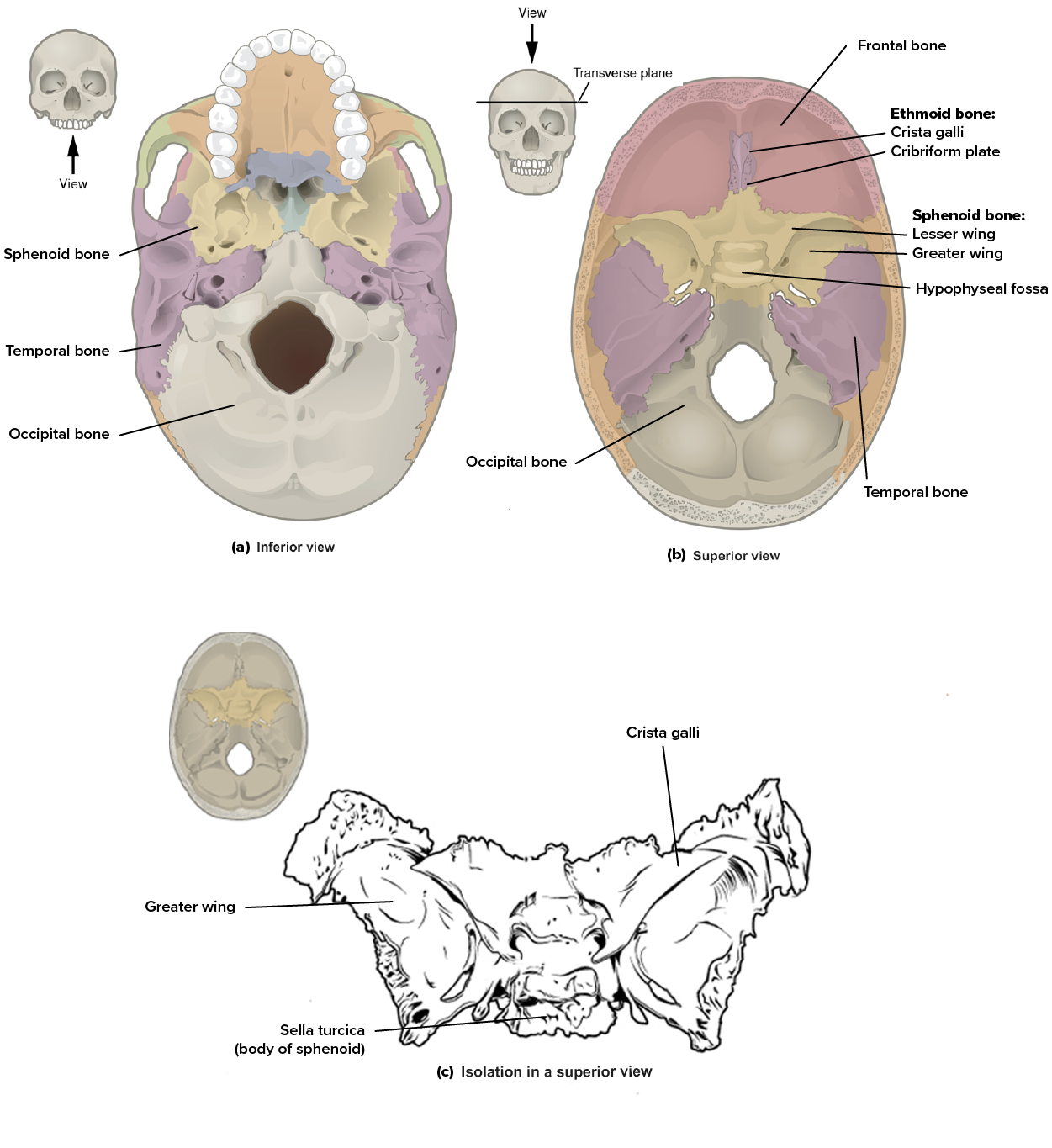
Sphenoid Bone - Shown in the (a) Inferior and (b) Superior views. The complex floor of the cranial cavity is formed by the frontal, ethmoid, sphenoid, temporal, and occipital bones. (c) Shown in isolation in a superior view. The sphenoid bone is a single midline bone that forms the anterior walls and floor of the middle cranial fossa. It has a pair of lesser wings and a pair of greater wings attached to the sella turcica.
The sphenoid bone is said to look like a butterfly (or moth) with two sets of wings spreading out from the body of the sphenoid bone. The body of the sphenoid bone is called the sella turcica, or Turkish saddle because of its resemblance to the horse saddles used by the Ottoman Turks with a high back and tall front. The lesser wings (right and left) of the sphenoid bone are the smaller and more anterior set of wings that form a prominent ridge. Below them are the greater wings (right and left) of the sphenoid bone which extend laterally to the outside of the lateral skull.
2f. Ethmoid Bone
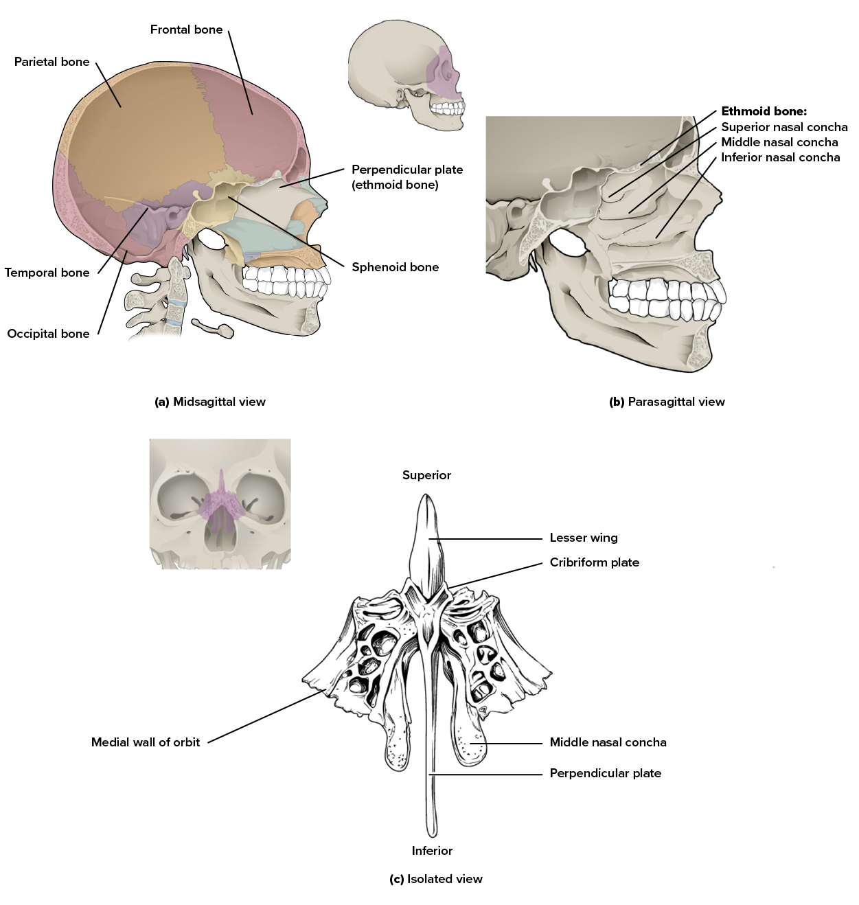
The ethmoid bone is a single irregular bone that forms the roof and lateral walls of the upper nasal cavity, the upper portion of the nasal septum, and contributes to the medial wall of the orbit. On the interior of the skull, the ethmoid also forms a portion of the floor of the anterior cranial cavity. The ethmoid bone has a number of bony landmarks.
The cribriform plate is the part of the ethmoid bone that separates the cranial and nasal cavities. It contains foramina (small holes) that allow sensory nerves for the sense of smell to travel through.
The crista galli, or “rooster’s crest,” is the superior midline projection into the cranial cavity.
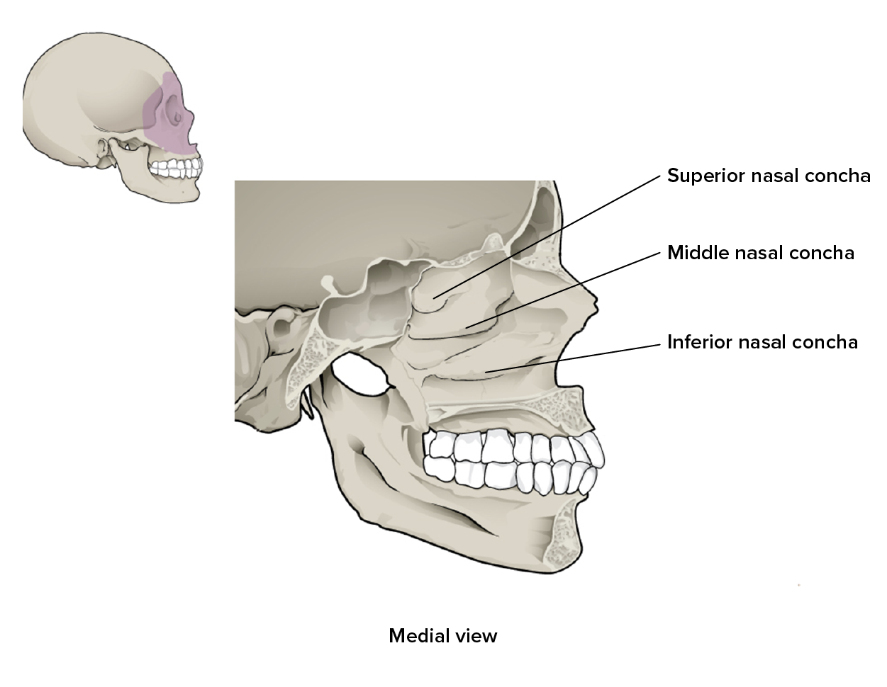
The ethmoid bone extends inferiorly to form the lateral walls of the nasal cavity. Extending from each lateral wall are the superior nasal concha and middle nasal concha, which are thin, curved projections that extend into the nasal cavity. You will learn about the inferior projection later in this lesson as it is formed by a separate facial bone. The tissues that form on the surface of the conchae serve to warm and moisturize air as it is breathed in through the nose.
The perpendicular plate is the inferior projection of the ethmoid bone that forms the superior portion of the nasal septum.
The lateral wall of the ethmoid bone forms the medial wall of the orbit (eye socket).
Ethmoid Bone - This midline view of the sagittally sectioned skull shows the (a) nasal septum and (b) nasal conchae. (c) The unpaired ethmoid bone is located at the midline within the central skull. It has an upward projection, the crista galli, and a downward projection, the perpendicular plate, which forms the upper nasal septum. The cribriform plates form both the roof of the nasal cavity and a portion of the anterior cranial cavity floor. The lateral sides of the ethmoid bone form the lateral walls of the upper nasal cavity, part of the medial orbit wall, and give rise to the superior and middle nasal conchae.
watch
View the following video for more information on this topic.
terms to know
Frontal Bone
The single flat bone that forms the anterior and part of the superior portion of the cranial cavity as well as the forehead.
Glabella
The slight depression along the anterior midline of the frontal bone, between the eyebrows.
Supraorbital Margin
The upper border of the orbit formed by the frontal bone.
Supraorbital Foramen
A hole in the supraorbital margin of the frontal bone that provides passage for a sensory nerve to the forehead.
Parietal Bone
One of a pair of flat bones that form most of the superior and lateral side of the brain case.
Occipital Bone
The single flat bone that forms the posterior skull and posterior base of the cranial cavity.
External Occipital Protuberance
A small projection at the posterior midline of the occipital bone.
Foramen Magnum
A large hole on the inferior side of the occipital bone and base of the skull which allows for passage of the spinal cord as it connects to the brain.
Occipital Condyle
One of two oval-shaped projections of the occipital bone located on either side of the foramen magnum that form a joint with the first vertebra.
Temporal Bone
The pair of flat bones that form the lower lateral side of the skull.
External Acoustic Meatus
A large opening in the lateral temporal bone that is associated with the ear.
Mandibular Fossa
A deep, oval-shaped depression of the temporal bone just in front of the external acoustic meatus which forms a joint with the mandible.
Temporomandibular Joint
A joint formed between the temporal bone and the mandible.
Zygomatic Process
An anterior bone projection of the temporal bone that extends towards the cheek (zygomatic) bone, forming the posterior portion of the zygomatic arch.
Mastoid Process
A large prominence of the temporal bone pointed inferiorly and located just posterior to the external acoustic meatus.
Sphenoid Bone
A single, irregular bone of the skull which forms much of the base of the central skull and also extends laterally to contribute to the sides of the skull.
Sella Turcica
The body of the sphenoid bone; ‘Turkish saddle.’
Lesser Wings
The smaller and more anterior set of wings of the sphenoid bone that form a prominent ridge.
Greater Wings
The larger and more posterior set of wings of the sphenoid bone which extend laterally to the outside of the lateral skull.
Ethmoid Bone
A single irregular bone that forms the roof and lateral walls of the upper nasal cavity, the upper portion of the nasal septum, and contributes to the medial wall of the orbit.
Cribriform Plate
The central portion of the ethmoid bone that separates the cranial and nasal cavities.
Crista Galli
The superior midline projection of the ethmoid bone into the cranial cavity; ‘roosters crest.’
Superior Nasal Concha
The upper most thin, curved projection of the ethmoid bone that extends into the nasal cavity.
Middle Nasal Concha
The middle thin, curved projection of the ethmoid bone that extends into the nasal cavity.
Perpendicular Plate
The inferior projection of the ethmoid bone that forms the superior portion of the nasal septum.
3. Sutures of the Skull
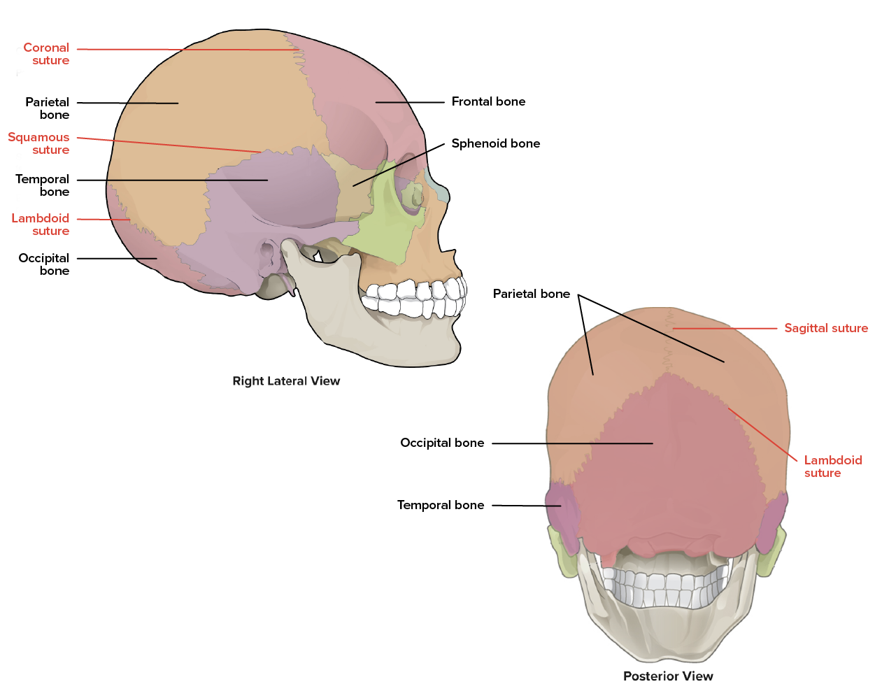
A suture is an immobile joint between adjacent bones of the skull. The narrow gap between the bones is filled with dense, fibrous connective tissue that unites the bones along irregular, tightly twisting paths. These twisting lines serve to tightly interlock the adjacent bones, thus adding strength to the skull for brain protection. There are four primary sutures in the skull.
The coronal suture is formed between the frontal and parietal bones. Recall that a frontal (or coronal) section splits the body into front and back pieces which is how this suture is named.
The sagittal suture is formed between the two parietal bones. Recall that a sagittal section splits the body into right and left pieces which is how this suture is named.
The lambdoid suture is formed between the parietal bones and the occipital bone. This suture, which forms an upside-down V, gets its name from the capital Greek letter lambda (Λ).
The squamous suture is formed between the parietal bones and the temporal bones.
Cranial Sutures - Cranial bones are joined to one another by joints called cranial sutures.
IN CONTEXT Disorders of the Skeletal System
Head and traumatic brain injuries are major causes of immediate death and disability, with bleeding and infections as possible additional complications. According to the Centers for Disease Control and Prevention (2010), approximately 30% of all injury-related deaths in the United States are caused by head injuries. The majority of head injuries involve falls. They are most common among young children (ages 0–4 years), adolescents (15–19 years), and the elderly (over 65 years). Additional causes vary, but prominent among these are automobile and motorcycle accidents.
Strong blows to the brain-case portion of the skull can produce fractures. These may result in bleeding inside the skull with subsequent injury to the brain. The most common is a linear skull fracture, in which fracture lines radiate from the point of impact. Other fracture types include a comminuted fracture, in which the bone is broken into several pieces at the point of impact, or a depressed fracture, in which the fractured bone is pushed inward. In a contrecoup (counterblow) fracture, the bone at the point of impact is not broken, but instead a fracture occurs on the opposite side of the skull. Fractures of the occipital bone at the base of the skull can occur in this manner, producing a basilar fracture that can damage the artery that passes through the carotid canal.
terms to know
Suture
An immobile joint between adjacent bones of the skull.
Coronal Suture
The joint formed between the frontal bone and the parietal bones.
Sagittal Suture
The joint formed between the two parietal bones.
Lambdoid Suture
The joint formed between the parietal bones and the occipital bone.
Squamous Suture
The joint formed between the parietal bones and temporal bones.
4. Facial Bones of the Skull
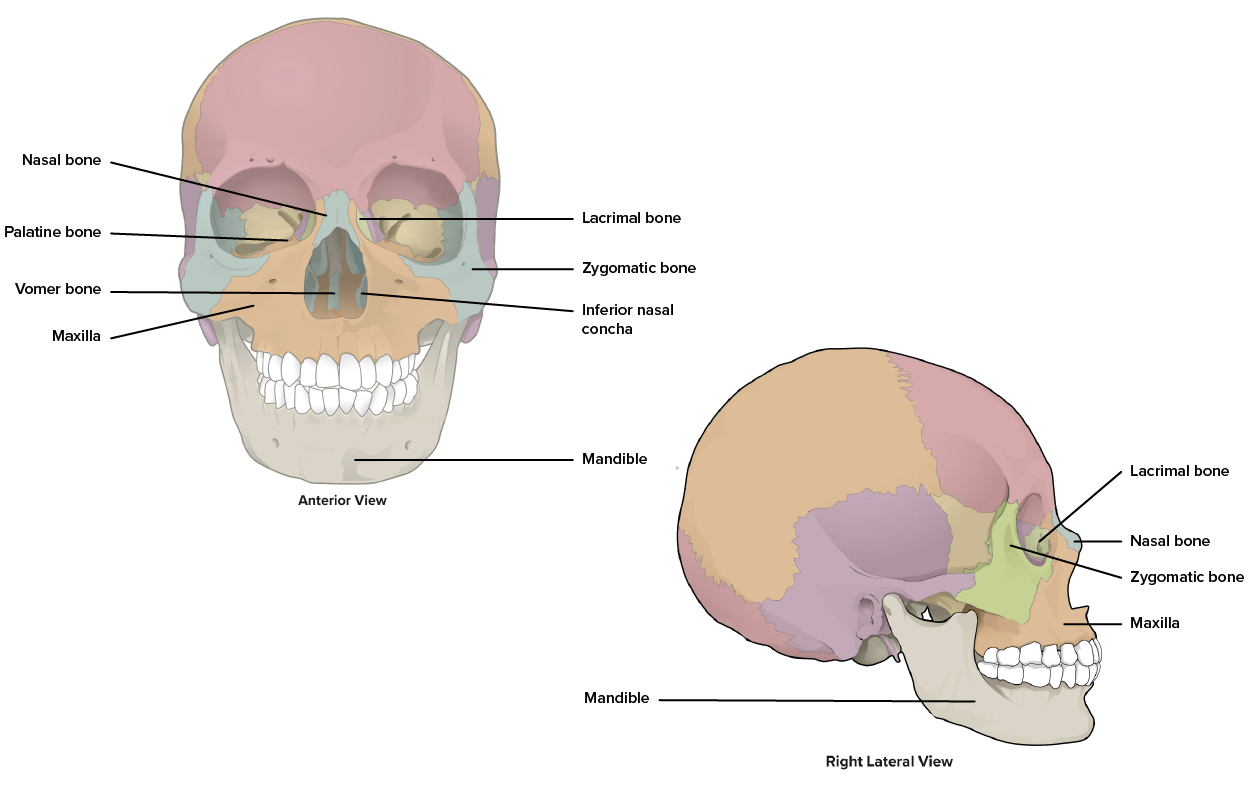
The facial bones of the skull form the upper and lower jaws, the nose, and portions of the nasal cavity, nasal septum, and orbit. The facial bones include 14 bones, with six paired bones and two unpaired bones. The paired bones are the maxilla, palatine, zygomatic, nasal, lacrimal, and inferior nasal conchae bones. The unpaired bones are the vomer and mandible bones. Although classified with the cranial bones, the ethmoid bone also contributes to the nasal septum and the walls of the nasal cavity and orbit.
Facial Bones - The facial structure is formed by fourteen bones.
4a. Zygomatic Bone
The zygomatic bone is an irregular facial bone also known as the cheekbone. Each of the paired zygomatic bones forms much of the lateral wall of the orbit. The lateral portion of the zygomatic bone is known as the temporal process of the zygomatic bone which extends posteriorly to meet the zygomatic process of the temporal bone to form the zygomatic arch.
Zygomatic Bone - This bone is also known as the cheekbone.
4b. Maxillary Bone
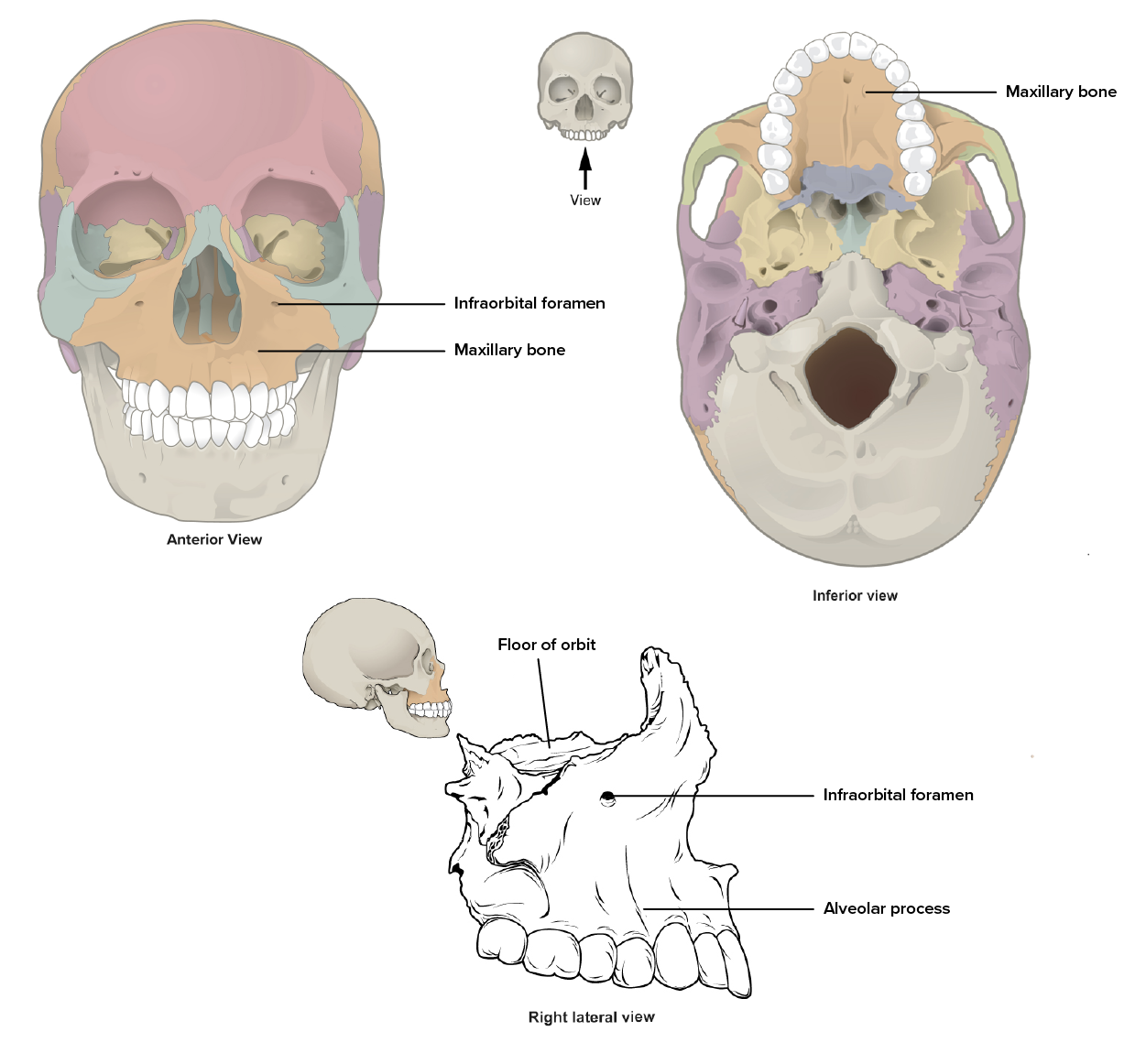
The maxillary bone, often referred to simply as the maxilla (plural, maxillae), is one of a pair of irregular bones that fuse together during development in the womb to form the upper jaw, much of the hard palate, the medial floor of the orbit, and the lateral base of the nose. The maxillary bone has a number of bony landmarks.
An alveolar process of the maxillary bone is a thickened bony projection that contains a tooth socket, also known as an alveolus (plural, alveoli) which houses a tooth.
The infraorbital foramen (infra, below) is a small hole below the eye socket which functions as a point of exit for a sensory nerve supplying regions of the face.
Inside the oral cavity, the maxillary bone projects posteriorly as the palatine process (towards the palatine bone) which forms a portion of the hard palate, a bony plate that forms the roof of the mouth and floor of the nasal cavity, separating the oral and nasal cavities.
Maxillary Bone - The maxillary bone forms the upper jaw and supports the upper teeth. Each maxilla also forms the lateral floor of each orbit and the majority of the hard palate.
IN CONTEXT Paranasal Sinuses
Four craniofacial bones, including the maxillary, frontal, sphenoid, and ethmoid bones, contain paranasal sinuses, or hollow, air-filled spaces that communicate by open passageways with the nasal cavity (paranasal, next to nasal cavity). These internal spaces are lined with nasal mucosa, a mucosal membrane. These spaces reduce bone mass to lighten the skull, add resonance to the voice, and as you can learn about in the respiratory system in A&P 2, they help warm and humidify incoming air while reabsorbing heat and humidity from outgoing air.
One obvious downside to the general function of the sinuses is when you have a cold or sinus congestion. The mucosal lining inflames and produces excess mucus, which can obstruct the narrow passageways between the sinuses and the nasal cavity, causing your voice to sound different to yourself and others. This blockage can also allow the sinuses to fill with fluid, with the resulting pressure producing pain and discomfort.
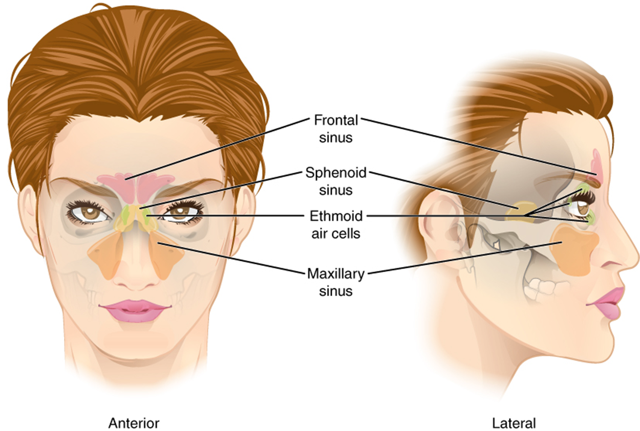
The paranasal sinuses are named for the skull bone that each occupies. Three exist in cranial bones (fronta, sphenoid, and ethmoid) while one exists in the maxillary bone. The frontal sinus is located just above the eyebrows, within the frontal bone. This irregular space may be divided at the midline into bilateral spaces, or these may be fused into a single sinus space. The frontal sinus is the most anterior of the paranasal sinuses. The largest sinus is the maxillary sinus. These are paired and located within the maxillary bones, where they occupy the area just below the orbits. The maxillary sinuses are most commonly involved during sinus infections. Because their connection to the nasal cavity is located high on their medial wall, they are difficult to drain. The sphenoid sinus is a single, midline sinus. It is located within the body of the sphenoid bone, just anterior and inferior to the sella turcica, thus making it the most posterior of the paranasal sinuses. The lateral aspects of the ethmoid bone contain multiple small spaces separated by very thin bony walls. Each of these spaces is called an ethmoid air cell. These are located on both sides of the ethmoid bone, between the upper nasal cavity and medial orbit, just behind the superior nasal conchae.
Paranasal Sinuses - The paranasal sinuses are hollow, air-filled spaces named for the skull bone that each occupies. The most anterior is the frontal sinus, located in the frontal bone above the eyebrows. The largest are the maxillary sinuses, located in the right and left maxillary bones below the orbits. The most posterior is the sphenoid sinus, located in the body of the sphenoid bone, under the sella turcica. The ethmoid air cells are multiple small spaces located in the right and left sides of the ethmoid bone, between the medial wall of the orbit and lateral wall of the upper nasal cavity.
4c. Palatine Bone
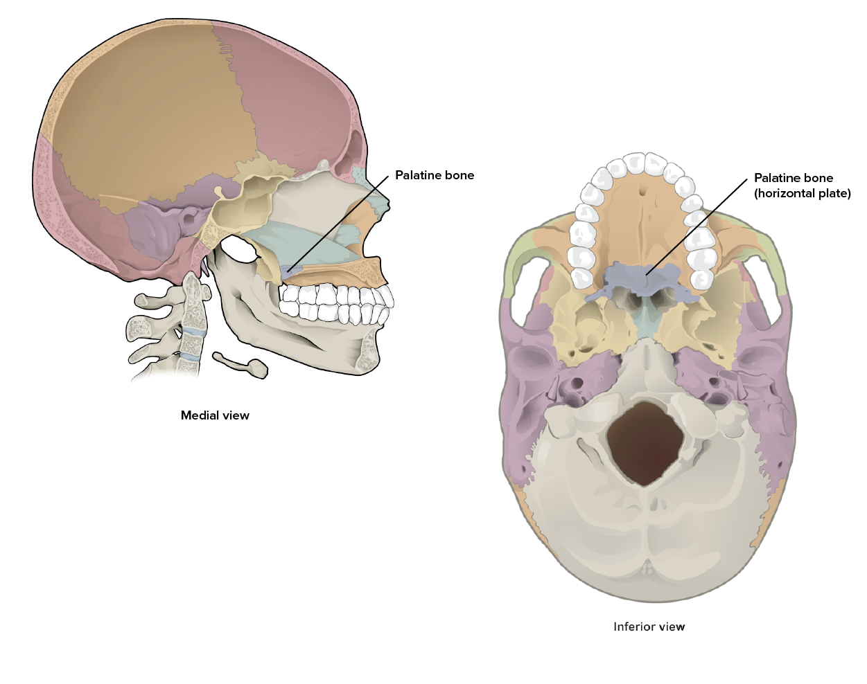
The palatine bone is one of a pair of irregular facial bones that contribute to the lateral walls of the nasal cavity, the medial wall of each orbit, and the posterior portion of the hard palate. The largest region of the palatine bone, known as the horizontal plate, projects anteriorly towards the palatine process of the maxillary bones. The plates from the right and left palatine bones join together at the midline to form the posterior quarter of the hard palate. Thus, the palatine bones are best seen in an inferior view of the skull and hard palate.
Palatine Bone - This bone forms parts of the nasal cavity, orbit, and hard palate.
IN CONTEXT Homeostatic Imbalances - Cleft Lip and Cleft Palate
During embryonic development, the right and left maxilla bones come together at the midline to form the upper jaw. At the same time, the muscle and skin overlying these bones join together to form the upper lip. Inside the mouth, the palatine processes of the maxilla bones, along with the horizontal plates of the right and left palatine bones, join together to form the hard palate. If an error occurs in these developmental processes, a birth defect of cleft lip or cleft palate may result.
Cleft lip is a common development defect that affects approximately 1:1000 births, most of which are male. This defect involves a partial or complete failure of the right and left portions of the upper lip to fuse together, leaving a cleft (gap).
A more severe developmental defect is cleft palate, which affects the hard palate. The hard palate is the bony structure that separates the nasal cavity from the oral cavity. It is formed during embryonic development by the midline fusion of the horizontal plates from the right and left palatine bones and the palatine processes of the maxilla bones. Cleft palate affects approximately 1 in 2500 births and is more common in females. It results from a failure of the two halves of the hard palate to completely come together and fuse at the midline, thus leaving a gap between them. This gap allows for communication between the nasal and oral cavities. In severe cases, the bony gap continues into the anterior upper jaw where the alveolar processes of the maxilla bones also do not properly join together above the front teeth. If this occurs, a cleft lip will also be seen. Because of the communication between the oral and nasal cavities, a cleft palate makes it very difficult for an infant to generate the suckling needed for nursing, thus leaving the infant at risk for malnutrition. Surgical repair is required to correct cleft palate defects.
4d. Nasal Bone
The nasal bone is one of two small flat bones that articulate (join) with each other to form the bony bridge of the nose. They also support the cartilages that form the lateral walls of the nose. These are the bones that are damaged when the nose is broken.
4e. Lacrimal Bone
Each lacrimal bone is one of two small, rectangular flat bones that forms the anteromedial wall of the orbit. This bone contains the nasolacrimal canal (naso, nasal; lacrimal, tears) which connects the eye to the nasal cavity allowing lacrimal fluid (tears) to drain.
4f. Inferior Nasal Conchae
Recall from earlier in this lesson that the nasal conchae are curved bony plates that project into the nasal cavity from the lateral wall and that there are three pairs - superior, middle, and inferior. The superior and middle nasal conchae are a part of the ethmoid bone. The inferior nasal conchae is the largest and most inferior projection.
Inferior Nasal Concha -This bone forms the largest and inferior most projection into the nasal cavity.
4g. Vomer Bone
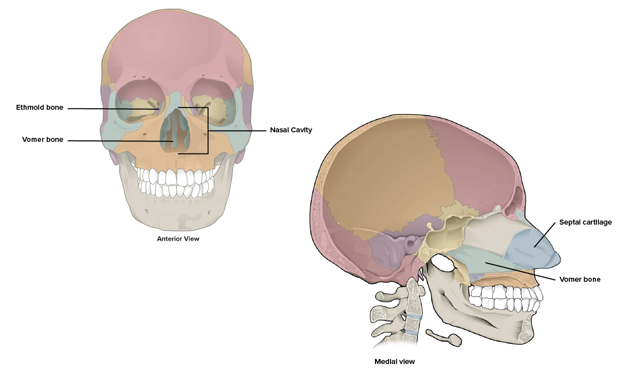
The vomer bone, often referred to simply as the vomer, is a triangular-shaped bone that forms the posterior-inferior part of the nasal septum. Recall that the anterior-superior portion of the nasal septum is formed by the perpendicular plate of the ethmoid bone. The anterior portion of the nasal septum between these two bones and extending anteriorly to separate the nostrils is formed by cartilage tissue (septal cartilage).
Vomer Bone - The vomer forms the posteroinferior portion of the nasal septum.
4h. Mandible
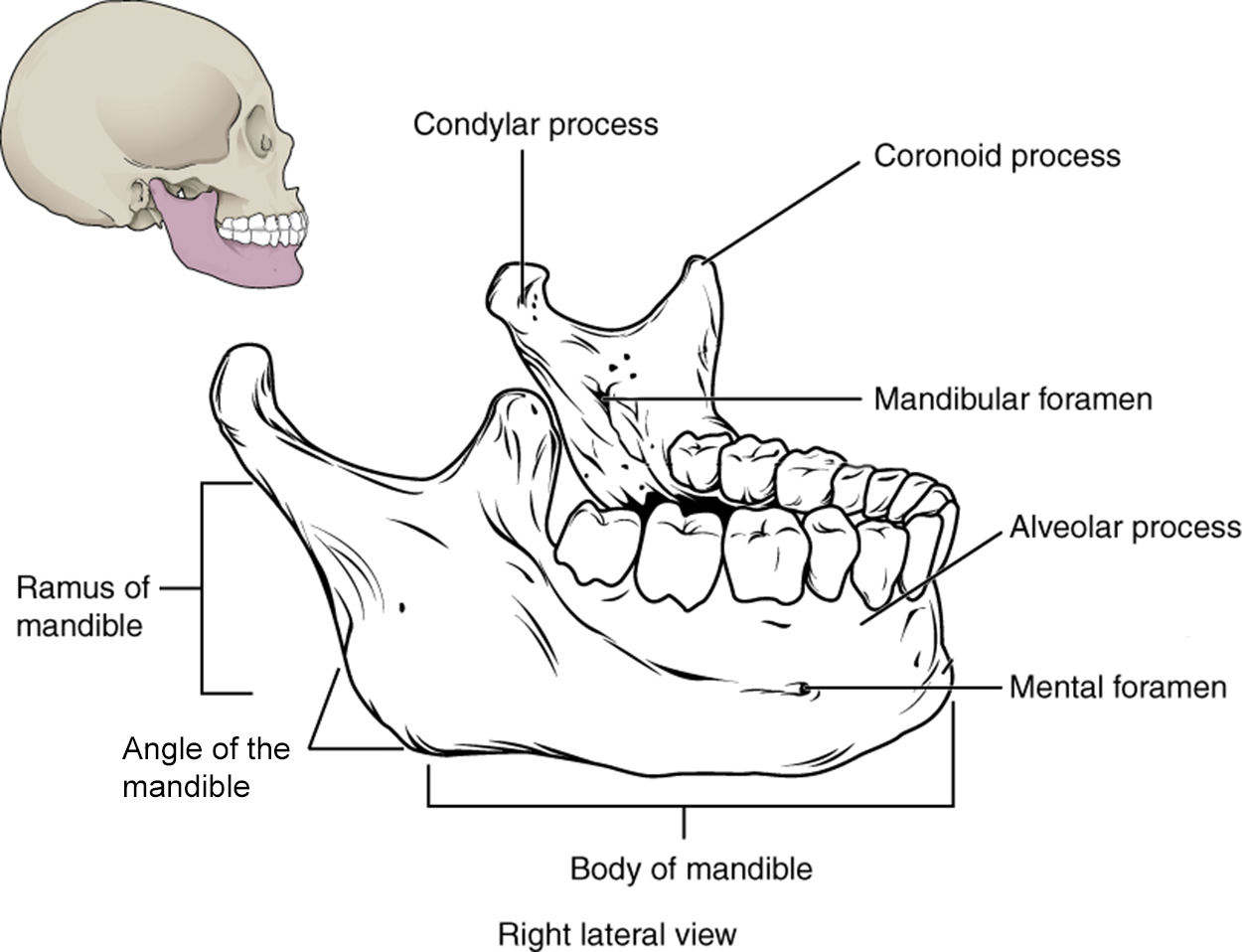
The mandible is an irregular facial bone, forms the lower jaw, and is the only moveable bone of the skull. At the time of birth, the mandible consists of paired right and left bones, but these fuse together during the first year to form the single U-shaped mandible of the adult skull.
Important landmarks for the mandible include the following:
The body of the mandible is the large horizontal, U-shaped section, the main part of the bone.
The ramus of the mandible (ramus, branch) is the wide upward projecting portion of the mandible.
The angle of the mandible is the corner where the body and ramus form a junction.
On top of the ramus of the mandible are two smaller projections. The condylar process of the mandible, also referred to as the mandibular condyle, is the posterior process and projects to the mandibular fossa of the temporal bone, forming the temporomandibular joint (TMJ). The coronoid process of the mandible is the anterior process and is flatter, allowing for attachment of muscles to aid in the chewing movement.
The alveolar process of the mandible is a thickened bony projection that contains a tooth socket, also known as an alveolus (plural, alveoli) which houses a tooth.
The mental foramen is a small hole located on each side of the anterior-lateral mandible, which is the exit site for a sensory nerve that supplies the chin.
Mandible - The mandible is the only moveable bone of the skull.
watch
Please watch the following video for more information on this topic.
terms to know
Zygomatic Bone
An irregular facial bone that forms the lateral wall of the orbit; the cheekbone.
Temporal Process of the Zygomatic Bone
Posterior projection forming the anterior portion of the zygomatic arch.
Zygomatic Arch
A bony arch of the lateral skull formed by the zygomatic process of the temporal bone and the temporal process of the zygomatic bone.
Maxillary Bone
One of a pair of irregular bones that together form the upper jaw and portions of the hard palate, orbit, and nose.
Alveolar Process of the Maxillary Bone
A thickened bony projection that contains a tooth socket.
Alveolus
A tooth socket.
Infraorbital Foramen
A small hole in the maxillary bone below the eye socket.
Palatine Process of the Maxillary Bone
The posterior projection extending towards the palatine bone that forms a portion of the hard palate.
Hard Palate
A bony plate that forms the roof of the mouth and floor of the nasal cavity, separating the oral and nasal cavities.
Palatine Bone
One of a pair of irregular facial bones that contribute to the nasal cavity, orbit, and hard palate.
Horizontal Plate
The anterior projection of the palatine bone that extends to the maxillary bones.
Nasal Bone
One of two small flat bones that articulate (join) with each other to form the bony bridge of the nose.
Lacrimal Bone
One of two small, rectangular flat bones that forms the anteromedial wall of the orbit.
Nasolacrimal Canal
A passageway in the lacrimal bone connecting the eye to the nasal cavity allowing lacrimal fluid (tears) to drain.
Inferior Nasal Concha
The largest and most inferior projection from the lateral nasal cavity wall.
Vomer Bone
A triangular-shaped bone that forms the posterior-inferior part of the nasal septum.
Mandible
An irregular facial bone that forms the lower jaw and is the only moveable bone of the skull.
Body of the Mandible
The large horizontal, U-shaped section of the lower jaw.
Ramus of the Mandible
The wide upward projecting portion of the mandible.
Angle of the Mandible
The corner where the body and ramus form a junction.
Condylar Process of the Mandible
The posterior process off the mandibular ramus that projects to the mandibular fossa of the temporal bone.
Coronoid Process of the Mandible
The flat, anterior process off the mandibular ramus.
Alveolar Process of the Mandible
A thickened bony projection that contains a tooth socket.
Mental Foramen
A small hole located on each side of the anterior-lateral mandible.
5. Hyoid Bone
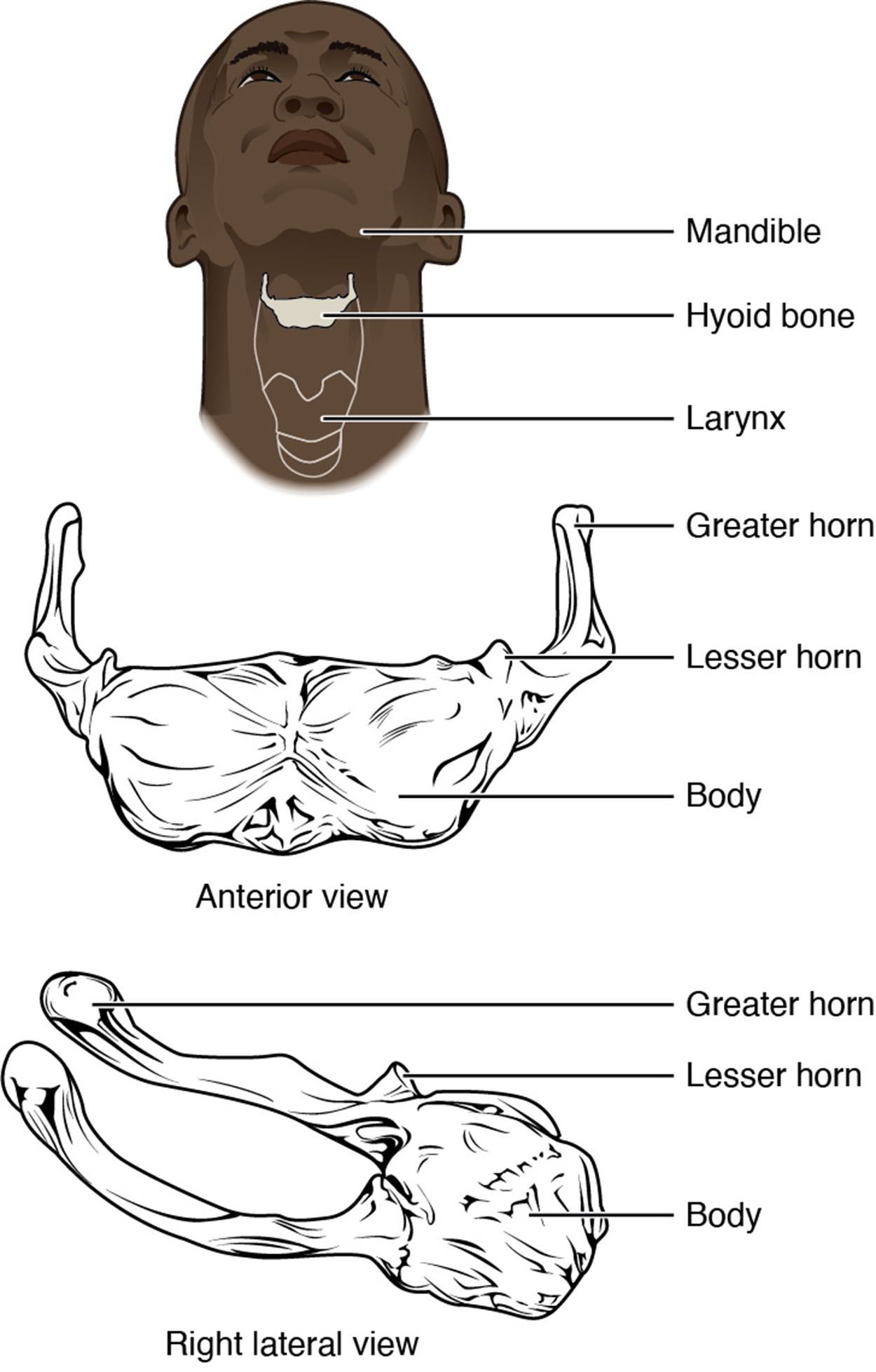
The hyoid bone is not a craniofacial bone. It is a single, independent, U-shaped irregular bone located in the upper throat. One unique feature of this bone is that it does not touch or make contact with any other bone of the body. The hyoid serves as the base for the tongue above, and is attached to the larynx (voicebox) below and the pharynx (throat) posteriorly. The hyoid is held in position by a series of small muscles that attach to it either from above or below. These muscles act to move the hyoid up/down or forward/back. Movements of the hyoid are coordinated with movements of the tongue, larynx, and pharynx during swallowing and speaking.
Hyoid Bone - The hyoid bone is located in the upper neck and does not join with any other bone. It provides attachments for muscles that act on the tongue, larynx, and pharynx.
term to know
Hyoid Bone
An irregular U-shaped bone in the upper neck.
summary
In this lesson, you learned how to subdivide the bones of the skull into cranial bones and facial bones. You learned the functions and select landmarks of the eight cranial bones - frontal, parietal, occipital, temporal, sphenoid, and ethmoid bones. You also learned to identify the sutures of the skull which connect the cranial bones. Then, you learned about the fourteen facial bones - the zygomatic, maxillary, palatine, nasal, lacrimal, inferior nasal conchae, vomer, and mandible bones. Lastly, you learned about one non-craniofacial bone, the hyoid bone.
A thickened bony projection that contains a tooth socket.
Alveolar Process of the Maxillary Bone
A thickened bony projection that contains a tooth socket.
Alveolus
A tooth socket.
Angle of the Mandible
The corner where the body and ramus form a junction.
Body of the Mandible
The large horizontal, U-shaped section of the lower jaw.
Condylar Process of the Mandible
The posterior process off the mandibular ramus that projects to the mandibular fossa of the temporal bone.
Coronal Suture
The joint formed between the frontal bone and the parietal bones.
Coronoid Process of the Mandible
The flat, anterior process off the mandibular ramus.
Cranial Bones
The bones of the skull that form the brain casing.
Craniofacial Bones
The bones of the skull.
Cranium
The skeletal structure of the head that supports the face and protects the brain.
Cribriform Plate
The central portion of the ethmoid bone that separates the cranial and nasal cavities.
Crista Galli
The superior midline projection of the ethmoid bone into the cranial cavity; ‘roosters crest.’
Ethmoid Bone
A single irregular bone that forms the roof and lateral walls of the upper nasal cavity, the upper portion of the nasal septum, and contributes to the medial wall of the orbit.
External Acoustic Meatus
A large opening in the lateral temporal bone that is associated with the ear.
External Occipital Protuberance
A small projection at the posterior midline of the occipital bone.
Facial Bones
The bones of the skull that underlie facial structure.
Foramen Magnum
A large hole on the inferior side of the occipital bone and base of the skull which allows for passage of the spinal cord as it connects to the brain.
Frontal Bone
The single flat bone that forms the anterior and part of the superior portion of the cranial cavity as well as the forehead.
Glabella
The slight depression along the anterior midline of the frontal bone, between the eyebrows.
Greater Wings
The larger and more posterior set of wings of the sphenoid bone which extend laterally to the outside of the lateral skull.
Hard Palate
A bony plate that forms the roof of the mouth and floor of the nasal cavity, separating the oral and nasal cavities.
Horizontal Plate
The anterior projection of the palatine bone that extends to the maxillary bones.
Hyoid Bone
An irregular U-shaped bone in the upper neck.
Inferior Nasal Concha
The largest and most inferior projection from the lateral nasal cavity wall.
Infraorbital Foramen
A small hole in the maxillary bone below the eye socket.
Lacrimal Bone
One of two small, rectangular flat bones that forms the anteromedial wall of the orbit.
Lambdoid Suture
The joint formed between the parietal bones and the occipital bone.
Lesser Wings
The smaller and more anterior set of wings of the sphenoid bone that form a prominent ridge.
Mandible
An irregular facial bone that forms the lower jaw and is the only moveable bone of the skull.
Mandibular Fossa
A deep, oval-shaped depression of the temporal bone just in front of the external acoustic meatus which forms a joint with the mandible.
Mastoid Process
A large prominence of the temporal bone pointed inferiorly and located just posterior to the external acoustic meatus.
Maxillary Bone
One of a pair of irregular bones that together form the upper jaw and portions of the hard palate, orbit, and nose.
Mental Foramen
A small hole located on each side of the anterior-lateral mandible.
Middle Nasal Concha
The middle thin, curved projection of the ethmoid bone that extends into the nasal cavity.
Nasal Bone
One of two small flat bones that articulate (join) with each other to form the bony bridge of the nose.
Nasal Cavity
A large space in the skull behind the nose.
Nasal Septum
The bony separation of the right and left sides of the nasal cavity.
Nasolacrimal Canal
A passageway in the lacrimal bone connecting the eye to the nasal cavity allowing lacrimal fluid (tears) to drain.
Occipital Bone
The single flat bone that forms the posterior skull and posterior base of the cranial cavity.
Occipital Condyle
One of two oval-shaped projections of the occipital bone located on either side of the foramen magnum that form a joint with the first vertebra.
Oral Cavity
A large space in the skull behind the jaws, also known as the mouth.
Orbit
The bony opening of the eye.
Palatine Bone
One of a pair of irregular facial bones that contribute to the nasal cavity, orbit, and hard palate.
Palatine Process of the Maxillary Bone
The posterior projection extending towards the palatine bone that forms a portion of the hard palate.
Parietal Bone
One of a pair of flat bones that form most of the superior and lateral side of the brain case.
Perpendicular Plate
The inferior projection of the ethmoid bone that forms the superior portion of the nasal septum.
Ramus of the Mandible
The wide upward projecting portion of the mandible.
Sagittal Suture
The joint formed between the two parietal bones.
Sella Turcica
The body of the sphenoid bone; ‘Turkish saddle.’
Sphenoid Bone
A single, irregular bone of the skull which forms much of the base of the central skull and also extends laterally to contribute to the sides of the skull.
Squamous Suture
The joint formed between the parietal bones and temporal bones.
Superior Nasal Concha
The upper most thin, curved projection of the ethmoid bone that extends into the nasal cavity.
Supraorbital Foramen
A hole in the supraorbital margin of the frontal bone that provides passage for a sensory nerve to the forehead.
Supraorbital Margin
The upper border of the orbit formed by the frontal bone.
Suture
An immobile joint between adjacent bones of the skull.
Temporal Bone
The pair of flat bones that form the lower lateral side of the skull.
Temporal Process of the Zygomatic Bone
Posterior projection forming the anterior portion of the zygomatic arch.
Temporomandibular Joint
A joint formed between the temporal bone and the mandible.
Vomer Bone
A triangular-shaped bone that forms the posterior-inferior part of the nasal septum.
Zygomatic Arch
A bony arch of the lateral skull formed by the zygomatic process of the temporal bone and the temporal process of the zygomatic bone.
Zygomatic Bone
An irregular facial bone that forms the lateral wall of the orbit; the cheekbone.
Zygomatic Process
An anterior bone projection of the temporal bone that extends towards the cheek (zygomatic) bone, forming the posterior portion of the zygomatic arch.