Table of Contents |
You have begun to learn about the anatomy of the skin, but now is an opportunity to explore it in more detail.
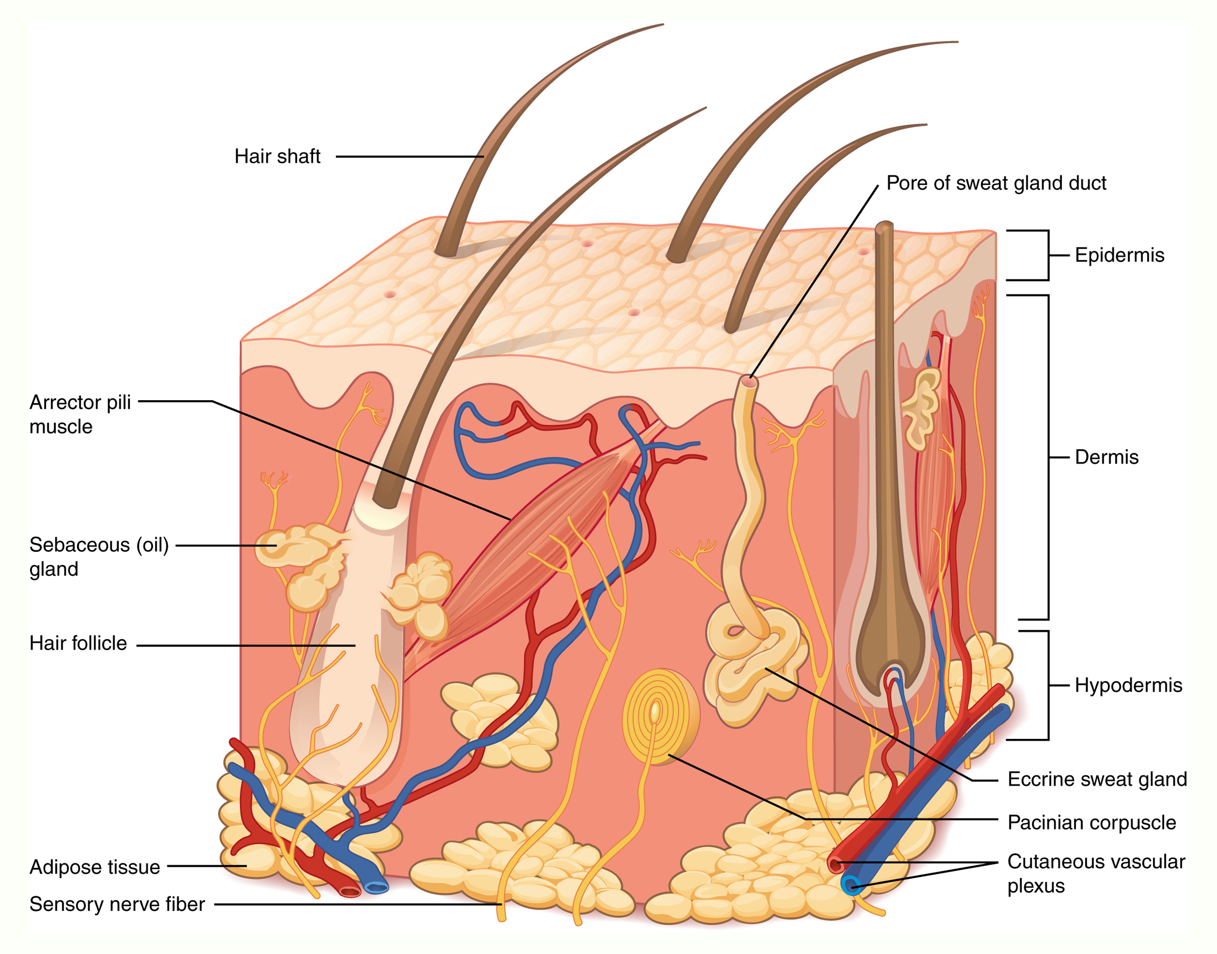
The skin has an upper epidermis with an underlying dermis. You can see these layers in the figure below, as well as part of a deeper hypodermis, or subcutaneous layer. The hypodermis is sometimes called the superficial fascia in gross anatomy and consists of connective tissue and adipose tissue.
The epidermis has multiple layers. Skin cells are produced by dividing cells deep in the skin, in a layer called the stratum basale. These cells migrate up toward the outer surface of the skin, becoming increasingly keratinized as they travel. The outer surface of skin, the stratum corneum, consists of dead cells that provide a tough outer barrier. The full set of skin layers, from deepest to most superficial, is the stratum basale, stratum spinosum, stratum granulosum, stratum lucidum, and stratum corneum. You can see these layers in the figure below.
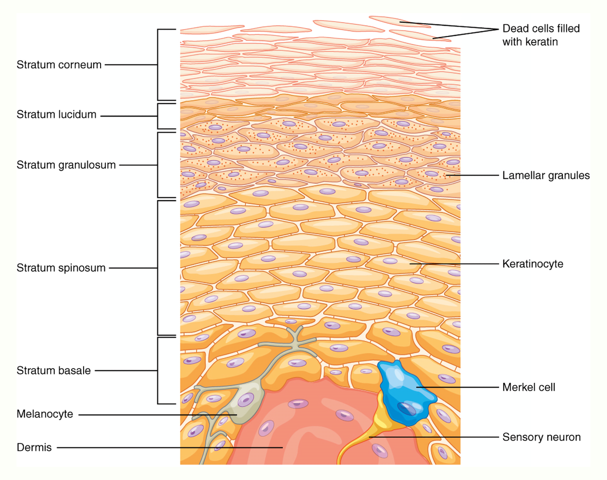
A double-layer membrane called the basement membrane separates the epidermis from the dermis. This membrane has an upper basal lamina (contributed by the epidermis) and a lower reticular lamina (contributed by the dermis).
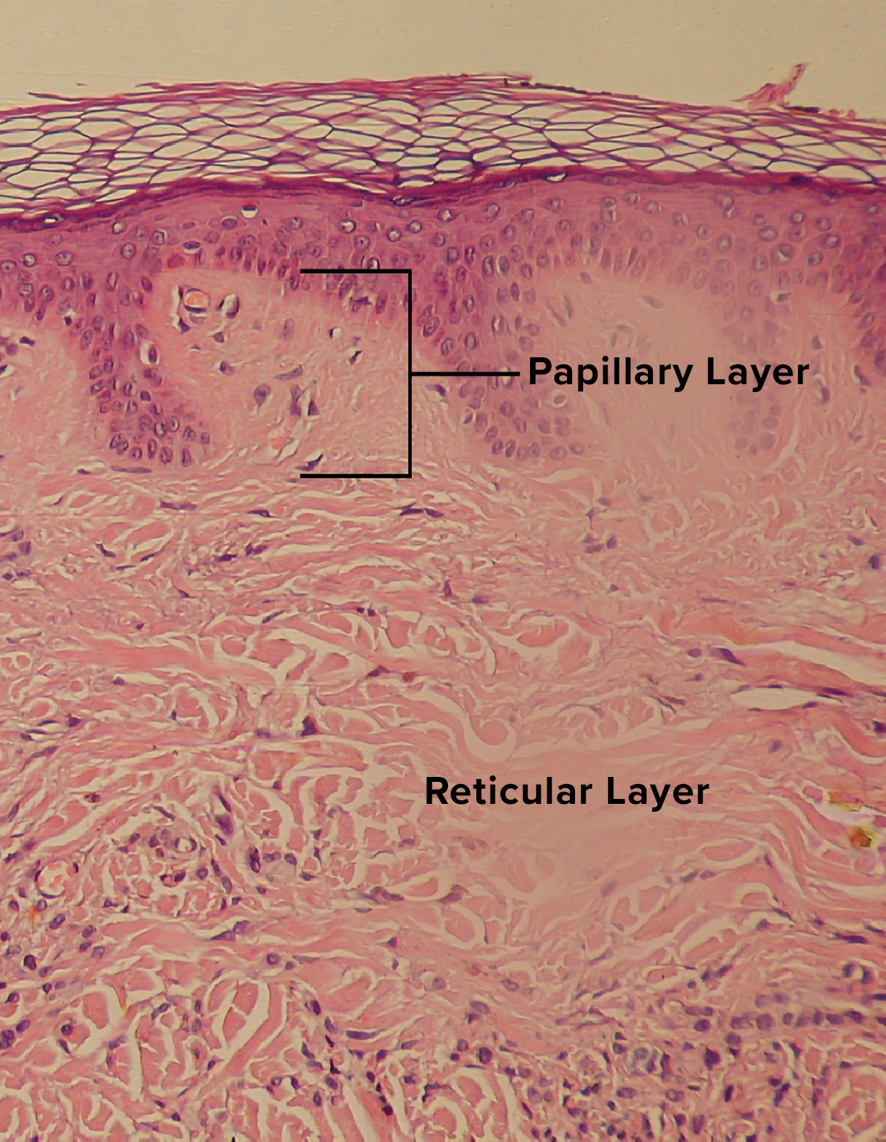
The dermis has two layers, as you can see in the figure below. The very top, darker layer in the figure is the epidermis. The region where the dermis folds up into the epidermis is the papillary layer. The deepest layer of dermis is the reticular layer.
The dermis contains sensory receptors, such as Meissner’s corpuscles (touch), Pacinian corpuscles (pressure), and Ruffini endings (small temperature variations and stretching). Nerve endings detect touch, temperature, and pain. The dermis also contains blood vessels and nerves.
The dermis contains collagen and elastin fibers. Collagen fibers provide structure and tensile strength, with strands of collagen extending into both the papillary layer and the hypodermis. In addition, collagen binds water to keep the skin hydrated. Elastin fibers provide some elasticity to the skin, enabling movement.
There are some common skin conditions related to the skin layers. These include:
Epidermolysis – Fragile skin due to genetic disorders.
Dermatosis – A general term for skin diseases.
Cellulitis – A bacterial skin infection in the dermis and hypodermis.
The skin has an important immune function. It is a barrier against infection because most pathogens cannot penetrate healthy skin. The skin also has a natural microbiota (the community of microbes that normally live somewhere). This microbiota helps to prevent colonization by pathogens. The most common microbes on the skin are Staphylococcus epidermidis, Staphylococcus aureus, and Propionibacterium acnes.
Common skin conditions include burns and wounds, which you will learn about in other lessons. Skin also experiences aging. As skin ages, collagen and elastin are lost, which leads to wrinkles and sagging.
Skin cancer can also develop. The risk of skin cancer increases with increased exposure to the sun. Regardless of your skin tone, pay attention to skin changes to detect anything that may need medical attention. Skin cancers can often be treated or removed if caught early, but melanoma in particular can be deadly if not caught quickly.
Skin accessory structures include the hair, nails, and glands. You have already learned some terms related to these structures.
The figure below shows a close-up of a hair follicle so that you can see details of hair follicle structure.
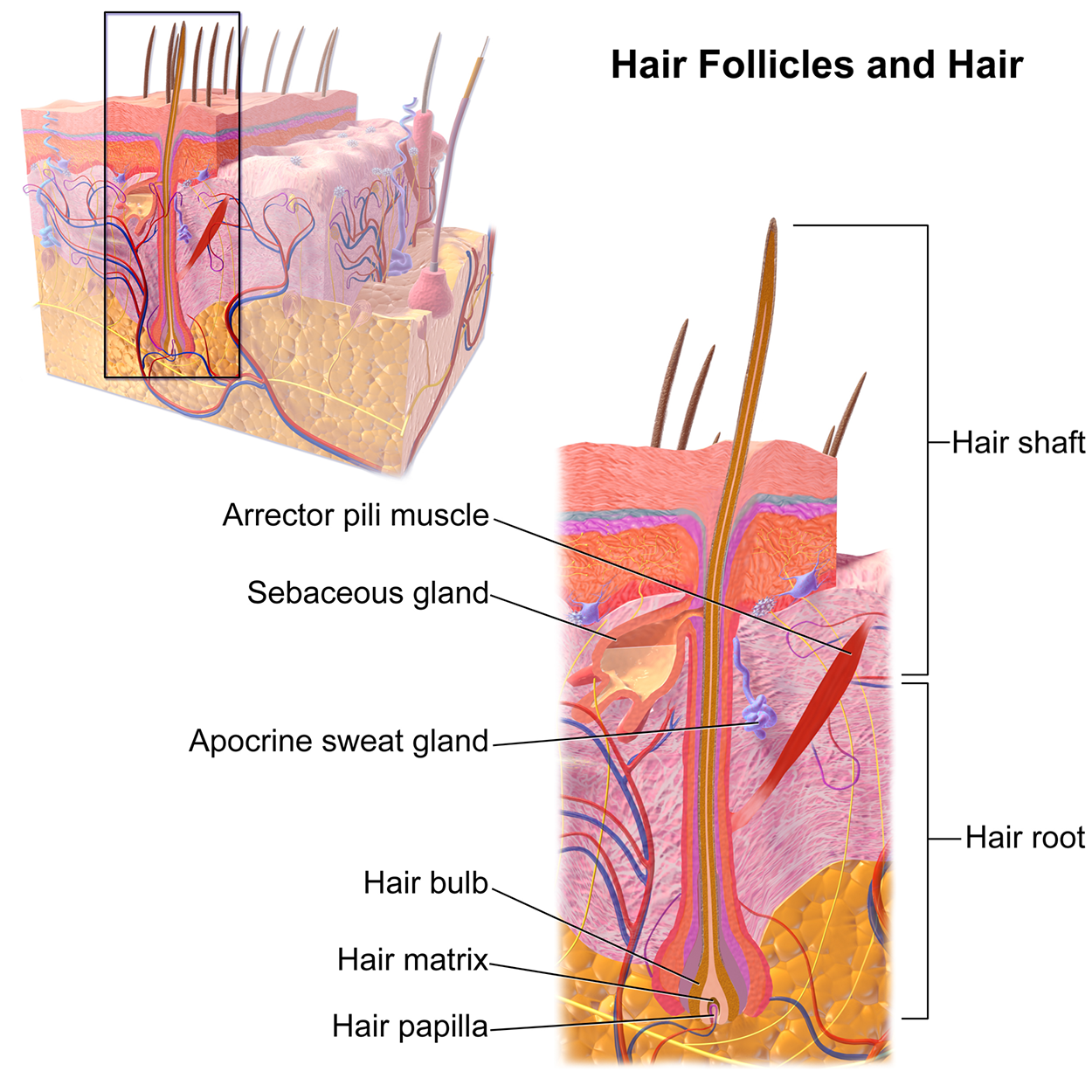
As you can see, the hair follicle has a hair bulb that extends down to where the epidermis and dermis meet. A small protrusion of dermis into the bulb is called the hair papilla. You can also see the distinction between the hair root and the hair shaft. The arrector pili muscle is a small muscle that can cause the hair to stand up, as may happen if you are cold or scared. You can also see a sebaceous gland on one side, producing sebum. There is also a coiled sweat gland.
Hair is not just about appearance. It also offers protection, aids in thermoregulation, and has a sensory function.
The sebum produced by sebaceous glands provides lubrication, waterproofing, and protection for the skin. Sebaceous glands are found all over the body, except on the palms and soles of the feet.
There are two types of sweat glands. Eccrine sweat glands are found all over the body and produce the sweat that helps you cool off. Apocrine glands produce a thicker sweat and are only located in the armpits and groin. Unlike eccrine glands, apocrine glands have ducts into hair follicles and can produce pheromones (chemicals used for signaling).
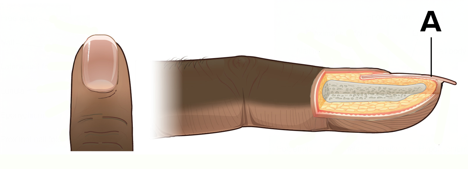
Nails are also part of the integumentary system. As you have already learned, nails have a nail plate, nail bed, lunula, and cuticle. Nails help to protect fingers and toes, but also help in touching and maneuvering objects.
SOURCE: THIS TUTORIAL HAS BEEN ADAPTED FROM (1) “OPEN RN | MEDICAL TERMINOLOGY – 2E” BY ERNSTMEYER & CHRISTMAN AT OPEN RESOURCES FOR NURSING (OPEN RN). (2) "ANATOMY AND PHYSIOLOGY 2E" AT OPENSTAX. ACCESS FOR FREE AT WTCS.PRESSBOOKS.PUB/MEDTERM/ AND OPENSTAX.ORG/DETAILS/BOOKS/ANATOMY-AND-PHYSIOLOGY-2E. LICENSING: CREATIVE COMMONS ATTRIBUTION 4.0 INTERNATIONAL.
REFERENCES
Harper, C.D. & Bermudez, R. (2023). Anhidrosis. In: StatPearls [Internet]. StatPearls Publishing. www.ncbi.nlm.nih.gov/books/NBK555988
Merriam-Webster. (n.d.). Idiopathic. In Merriam-Webster.com dictionary. Retrieved 6 April 2025, from www.merriam-webster.com/dictionary/idiopathic