Table of Contents |
The developments that led to the first microscopes began in the 1500s. During these years, eyeglass makers in the Netherlands began to develop the techniques that would later be used in microscopes. Italian astronomer Galileo Galilei (1564–1642) developed a compound microscope to examine insect parts. In compound microscopes, light passes through two lenses. In contrast, light passes through only one lens in a simple microscope.
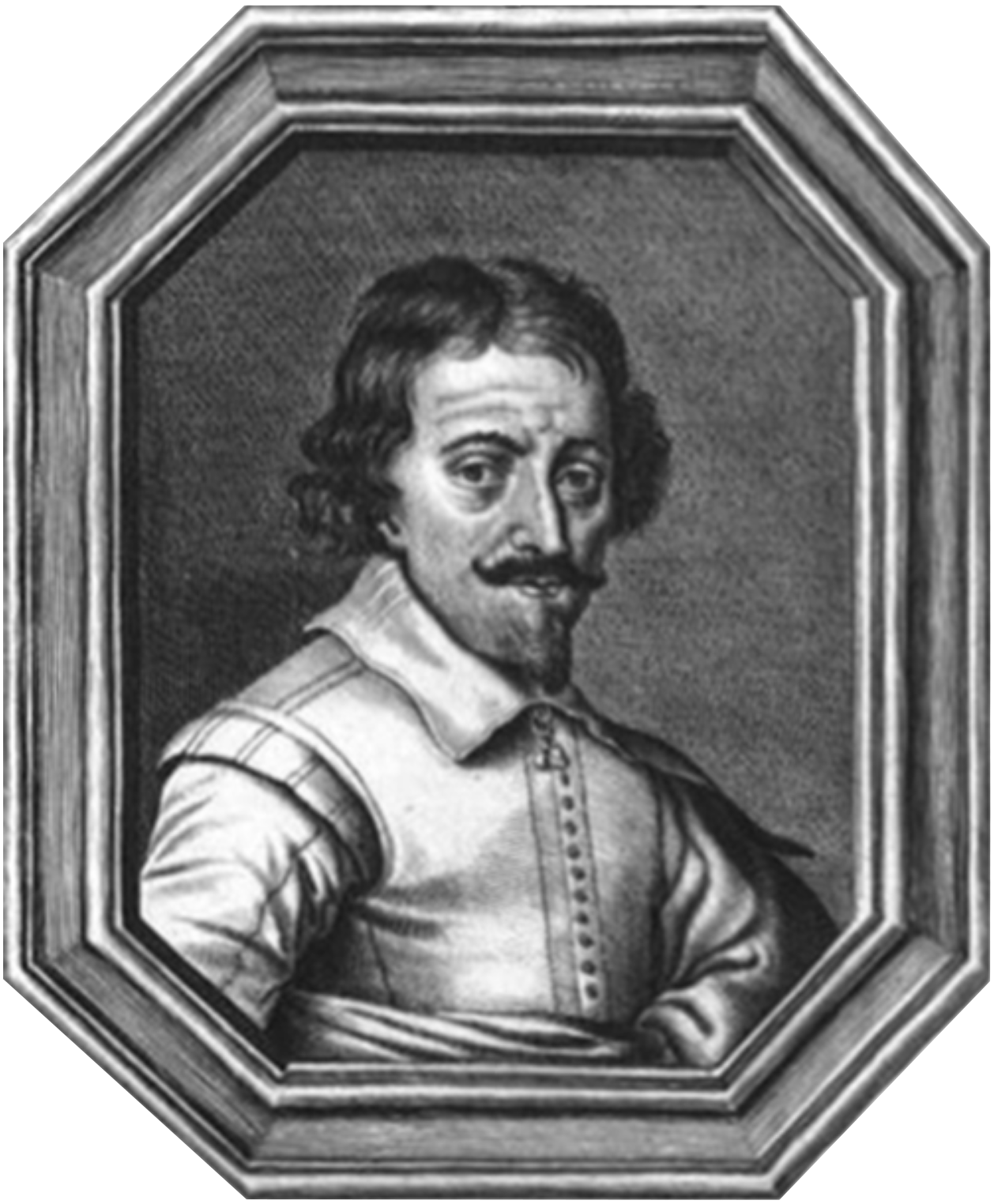 There is uncertainty about exactly who developed the very first microscope. Dutch spectacle makers Zacharias Janssen, pictured right, along with his father Hans, may have invented the telescope, the simple microscope, and the compound microscope during the late 1500s or early 1600s. The historical evidence is inconclusive, which has led to uncertainty about exactly who developed the very first microscope. Zacharias and Hans worked secretively to develop microscopes and telescopes. Because they did not publish their work, it is unclear exactly what they were able to view. Even their dates of birth and death are unknown.
At about the same time as the Janssens, their neighbor Hans Lippershey also worked on developing microscopes and telescopes. Lippershey often receives credit for inventing the first telescope. However, he also did not publish his work and, therefore, the details of his work are fuzzy.
More is known about individuals who published and disseminated their work widely. The earliest known clearly identifiable drawings of microscopic organisms were made by Antonie van Leeuwenhoek (1632–1723). Van Leeuwenhoek was a Dutch fabric seller. He became interested in making lenses, possibly to view thread. He was able to invent a simple microscope that was powerful enough that many of the organisms he viewed, including bacteria, were clearly identifiable. When he sent a series of letters describing his observations to the Royal Society of London in 1674, people were initially skeptical. He became well known as people realized that his observations of previously unknown organisms were real. As a result, he is sometimes called the “Father of Microbiology.”
Antonie van Leeuwenhoek (1632–1723), pictured in image (a) below, is often given credit for developing a microscope through which he could successfully view microbes, although it is not certain whether he was definitely the first. He called these organisms “animalcules” and “wee little beasties.” Even though van Leeuwenhoek’s microscopes were simple microscopes, as seen in this replica in image (b), they were more powerful and provided a better resolution than the compound microscopes of his day. Though more famous for developing the telescope, Galileo Galilei (1564–1642), pictured in image (c), was also one of the pioneers of microscopy.

Robert Hooke (1635–1703) also published his work on microscopy through the Royal Society. His bestselling book, published in 1865, was called Micrographia. Hooke developed a compound microscope, shown in image (a) below. He viewed cork cells, shown in image (b) below, and named them “cells.” These cork cells are dead plant cells with stiff cell walls surrounding empty regions that appear to be filled with air. Hooke described cork cells as “small Boxes or Bladders of Air” and noted that they resembled “Honey-comb.” He also observed that each “Cavern, Bubble, or Cell” was distinct.

| Italian astronomer Galileo Galilei (1564–1642) | Developed a compound microscope. |
| Dutch spectacle makers Hans Janssen (1601–1645) and Zacharias Janssen (1585–1638) | May have invented the telescope, the simple microscope, and the compound microscope in the late 1500s or early 1600s. |
| Dutch spectacle makers Hans Lippershey (1570–1619) | Developed microscopes and telescopes around the same time as the Janssens and is credited with developing the telescope. |
| Dutch fabric maker Antonie van Leeuwenhoek (1632–1723) | Observed and reported small organisms viewed using a simple microscope. |
| English physicist Robert Hooke (1635–1703) | Viewed and named cork cells; published Micrographia. |
| British surgeon Joseph Jackson Lister (1827–1912) | Developed the first essentially modern light microscope in 1830 (also developed techniques to reduce surgical infections). |
In the twentieth century, new microscopes were developed that used nonvisible light or energy in the form of electrons. This opened up possibilities to view very small specimens. The smaller the wavelength, the higher the possible resolution. This is why microscopes that use electrons instead of light can produce a far higher magnification with good resolution. You will learn more about these types of microscopes in the lesson on types of microscopy.
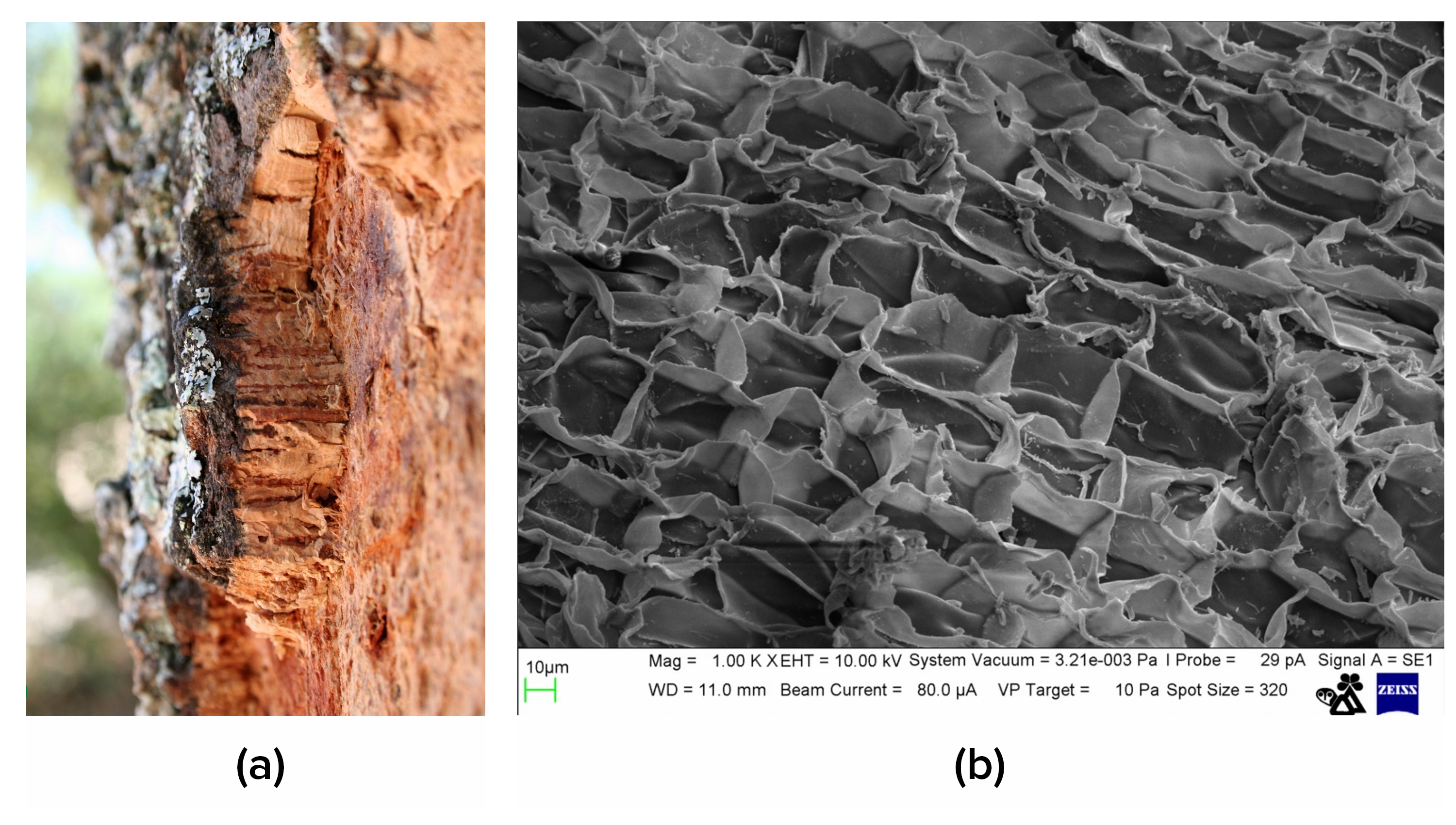
Image (a) below shows some cork on a tree with no magnification. Image (b), depicting a scanning electron micrograph, shows cork cells at a much higher magnification compared with the early engraving of cork cells viewed through a light microscope by Robert Hooke.
The first microscopes were light microscopes. Brightfield microscopes are probably the most commonly used and widely available light microscopes at present, and they are the focus of this lesson. You will learn more about other types of light microscopes in the lesson on types of microscopes.
Brightfield microscopes are compound microscopes. Light passes through a specimen and through at least two lenses to reach the viewer’s eyes. The light that does not pass through the specimen is bright because it passes only through clear lenses on its way to the viewer. As a result, the specimen is dark and the background is bright. The appearance of the image results from the ways in which structures in the specimen differentially transmit, absorb, reflect, and refract light.
To see the specimen, the viewer looks through one or two eyepieces that each contain an ocular lens (1 in the image below) that magnifies the specimen. Brightfield microscopes with one eyepiece are called monocular, and those with two eyepieces are called binocular.
The image is magnified by the ocular lens and by an objective lens that varies in magnification. The viewer turns a revolving nosepiece (2 in the image below) to change the magnification by moving the cylindrical objective containing the desired objective lens over the specimen. Objective lenses are generally labeled on the outside with their magnification, and longer objectives magnify more than shorter objectives.
When a brightfield microscope is used, the specimen to be viewed is placed on a slide. This can involve several steps, and you will learn more about slide preparation in other lessons.
To view a slide using a brightfield microscope, the slide must be placed on a platform called the stage (6 in the image below). Some stages have mechanical devices with knobs that allow the specimen to be moved very precisely by turning the dials.
Once the slide is in place, the coarse focusing knob (4 in the image below) and fine focusing knob (5 in the image below) are used to bring the image into focus. The coarse focusing knob is used to move the stage of a microscope over relatively large distances at low magnifications. This is used for large-scale movements with 4x and 10x objectives (3 in the image below). The fine focusing knob is used to move the stage of a microscope over relatively small distances. This is used for small-scale movements to focus precisely on a specimen. It is the only focusing knob that should be used at magnifications above 10x.
The image can be improved by adjusting the light reaching the specimen. The image is formed by light that travels upward through the specimen from an illuminator. The illuminator is often a high-intensity light bulb below the stage. There is a small opening in the stage that allows light to reach the specimen. Below this opening, there is a condenser lens (8 in the image below) that focuses light from the illuminator on the specimen resting on the stage above. A knob can be used to move the condenser as needed. Between the condenser lens and the stage, there is a diaphragm (8 in the image below) that can be opened or closed to adjust the amount of light reaching the specimen.
It is often helpful to adjust the amount of light reaching the specimen when the magnification is changed because images become dimmer as the magnification is increased. This is because the amount of light per unit area of the image decreases with increasing magnification. Additionally, adjusting the light can improve contrast. Brightness can also be adjusted using a rheostat that controls the intensity of the illuminator.
IN CONTEXT
As light passes through the specimen, it interacts with chromophores. These are pigments that absorb and reflect particular pigments of light. Stains are chemicals used to artificially add chromophores to a specimen. Particular stains bind preferentially to different structures and can be chosen to increase the contrast and visibility of structures of interest. You will learn more about stains in the lesson on staining microorganisms.

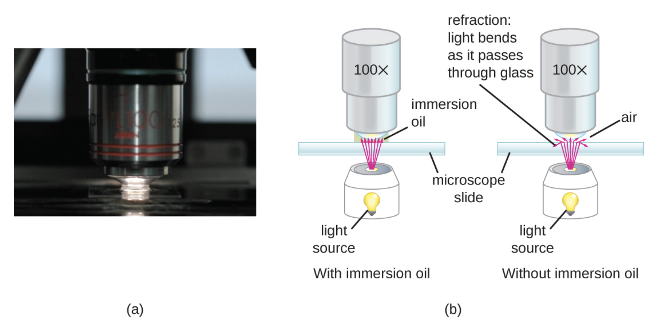
Because glass and air have different refractive indices, some light can be lost as it moves through the glass of a slide, then through the air, and then through the glass of the objective lens. Resolution can be improved by using an oil immersion lens and immersion oil. Immersion oil, with a similar refractive index to glass, can be placed between the slide and the objective to reduce the amount of light lost because of refraction. A variety of oils can be used for different types of light.
Oil immersion lenses like the one in image (a) below are used to improve resolution. Image (b) shows that because immersion oil and glass have very similar refractive indices, there is minimal refraction before the light reaches the lens. Without immersion oil, light scatters as it passes through the air above the slide, degrading the resolution of the image.
To calculate the total magnification of an image, the magnification of the ocular (usually 10x) is multiplied by the magnification of the chosen objective.
EXAMPLE
A specimen viewed through an ocular lens of 10x and an objective lens of 100x would be magnified (10x)(100x) = 1,000x.As the magnification is increased, the area of the slide viewed through the microscope decreases. Brightfield microscopes are often parfocal, meaning that the image stays basically in focus as the objective is changed. This means that it is easier to find a specimen by scanning the slide at low magnification to quickly view a large area before raising the magnification.
Source: THIS TUTORIAL HAS BEEN ADAPTED FROM OPENSTAX "MICROBIOLOGY." ACCESS FOR FREE AT openstax.org/details/books/microbiology. LICENSE: CC ATTRIBUTION 4.0 INTERNATIONAL. Accessed by August 2022.
REFERENCES
Merriam-Webster Dictionary. 2022. Parfocal. Retrieved July 31, 2022, from www.merriam-webster.com/dictionary/parfocal.
Nicola Angeli/MUSEThis file was uploaded by MUSE - Science Museum of Trento in cooperation with Wikimedia Italia. - MUSE, CC BY-SA 3.0, commons.wikimedia.org/w/index.php?curid=48378572.
Parker, N., Schneegurt, M., Thi Tu, A.-H., Lister, P., & Forster, B. (2016). Microbiology. OpenStax. Access for free at openstax.org/books/microbiology/pages/1-introduction.