Use Sophia to knock out your gen-ed requirements quickly and affordably. Learn more
×

Muscle Tissue and Motion

Author: Sophia
what's covered
In this lesson, you will explore the structure and function of the third of the four primary tissue categories, muscle tissue. Specifically, this lesson will cover:

Table of Contents

1. Characteristics of Muscle Tissue

Muscle tissue functions to allow for movement. In order to provide this function, all muscle cells share several characteristics:

  • Excitable: A muscle cell can respond to an electrochemical stimulus from nervous tissue and propagate an electrochemical stimulus along the cell.
  • Elastic: A muscle can return to its original length when relaxed.
  • Extendible: A muscle cell can stretch or extend.
  • Contractile: A muscle cell can shorten and generate a pulling force.

EXAMPLE

Some muscle movement is voluntary, which means it is under conscious control. A person decides to open a book and read a chapter on anatomy. Other movements are involuntary, meaning they are not under conscious control, such as the contraction of the pupil in your eye in bright light. Muscle tissue is classified into three types according to structure and function: skeletal, cardiac, and smooth.

terms to know
Excitable
The ability to respond to be stimulated by an electrochemical signal.
Elastic
The ability to return to an original shape after being stretched or compressed.
Extendible
The ability to be stretched or extended.
Contractile
The ability to shorten and generate a pulling force.

2. Muscle Tissue

Comparison of Structure and Properties of Muscle Tissue Types

Tissue Histology Function Location
Skeletal Long cylindrical fiber, striated, many peripherally located nuclei Voluntary movement, produces heat, protects organs Attached to bones and around entrance points to the body (e.g., mouth, anus)
Cardiac Short, branched, striated, single central nucleus Involuntary movement, contracts to pump blood Heart
Smooth Short, spindle-shaped, no evident striation (smooth appearance), a single nucleus in each fiber Involuntary movement, moves food, involuntary control of respiration, moves secretions, regulates the flow of blood in arteries by contraction Walls of major organs and passageways

Skeletal muscle is a voluntary muscle tissue that is attached to bones (i.e., the skeleton) as well as skin. Contraction of skeletal muscle makes locomotion, facial expressions, and posture possible. As a byproduct of their contraction, skeletal muscles generate heat thus contributing to the body's thermal homeostasis. When the body temperature drops, muscles respond involuntarily by shivering—tiny contractions which help increase body temperature.

key concept
Each muscle is composed of many muscle cells, also called muscle fibers or myocytes. The number of myocytes in a muscle remains relatively constant throughout life—numbers do not increase, but due to cell death, they can decrease. Under the light microscope, muscle cells appear striated (striped) with many nuclei just underneath the membrane. The striation is due to the regular alternation of the contractile proteins actin and myosin. Skeletal muscle cells are multinucleated as a result of the fusion of the early progenitor cells to form each long muscle fiber.

Cardiac muscle is an involuntary muscle tissue that forms the contractile walls of the heart. The cells of cardiac muscle, known as cardiomyocytes, also appear striated under the microscope. Unlike skeletal muscle fibers, cardiomyocytes are single cells typically with a single centrally located nucleus. A principal characteristic of cardiomyocytes is that they contract on their own intrinsic rhythms without any external stimulation. Cardiomyocytes attach to one another with specialized cell junctions called intercalated discs. Intercalated discs have both anchoring junctions and gap junctions. Attached cells form long, branching cardiac muscle fibers that are, essentially, a mechanical and electrochemical syncytium allowing the cells to synchronize their actions. The cardiac muscle pumps blood through the body and is under involuntary control. The attachment junctions hold adjacent cells together across the dynamic pressure changes of the cardiac cycle.

Smooth muscle tissue contraction is responsible for involuntary movements in the internal organs. It forms the contractile component of the digestive, urinary, and reproductive systems as well as the airways and arteries. Each cell is spindle-shaped with a single nucleus and no visible striations which creates its smooth cellular appearance.
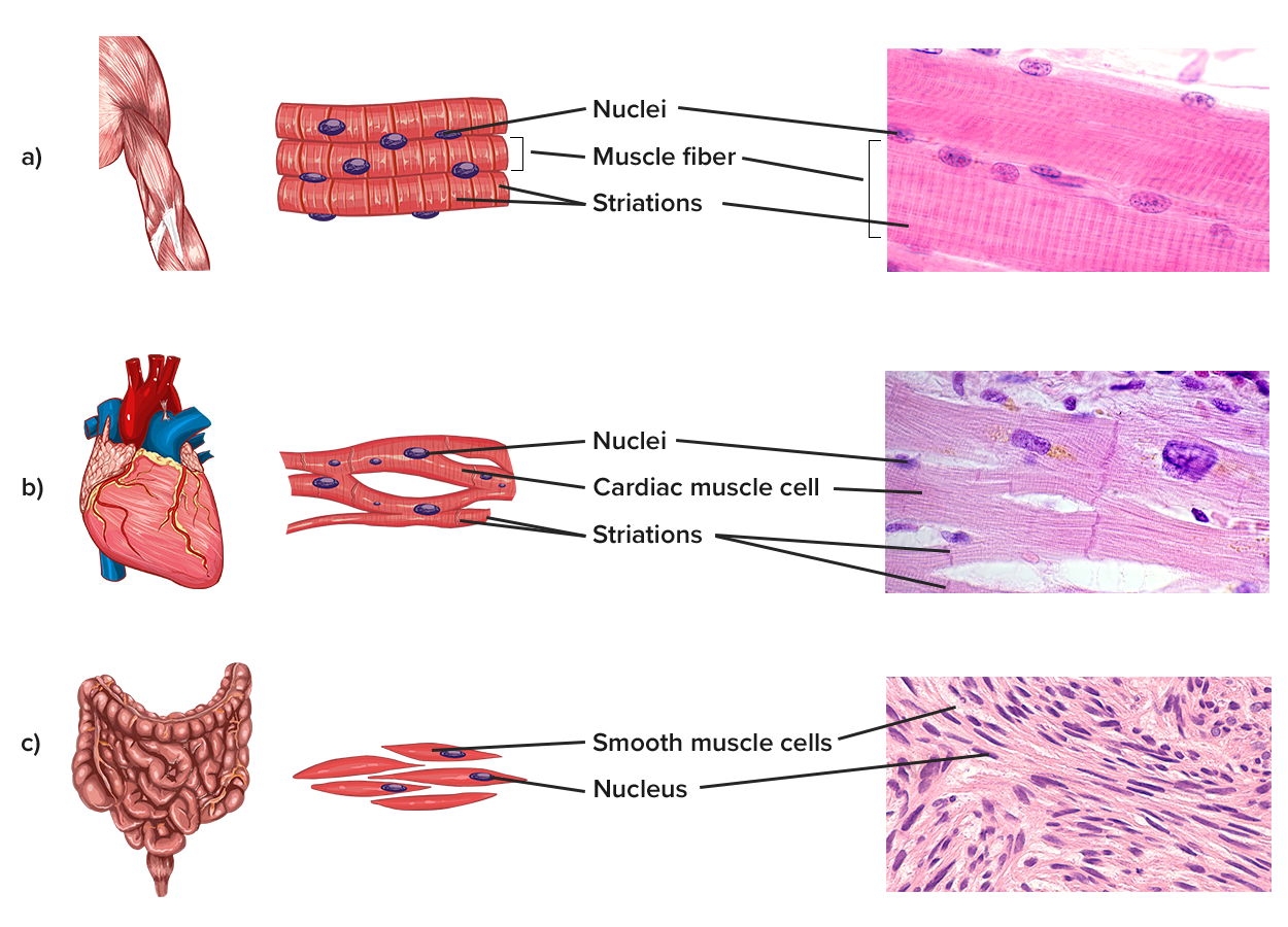
Image shows diagrams and micrographs of the three main muscle types. Skeletal muscle is highly organized, with parallel fibers, multiple nuclei, and striations along the length of each cell. Cardiac muscle is similar and striated, but mono-nucleated, less densely packed, and somewhat less parallel. Cells are oriented in the same direction, meandering like a stream. Smooth muscle is the most different, with muscle cells running in all directions, and much less apparent overall organization. Cells are non-striated and mono-nucleated.
Muscle Tissue - (a) Skeletal muscle cells are long, cylindrical cells with multiple nuclei and striations. This tissue provides voluntary movement as well as protection for entrances and exits into or out of the body and internal organs. (b) Cardiac muscles form branching networks with cells that have striations and one nucleus. This tissue forms the involuntary contractile tissue of the heart. (c) Smooth muscle cells are fusiform (tapered) cells with one nucleus and no striations. This tissue provides involuntary movements within internal organs.

terms to know
Skeletal Muscle Tissue
A muscle tissue that provides voluntary movement and protection of internal organs.
Myocyte
A muscle cell.
Striation
The stripes visible on skeletal and cardiac muscle tissue due to the regular alternation of the internal contractile proteins.
Cardiac Muscle Tissue
A muscle tissue that provides involuntary contraction of the heart.
Smooth Muscle Tissue
A muscle tissue that provides involuntary contraction of the internal organs.


summary
In this lesson, you learned to identify the common characteristics of muscle tissue. You also learned to identify the three types of muscle tissue based on cell shape, structure, and function.

Source: THIS TUTORIAL HAS BEEN ADAPTED FROM OPENSTAX “ANATOMY AND PHYSIOLOGY 2E.” ACCESS FOR FREE AT HTTPS://OPENSTAX.ORG/DETAILS/BOOKS/ANATOMY-AND-PHYSIOLOGY-2E. LICENSE: CC ATTRIBUTION 4.0 INTERNATIONAL.

Terms to Know
Cardiac Muscle Tissue

A muscle tissue that provides involuntary contraction of the heart.

Contractile

The ability to shorten and generate a pulling force.

Elastic

The ability to return to an original shape after being stretched or compressed.

Excitable

The ability to respond to be stimulated by an electrochemical signal.

Extendible

The ability to be stretched or extended.

Myocyte

A muscle cell.

Skeletal Muscle Tissue

A muscle tissue that provides voluntary movement and protection of internal organs.

Smooth Muscle Tissue

A muscle tissue that provides involuntary contraction of the internal organs.

Striation

The stripes visible on skeletal and cardiac muscle tissue due to the regular alternation of the internal contractile proteins.