Table of Contents |
The epidermis is the superficial layer of the integument (skin).
 is composed of the superficial epidermis and deep dermis. These layers are held to underlying structures by the deeper hypodermis.")
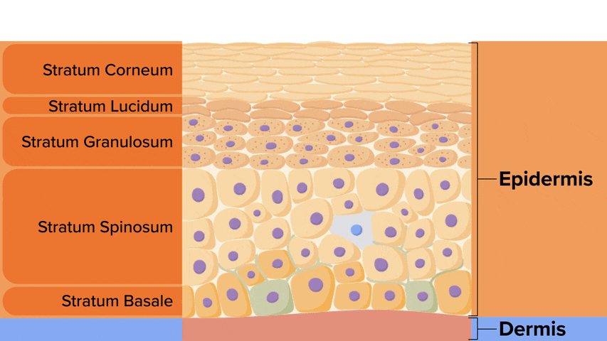
As you previously learned, the epidermis is composed of keratinized, stratified squamous epithelium. The epidermis is organized into four or five layers of epithelial cells, depending on its location in the body. Skin that has four layers of cells is referred to as “thin skin.” Most of the skin can be classified as thin skin. “Thick skin” is found only on the palms of the hands and the soles of the feet and has a unique fifth layer. From superficial (close to the surface of the body) to deep (away from the surface of the body), the layers of the epidermis are listed below. The term “stratum” is Latin for layer:
 contains four layers in the epidermis and makes up the majority of the skin on the body. Thick skin (right) contains five layers in the epidermis and is only found in the palms of the hands and soles of the feet.")
The cells in all of the layers except the stratum basale are called keratinocytes. A keratinocyte is a cell that manufactures and stores the protein keratin. Keratin is a water-resistant protein found in keratinized stratified squamous epithelial tissue. This protein gives hair, nails, and skin their hardness and water-resistant properties.
The stratum basale is the deepest epidermal layer and is a single layer of cells primarily made of basal cells. A basal cell is a cuboidal-shaped stem cell that is a precursor of the keratinocytes of the epidermis. All of the keratinocytes are produced from this single layer of cells, which are constantly going through mitosis to produce new cells. As new cells are formed, the existing cells are pushed superficially away from the stratum basale and eventually shed at the superficial surface. Another cell found dispersed among the basal cells in the stratum basale is a melanocyte, a cell that produces the pigment melanin. Melanin is a yellow, brown, or black pigment that gives hair and skin their color and is also able to block ultraviolet (UV) radiation from the sun or other sources that might damage underlying tissues.
The stratum basale attaches the epidermis to the underlying dermis. Epithelial tissues connect to underlying tissues through a basement membrane.
Recall that thick skin (palms of the hands and soles of the feet) contains five epidermal layers. Due to its increased thickness, the formation of dermal papillae in thick skin forms ridges on the superficial surface of the epidermis called epidermal ridges. On the fingers, these are collectively known as fingerprints and serve to increase friction, making it easier to hold a glass jar without it slipping. Since these are formed based on movement within the womb, every individual person’s epidermal ridges are unique; even identical twins will be different. However, certain general patterns (swirl, loop, etc.) are inherited. A person’s epidermal ridges do not change with growth or age but can be altered by injuries that alter the epidermal–dermal boundary. Epidermal ridges also exist on the palm of the hand, sole of the foot, and toes.
. These form unique identifying patterns, commonly referred to as fingerprints.")
The stratum spinosum is the second layer of the epidermis, superficial to the stratum basale, and is named based on the spiny shape of the cells. However, this shape is an artifact of the removal from the tissue and staining for microscopy. Unstained epidermis samples do not exhibit this characteristic appearance. The stratum spinosum is composed of 8 to 10 layers of keratinocytes, formed as a result of cell division in the stratum basale. Interspersed among the keratinocytes of this layer is a type of cell with long extensions called the Langerhans cell, which performs phagocytosis, engulfing bacteria, foreign particles, and damaged cells that occur in this layer.
The keratinocytes in the stratum spinosum begin the synthesis of keratin and release a water-repelling glycolipid that helps prevent water loss from the body, making the skin relatively waterproof. As new keratinocytes are produced atop the stratum basale, the keratinocytes of the stratum spinosum are pushed into the stratum granulosum.
The stratum granulosum is the third layer of the epidermis and is named for its grainy appearance due to increased production and accumulation of keratin. These three to five layers of cells become flatter, and their cell membranes thicken. The nuclei and other cell organelles disintegrate as the cells begin to die, leaving behind the keratin and cell membranes that will form the stratum lucidum, the stratum corneum, and the accessory structures of hair and nails.
The stratum lucidum is a thin layer of the epidermis only present in thick skin. This layer is named based on being seemingly translucent (lucidum, clear). The keratinocytes that compose the stratum lucidum are dead and flattened.
The stratum corneum is the most superficial layer of the epidermis and is the layer exposed to the outside environment. The increased keratinization (also called cornification) of the cells in this layer gives it its name. There are usually 15 to 30 layers of cells in the stratum corneum. This dry, dead layer helps prevent the penetration of microbes and dehydration of underlying tissues and provides mechanical protection against abrasion for the more delicate, underlying layers. Cells in this layer are shed periodically and are replaced by cells pushed up from the stratum granulosum (or stratum lucidum in the case of the palms and soles of feet). The entire layer is replaced during a period of about 4 weeks. Cosmetic procedures, such as microdermabrasion, help remove some of the dry, upper layers and aim to keep the skin looking “fresh” and healthy.
The dermis might be considered the “core” of the integumentary system (derma, skin) and is distinct from the epidermis (epi, upon/over) and hypodermis (hypo, below). It contains blood vessels, lymph vessels, nerves, and other structures, such as hair follicles and sweat glands. The dermis is made of two layers of connective tissue that compose an interconnected mesh of elastin and collagenous fibers, produced by fibroblasts.

The papillary layer is the superficial layer of the dermis and is made of loose, areolar connective tissue. Areolar tissue forms a mesh of collagen, elastic, and reticular fibers among fibroblasts, a small number of fat cells (adipocytes), and an abundance of small blood vessels. This layer fills in space and provides support for the superficial epidermis. In addition, the papillary layer contains phagocytes, which are defensive cells that help fight bacteria or other infections that have breached the skin, and sensory nerve fibers.
Deep to the papillary layer is the much thicker reticular layer, composed of dense, irregular connective tissue. Dense, irregular tissue contains tightly packed, randomly oriented collagen fibers, providing strength in all directions. Therefore, the reticular layer provides strength.
This layer has a rich nerve supply and is well vascularized. The subpapillary plexus is a network of vessels that provides blood to the stratum basale of the epidermis and the papillary layer of the dermis. This plays a role in thermoregulation. As the body increases blood flow to the skin, this allows for the transfer of heat from the body to the environment.
The reticular layer appears reticulated (net-like) due to a tight meshwork of fibers. Elastic fibers provide some elasticity to the skin, enabling movement and recoil. Collagen fibers provide structure and tensile strength, with strands of collagen extending into both the papillary layer and the hypodermis. In addition, collagen binds water to keep the skin hydrated.
In the reticular layer is also the cutaneous plexus. This is a network of arteries along the hypodermis border, and this provides blood to the reticular layer.
The hypodermis (also called the subcutaneous layer or superficial fascia) is a layer deep in the dermis and serves to connect the integument to the underlying tissue of bones and muscles. It is not strictly a part of the skin, although the border between the hypodermis and dermis can be difficult to distinguish. The hypodermis consists of well-vascularized, loose, areolar connective tissue and adipose tissue, which functions as a mode of lipid storage, providing energy storage, insulation, and cushioning.
IN CONTEXT
Everyday Connection—Lipid Storage
The hypodermis is home to most of the fat that concerns people when they are trying to keep their weight under control. Adipose tissue present in the hypodermis consists of fat-storing cells called adipocytes. This stored fat can serve as an energy reserve, insulate the body to prevent heat loss, and act as a cushion to protect underlying structures from trauma.
Where the fat is deposited and accumulates within the hypodermis depends on hormones (testosterone, estrogen, insulin, glucagon, leptin, and others), as well as genetic factors. Fat distribution changes as our bodies mature and age. Males tend to accumulate fat in different areas (neck, arms, lower back, and abdomen) than do females (breasts, hips, thighs, and buttocks). The body mass index (BMI) is often used as a measure of fat, although this measure is, in fact, derived from a mathematical formula that compares body weight (mass) to height. Therefore, its accuracy as a health indicator can be called into question in individuals who are extremely physically fit.
In many animals, there is a pattern of storing excess calories as fat to be used in times when food is not readily available. In much of the developed world, insufficient exercise coupled with the ready availability and consumption of high-calorie foods have resulted in unwanted accumulations of adipose tissue in many people. Although periodic accumulation of excess fat may have provided an evolutionary advantage to our ancestors, who experienced unpredictable bouts of famine, it is now becoming chronic and considered a major health threat.
SOURCE: THIS TUTORIAL HAS BEEN ADAPTED FROM OPENSTAX “ANATOMY AND PHYSIOLOGY 2E”. ACCESS FOR FREE AT OPENSTAX.ORG/BOOKS/ANATOMY-AND-PHYSIOLOGY-2E/PAGES/1-INTRODUCTION. LICENSE: CREATIVE COMMONS ATTRIBUTION 4.0 INTERNATIONAL.
REFERENCES
Millisky, R. (2022, August 9). The true story of paul karason, the man whose skin slowly turned blue. MSN. Retrieved November 11, 2022, from www.msn.com/en-us/health/wellness/the-true-story-of-paul-karason-the-man-whose-skin-slowly-turned-blue/ar-AA10tWT5
Brown University. (2012, October 30). How silver turns people blue. ScienceDaily. Retrieved November 11, 2022 from www.sciencedaily.com/releases/2012/10/121030143029.htm