Table of Contents |
The urinary system, also referred to as the renal system or urinary tract, consists of the kidneys, ureters, bladder, and urethra. The kidneys form urine and perform other functions attributed to the urinary system. The ureters carry the urine away from the kidneys to the urinary bladder, where it is stored until it is expelled during urination. The urethra carries the urine from the urinary bladder to the outside of the body during urination.
The kidneys are a pair of organs that filter waste from the blood and produce urine. Kidneys lie on either side of the spine behind the abdominal cavity, well protected by muscle, fat, and ribs (they are retroperitoneal). They are roughly the size of your fist. The male kidneys are typically a bit larger than the female kidneys. The kidneys are well-vascularized, receiving about 25 percent of the blood pumped out of the heart with each heartbeat.
The outer region of the kidney is called the renal cortex, and the inner region is called the renal medulla. The renal hilum is the entry and exit site for vessels, nerves, and ureters. The renal arteries (colored red) come directly from the descending aorta, and the renal veins (colored blue) return cleansed blood to the inferior vena cava.
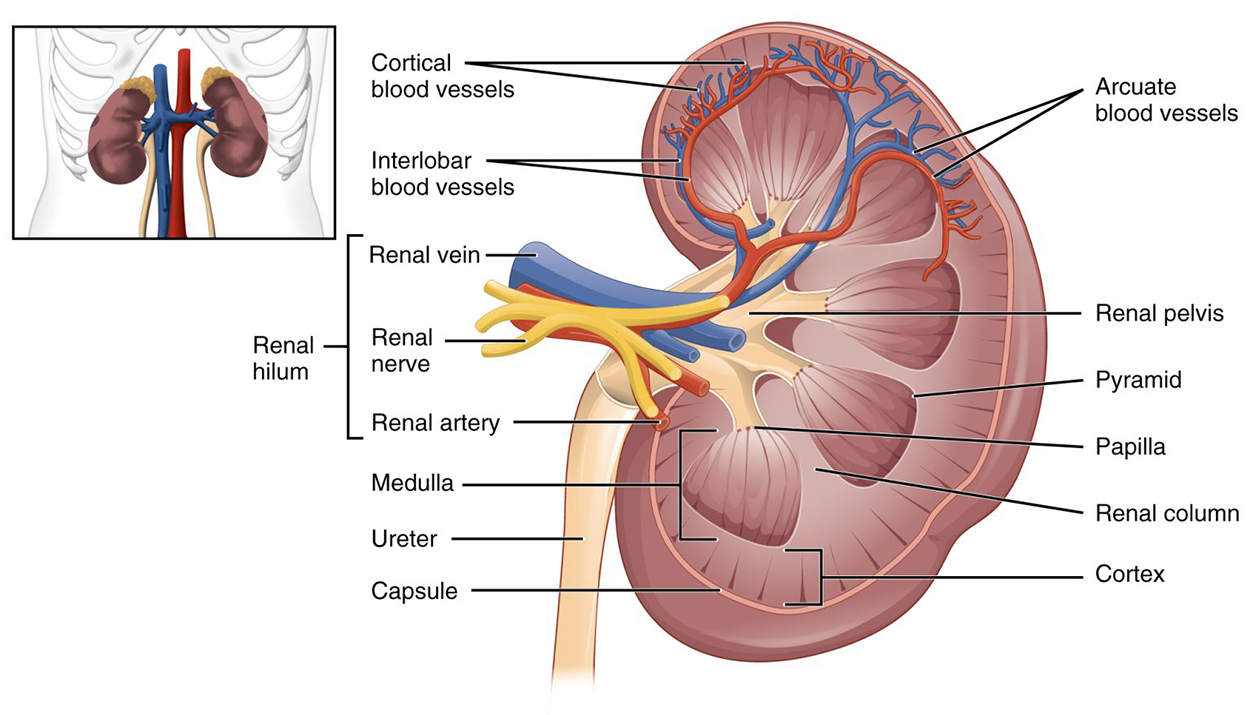
The figure above shows major structures in the kidney. The kidney has an outer capsule with a cortex below. The innermost region is the medulla. The kidney has a kidney bean shape and the indentation on one side, the hilum, through which the renal vein, renal nerve, and renal artery pass. The renal artery branches into interlobar blood vessels, which further branch into arcuate and cortical blood vessels. Pyramidal structures (pyramids or renal pyramids) are arranged in the kidney so that their wider bases face the outside and each has a narrower end that joins to a tubular renal pelvis that drains into the ureter. The structures between the pyramids are renal columns. Where the tip of each pyramid reaches a renal pelvis, there is a papilla.
The renal artery branches into small arteries called afferent arterioles that bring blood to the nephron. The nephron is the functional unit of the kidney and filters the blood, removes wastes, and balances fluid and electrolyte levels. Afferent arterioles supply blood to about 1.3 million nephrons in each kidney.
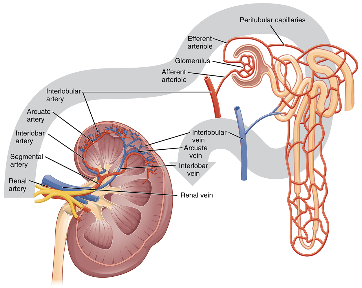
The figure below shows the path of blood flow around a kidney. Note that the renal artery branches to a segmental artery that further branches to the interlobar artery.
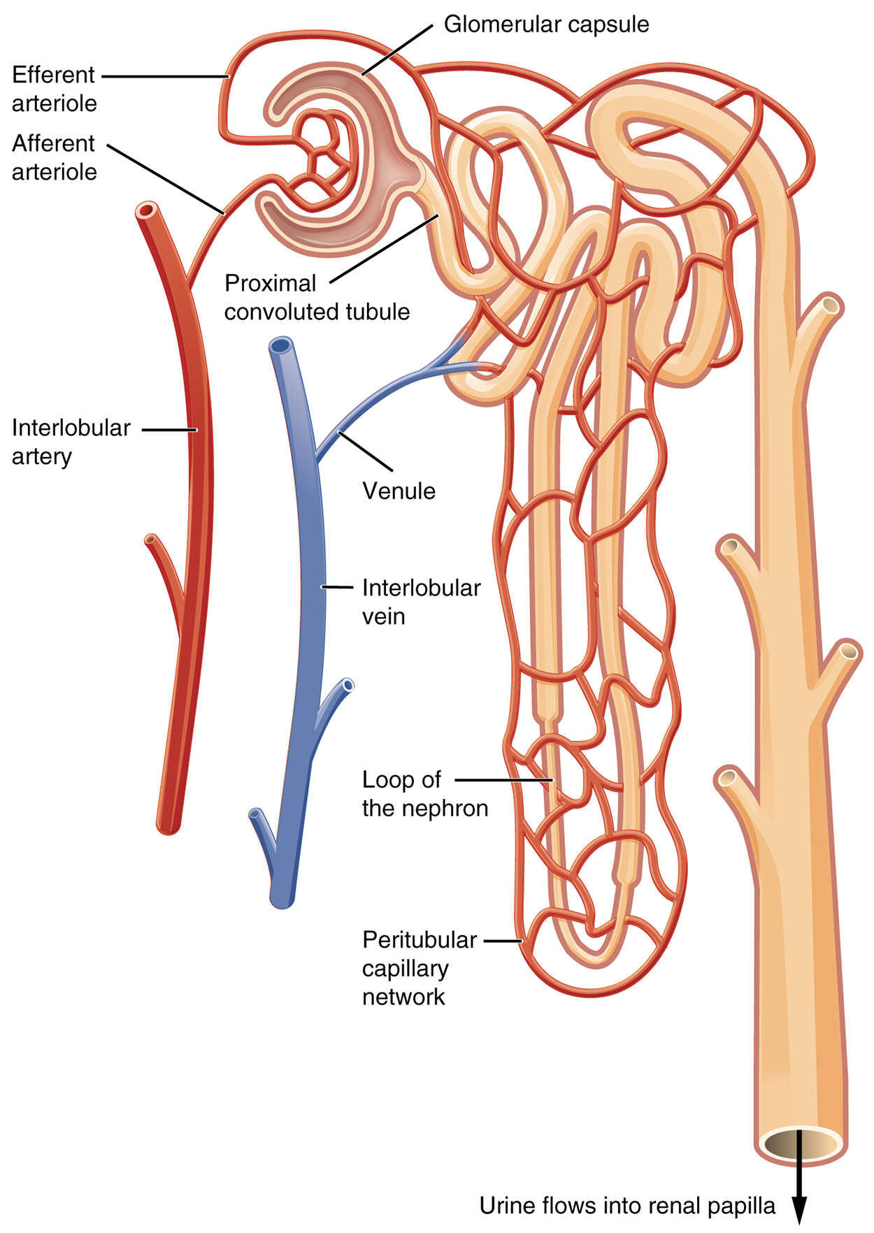
A nephron has two main sections, called a renal corpuscle and a renal tubule. The renal corpuscle consists of a cluster of high-pressure capillaries, called the glomerulus, surrounded by a glomerular capsule. Blood in the glomerulus is filtered as substances move into the filtrate within the capsule. This filtrate is mostly water, amino acids, glucose, and ions. The filtrate then flows through the renal tubule, beginning with the proximal tubule, a long loop-like structure called the nephron loop (or loop of Henle), and the distal convoluted tubule. Different portions of the renal tubule have different permeabilities for solutes (dissolved substances in solution) and water, and efferent arterioles recover most of the water and electrolytes and return them back into the circulation (Merriam-Webster, n.d.). The remaining wastes pass as urine from the nephrons into the collecting ducts and eventually into the ureters for elimination from the body.
The illustration below shows details of the nephron structure. The interlobar artery branches to an afferent arteriole that branches to the glomerulus, which is enclosed by the glomerular capsule. The efferent arteriole leaves the glomerulus and branches to form the peritubular capillaries that surround the nephron. A venule extends from the peritubular capillary network to the interlobar vein. Note that the loop of Henle is labeled as the loop of the nephron in this illustration.
As urine is formed, it drains into the calyces of the kidney, which merge to form the funnel-shaped renal pelvis in the hilum of each kidney. The hilum narrows to become the ureter. As urine passes through the ureter, it does not passively drain into the bladder, but rather is propelled by wave-like contractions called peristalsis. The ureters are approximately 30 cm long (about 12 inches).
The bladder collects urine from both ureters. The bladder lies anterior to the uterus in females, posterior to the pubic bone, and anterior to the rectum. During late pregnancy, bladder capacity is reduced due to compression by the enlarging uterus, resulting in increased frequency of urination. In males, the anatomy is similar, except for the absence of a uterus and the addition of the prostate inferior to the bladder. The bladder can project into the abdomen when it becomes distended (stretched out) with urine. The bladder can extend because it has folds in its walls called rugae (NIH, n.d.).
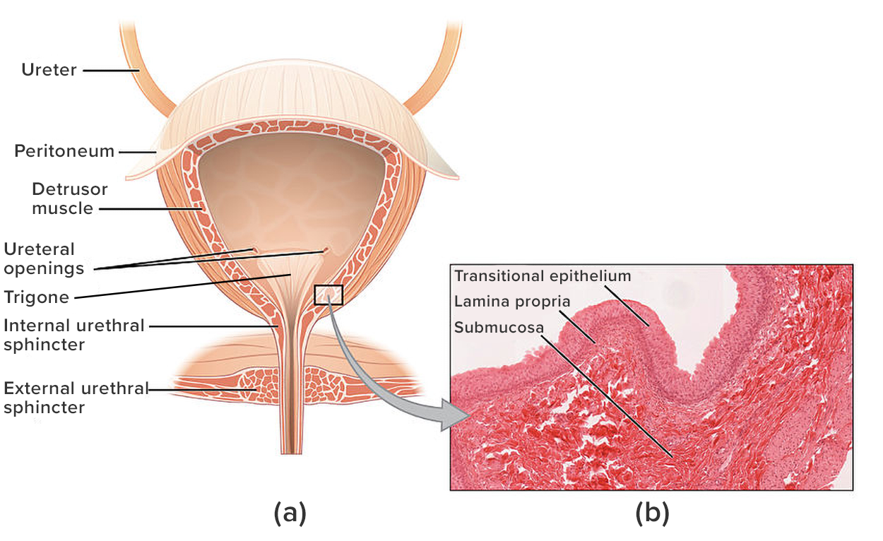
The figure above shows the structure of the bladder. Note that there is a triangular region at the bottom above the internal urethral orifice. This triangular region, which has vertices at the opening of the urethra and each ureter, is called the trigone (NIH, n.d.).
The urethra transports urine from the bladder to the outside of the body during urination, where urine exits through the urinary meatus. The urethra is the only urologic organ that is significantly different between males and females; all other urine transport structures are identical. In females, the urethra is shorter, which causes an increased risk for urinary tract infections.
Urinate means to pass urine, also referred to as void or micturate. A healthy adult with normal functioning kidneys produces an average of 800–2,000 mL of urine per day, depending on their fluid intake and other physiological processes. The adult bladder typically holds about 360–480 mL of urine.
Urination is regulated by the internal and external urinary sphincters, circular muscles constricting an orifice (opening). As the bladder fills to about 150 mL (5 ounces), it sends signals to the brain to create an urge to urinate. The internal and external urinary sphincters work together to close off the urethra to keep urine in the bladder until the brain sends signals that it is time to urinate. As the bladder continues to fill, subsequent urges become harder to ignore. If voluntary voiding does not occur and the bladder overfills, voluntary control fails, resulting in urinary incontinence (incontinence can be used more generally to reflect the inability to control either urination or defecation, leading to the involuntary loss of urine or feces).
Frequency of urination depends on how quickly the kidneys produce urine and how much urine a person’s bladder can comfortably hold. Normal urine is typically clear or pale to light yellow in color with little to no odor. However, some foods (like asparagus) or medications (such as antibiotics) may change the smell or color of urine.
Source: THIS TUTORIAL HAS BEEN ADAPTED FROM “OPEN RN | MEDICAL TERMINOLOGY – 2e” BY ERNSTMEYER & CHRISTMAN AT OPEN RESOURCES FOR NURSING (Open RN). ACCESS FOR FREE AT https://wtcs.pressbooks.pub/medterm/ LICENSING: CREATIVE COMMONS ATTRIBUTION 4.0 INTERNATIONAL.
REFERENCES
Merriam-Webster. (n.d.). Solute. In Merriam-Webster.com dictionary. Retrieved July 24, 2025, from www.merriam-webster.com/dictionary/solute
NIH. (n.d.). Urinary Bladder. National Institutes of Health (NIH) SEER Training Modules https://www.training.seer.cancer.gov/anatomy/urinary/components/bladder.html