Table of Contents |
Joints, also called articulations, are places where two bones or bone and cartilage come together and form a connection. Dislocation refers to the displacement of a bone from its normal position in a joint.
Some word parts related to joints include:
- Arthr/o (joint)
- Articul/o (joint or articulation)
- Synovi/o (synovial fluid)
- Ligament/o (ligaments)
- Chondr/o (cartilage)
Joints are categorized based on their form and amount of movement and are referred to as fibrous joints, cartilaginous joints, or synovial joints.
Fibrous joints, also called synarthrosis or immovable/nonmoveable joints, occur when bones are attached by fibrous connective tissue. There are three types of fibrous joints: suture, gomphosis, and syndesmosis. A suture is the narrow fibrous joint found between most bones of the skull (cranial joints). A gomphosis is the narrow fibrous joint between the roots of a tooth and the bony socket in the jaw into which the tooth fits. A syndesmosis is a joint held together by a ligament, a type of connective tissue that connects bones at joints.
Cartilaginous joints, also called amphiarthrosis or slightly moveable joints, occur when two bones are connected by cartilage, a tough but flexible type of connective tissue. Cartilage has varied roles in the skeletal system; it can provide a template from which bones form and cushions joints. Examples of cartilaginous joints include the pubic symphysis and intervertebral disks.
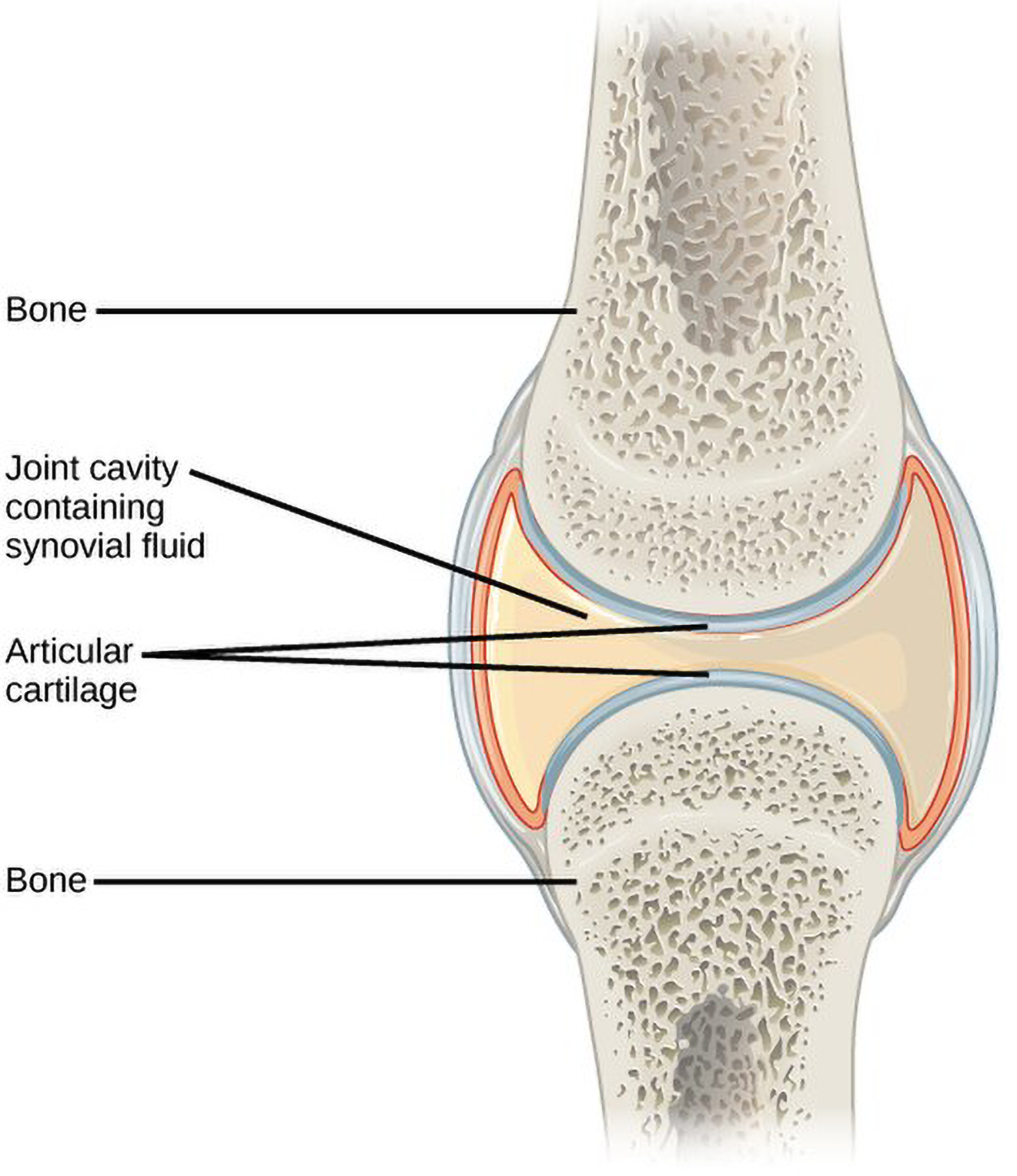
Synovial joints, also called diarthroses or fully movable joints, have a fluid-filled space where two bones come together, called a joint cavity. Because the bones in a synovial joint are not directly connected to each other with fibrous connective tissue or cartilage, they are able to move freely against each other, allowing for increased joint mobility. Synovial joints are the most common type of joint in the body. A synovial membrane is the lining or covering of synovial joints, and synovial fluid is the lubricating fluid found between synovial joints.
The figure below shows an example of a synovial joint. Note that a cavity containing synovial fluid separates the two bones.
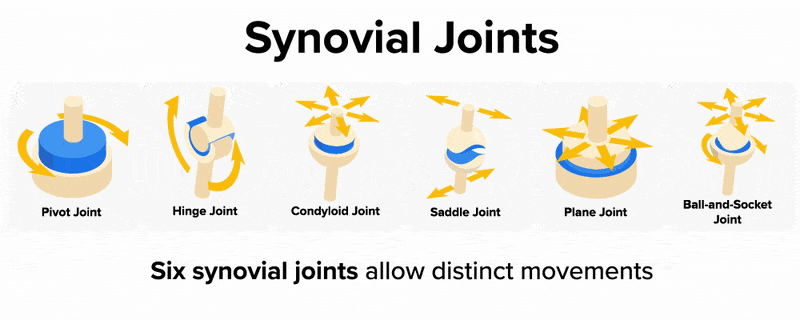
Synovial joints are categorized based on the shapes of the articulating surfaces of the bones that form each joint. The six types of synovial joints are pivot, hinge, condyloid, saddle, plane, and ball-and-socket joints, which are summarized in the figure below and then described in more detail. When looking at the figure, note how each type of joint has a specific range of motion. Pay attention to the arrows in the figures showing how each joint moves.
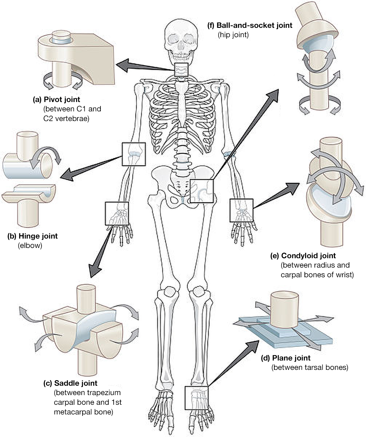
At a pivot joint, a rounded portion of a bone is enclosed within a ring formed partially by the articulation with another bone and partially by a ligament. The bone rotates within this ring. Since the rotation is around a single axis, pivot joints are functionally classified as a uniaxial diarthrosis type of joint. An example of a pivot joint is the atlantoaxial joint, found between the C1 (atlas) and C2 (axis) vertebrae. Here, an upward-projecting structure of the axis articulates with the inner aspect of the atlas, where it is held in place by a ligament. Rotation at this joint allows you to turn your head from side to side. A second pivot joint is found at the proximal radioulnar joint. Here, the head of the radius is largely encircled by a ligament that holds it in place as it articulates with the radial notch of the ulna. Rotation of the radius allows for forearm movements.
In a hinge joint, the convex end of one bone articulates with the concave end of the adjoining bone. This type of joint allows only for bending and straightening motions along a single axis, and thus hinge joints are functionally classified as uniaxial joints. A good example is the elbow joint, with the articulation between the trochlea of the humerus and the trochlear notch of the ulna. Other hinge joints of the body include the knee, ankle, and interphalangeal joints between the phalanx bones of the fingers and toes.
At a condyloid joint (ellipsoid joint), the shallow depression at the end of one bone articulates with a rounded structure from an adjacent bone or bones. The knuckle (metacarpophalangeal) joints of the hand, between the distal end of a metacarpal bone and the proximal phalanx bone, are condyloid joints. Another example is the radiocarpal joint of the wrist, between the shallow depression at the distal end of the radius bone and the rounded scaphoid, lunate, and triquetrum carpal bones. In this case, the articulation area has a more oval (elliptical) shape. Functionally, condyloid joints are biaxial joints that allow for two planes of movement. One movement involves the bending and straightening of the fingers or the anterior-posterior movements of the hand. The second movement is a side-to-side movement, which allows you to spread your fingers apart and bring them together, or to move your hand in a medial-going or lateral-going direction.
At a saddle joint, both of the articulating surfaces for the bones have a saddle shape, which is concave in one direction and convex in the other. This allows the two bones to fit together like a rider sitting on a saddle. Saddle joints are functionally classified as biaxial joints. The primary example is the first carpometacarpal joint, between the trapezium (a carpal bone) and the first metacarpal bone at the base of the thumb. This joint provides the thumb the ability to move away from the palm of the hand along two planes. Thus, the thumb can move within the same plane as the palm of the hand, or it can jut out anteriorly, perpendicular to the palm. This movement of the first carpometacarpal joint is what gives humans their distinctive “opposable” thumbs. The sternoclavicular joint is also classified as a saddle joint.
At a plane joint (gliding joint), the articulating surfaces of the bones are flat or slightly curved and of approximately the same size, which allows the bones to slide against each other. The motion at this type of joint is usually small and tightly constrained by surrounding ligaments. Based only on their shape, plane joints can allow multiple movements, including rotation. Thus, plane joints can be functionally classified as multiaxial joints. However, not all of these movements are available to every plane joint due to limitations placed on it by ligaments or neighboring bones. Thus, depending upon the specific joint of the body, a plane joint may exhibit only a single type of movement or several movements. Plane joints are found between the carpal bones (intercarpal joints) of the wrist or tarsal bones (intertarsal joints) of the foot, between the clavicle and acromion of the scapula (acromioclavicular joint), and between the superior and inferior articular processes of adjacent vertebrae (zygapophysial joints).
The joint with the greatest range of motion is the ball-and-socket joint. At these joints, the rounded head of one bone (the ball) fits into the concave articulation (the socket) of the adjacent bone. The hip joint and the glenohumeral (shoulder) joint are the only ball-and-socket joints of the body. At the hip joint, the head of the femur articulates with the acetabulum of the hip bone, and at the shoulder joint, the head of the humerus articulates with the glenoid cavity of the scapula.
Ball-and-socket joints are classified functionally as multiaxial joints. The femur and the humerus are able to move in both anterior-posterior and medial-lateral directions, and they can also rotate around their long axis. The shallow socket formed by the glenoid cavity allows the shoulder joint an extensive range of motion. In contrast, the deep socket of the acetabulum and the strong supporting ligaments of the hip joint serve to constrain movements of the femur, reflecting the need for stability and weight-bearing ability at the hip.
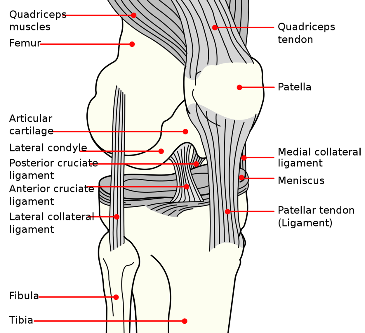
A few synovial joints of the body have a fibrocartilage structure located between the articulating bones called an articular disk or meniscus. Articular disks are generally small and oval-shaped whereas a meniscus is larger and C-shaped. For example, the knee connects the femur (upper leg bone) to the tibia (one of the lower leg bones) with a piece of fibrous cartilage called the meniscus.
In addition to the meniscus, the knee also contains ligaments, tendons, and additional cartilage. Ligaments in the knee include the anterior cruciate ligament (ACL), lateral collateral ligament (LCL), medial collateral ligament (MCL), and posterior cruciate ligament (PCL). You can see these structures in the figure below.
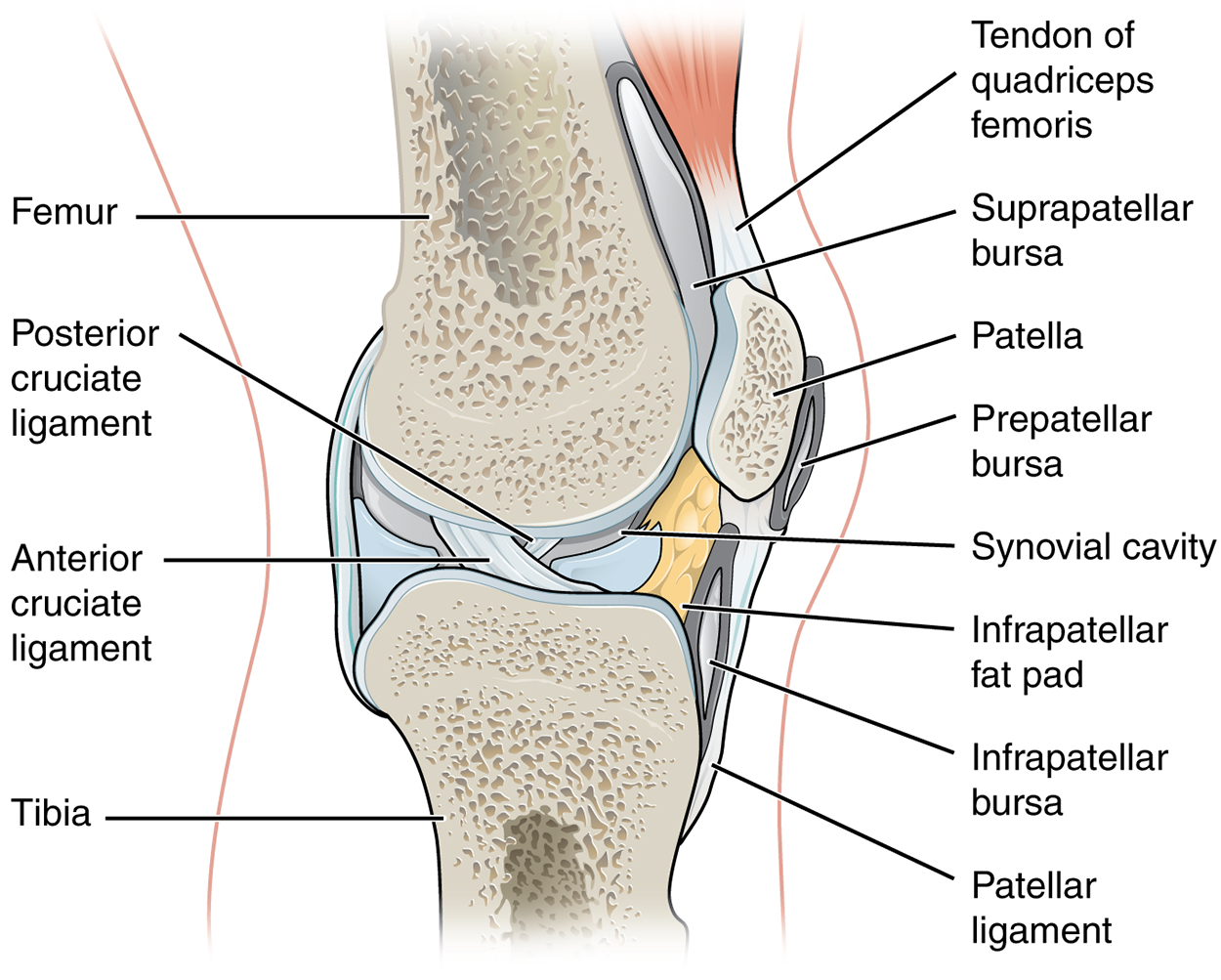
An additional structure located outside of a synovial joint is a bursa. A bursa is a thin connective tissue sac filled with lubricating fluid located in regions where skin, ligaments, muscles, or tendons rub against each other. Bursae are classified by their location.
EXAMPLE
A subcutaneous bursa is located between the skin and an underlying bone and allows skin to move smoothly over the bone (remember that sub- means below and cutane/o means skin).EXAMPLE
Examples of a subcutaneous bursa are the suprapatellar bursa, prepatellar bursa, and infrapatellar bursa located in the knee, which are shown in the figure above.Note that the prepatellar (“before patella”) bursa runs in front of the lower to middle patella; the suprapatellar (“above patella”) bursa runs between the femur and upper left of the patella, continuing up behind the tendon of the quadriceps femoris; and the infrapatellar (“below patella”) bursa runs from the upper right of the tibia to the right of the infrapatellar fat pad and below the patella.
There are a variety of common joint conditions and injuries.
Conditions include various types of arthritis, such as osteoarthritis, rheumatoid arthritis (RA), and gout.
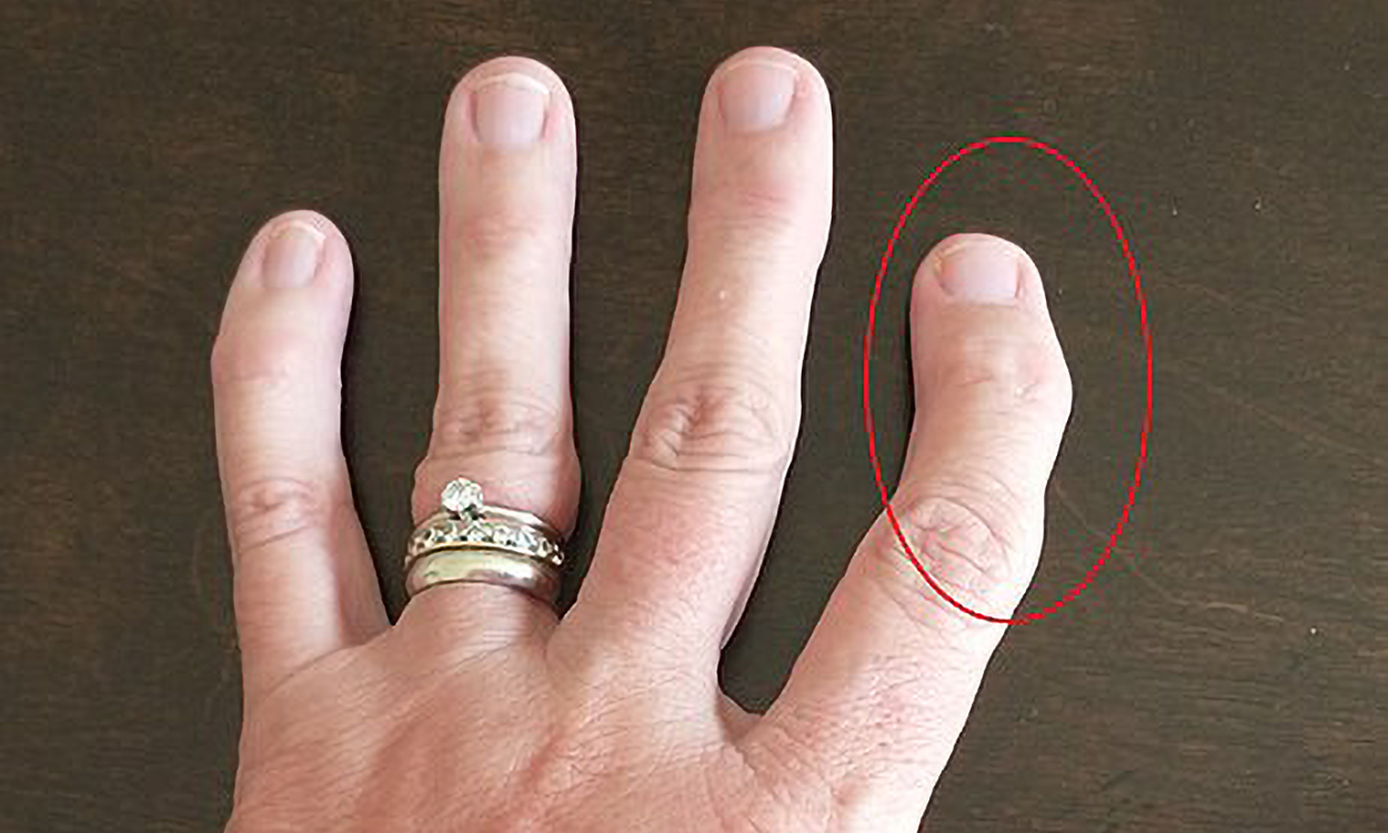
Osteoarthritis is the most common type of arthritis and commonly occurs as people age. It is a degenerative joint disease in which the tissues in the joint break down over several years. It is the most common type of arthritis and frequently occurs as people age. People with osteoarthritis usually have joint pain or stiffness after rest or inactivity for a short period of time. The most commonly affected joints include the hands, knees, hips, neck, and lower back. Osteoarthritis affects each person differently. For some people, osteoarthritis is relatively mild and does not affect day-to-day activities. For others, it causes significant pain and disability.
Crepitus describes a popping, clicking, or crackling sound when moving a joint that is associated with osteoarthritis. It typically reflects air movement in the joint and is harmless.
The figure below shows the hand of a person with osteoarthritis in their finger.
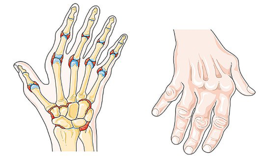
Rheumatoid arthritis (RA) is a chronic autoimmune disease that affects the joints. RA occurs in a symmetrical pattern, meaning that if one knee or hand has the condition, the other hand or knee is also affected. It can affect the joints in the wrists, hands, elbows, shoulders, feet, spine, knees, and jaw. RA causes pain, swelling, stiffness, deformity, and loss of function.
The figure below shows rheumatoid arthritis in the hands causing a common “swan-like” deformity.
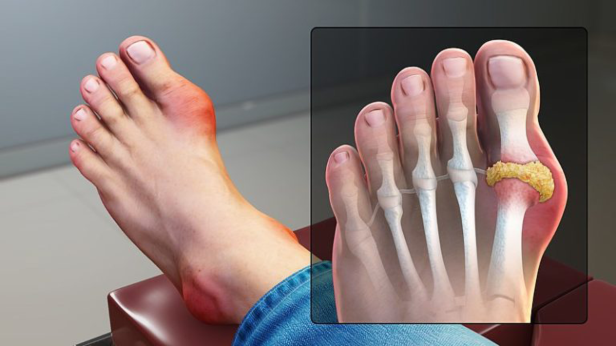
Gout is a type of inflammatory arthritis that causes pain and swelling in the joints, usually as flares that last for one or two weeks and then resolve. Gout flares often begin in the big toe or a lower limb. Gout occurs when high levels of serum uric acid build up in the body, which can form needle-shaped crystals in and around the joints and cause inflammation. Uric acid is a waste product produced by the breakdown of purine, a type of molecule found in many foods we eat. Gout is diagnosed with blood tests for uric acid. It is commonly treated with medications like nonsteroidal anti-inflammatories (NSAIDs) and colchicine.
The figure below shows the foot of an individual with gout.
Joints can also be physically injured. Luxation is another term for dislocation, and subluxation means a partial dislocation (Cleveland Clinic, 2023).
Additional terms include:
- Synovectomy is removal/excision of the synovial membrane.
- Meniscitis is inflammation of the meniscus.
- Meniscectomy is excision of the meniscus.
- Bursitis is inflammation of a bursa, typically in the knee, elbow, or shoulder.
- Bursectomy is excision of a bursa.
- Arthroplasty is the surgical repair of a joint.
- Tenorrhaphy is the suturing of a tendon (-rrhaphy is suturing).
SOURCE: THIS TUTORIAL HAS BEEN ADAPTED FROM (1) “OPEN RN | MEDICAL TERMINOLOGY – 2E” BY ERNSTMEYER & CHRISTMAN AT OPEN RESOURCES FOR NURSING (OPEN RN). (2) "ANATOMY AND PHYSIOLOGY 2E" AT OPENSTAX. ACCESS FOR FREE AT WTCS.PRESSBOOKS.PUB/MEDTERM/ AND OPENSTAX.ORG/DETAILS/BOOKS/ANATOMY-AND-PHYSIOLOGY-2E. LICENSING: CREATIVE COMMONS ATTRIBUTION 4.0 INTERNATIONAL.
REFERENCES
Dislocation. (n.d.) Cleveland Clinic. Retrieved 19 March 2025, from my.clevelandclinic.org/health/diseases/17873-dislocation
Mattu, A. T., Ghali, B., Linton, V., Zheng, A., & Pike, I. (2022). Prevention of Non-Contact Anterior Cruciate Ligament Injuries among Youth Female Athletes: An Umbrella Review. International journal of environmental research and public health, 19(8), 4648. doi.org/10.3390/ijerph19084648