Table of Contents |
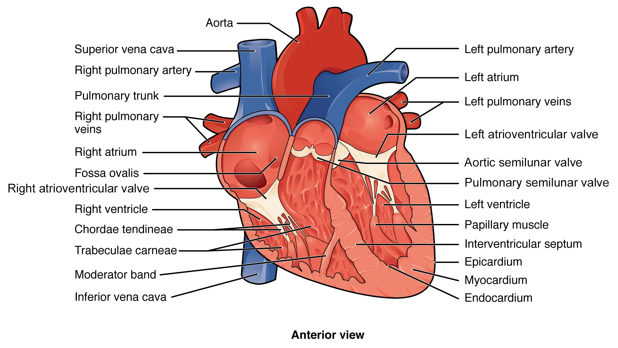
The septa are physical extensions of the myocardium lined with endocardium. Separating the two atria is the interatrial septum. Normally in an adult heart, the interatrial septum bears an oval-shaped depression known as the fossa ovalis (fossa, depression; ovalis, egg), a remnant of an opening in the fetal heart known as the foramen ovale (foramen, hole). During fetal development, the foramen ovale allows blood to pass directly from the right atrium to the left atrium, allowing some blood to bypass the lungs. Within seconds after birth, a flap of tissue known as the septum primum that previously acted as a valve for the foramen ovale closes over it and establishes the typical adult cardiac circulation pattern you learned previously.
Between the two ventricles is a second septum known as the interventricular septum. Unlike the interatrial septum, the interventricular septum is normally intact after its formation during fetal development. It is substantially thicker than the interatrial septum since the ventricles generate far greater pressure when they contract.
The septum between the atria and ventricles is known as the atrioventricular septum. It is marked by the presence of four openings that allow blood to move from the atria into the ventricles and from the ventricles into the pulmonary trunk and aorta. Located in each of these openings between the atria and ventricles is a valve, a specialized structure that ensures one-way flow of blood. The valves between the atria and ventricles are known generically as atrioventricular valves. The valves at the openings that lead to the pulmonary trunk and aorta are known generically as semilunar valves. Since these openings and valves structurally weaken the atrioventricular septum, the remaining tissue is heavily reinforced with dense connective tissue called the cardiac skeleton, or skeleton of the heart. It includes four rings that surround the openings between the atria and ventricles, and the openings to the pulmonary trunk and aorta, and serve as the point of attachment for the heart valves. The cardiac skeleton also provides an important boundary in the heart electrical conduction system.

| Term | Pronunciation | Audio File |
|---|---|---|
| Septum | sep·tum |
|
| Interatrial | in·ter·a·tri·al |
|
| Fossa Ovalis | fo·ssa o·val·is |
|
| Foramen Ovale | fo·ra·men o·va·le |
|
| Interventricular | in·ter·ven·tri·cu·lar |
|
| Atrioventricular | a·tri·o·ven·tri·cu·lar |
|
| Semilunar | sem·i·lu·nar |
|
The right atrium serves as the receiving chamber for blood returning to the heart from the body. The two major systemic veins, the superior and inferior venae cavae, empty into the right atrium. The superior vena cava drains blood from regions superior to the diaphragm: the head, neck, upper limbs, and thoracic region. It empties into the superior and posterior portions of the right atrium. The inferior vena cava drains blood from areas inferior to the diaphragm: the lower limbs and abdominopelvic region of the body. It, too, empties into the posterior portion of the atria, but inferior to the opening of the superior vena cava.
While the bulk of the internal surface of the right atrium is smooth, the depression of the fossa ovalis is medial, and the anterior surface has prominent parallel ridges of muscle called the pectinate muscles (pecten, comb) which resemble the teeth of a comb. The right auricle also has pectinate muscles. The left atrium does not have pectinate muscles except in the auricle.
The atria receive venous blood on a nearly continuous basis, preventing venous flow from stopping while the ventricles are contracting. While most ventricular filling occurs while the atria are relaxed, they do demonstrate a contractile phase and actively pump blood into the ventricles just prior to ventricular contraction. The opening between the atrium and ventricle is guarded by the right atrioventricular valve.
| Term | Pronunciation | Audio File |
|---|---|---|
| Pectinate | pec·ti·nate |
|
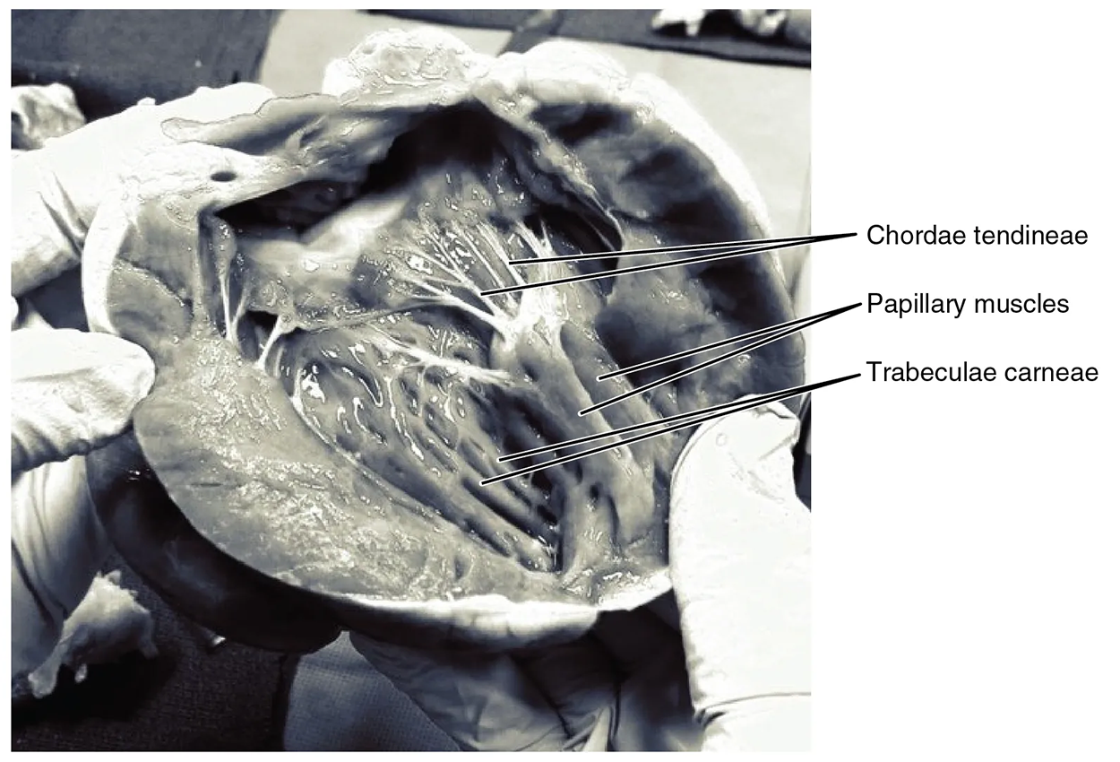
The right ventricle receives blood from the right atrium through the right atrioventricular valve, also known as the right AV valve or tricuspid valve (contains three cusps or flaps). Each flap of the valve is attached to strong strands of connective tissue, the chordae tendineae, literally “tendinous cords,” or sometimes more poetically referred to as “heart strings.” There are several chordae tendineae associated with each of the flaps. They are composed of approximately 80% collagenous fibers with the remainder consisting of elastic fibers and endothelium. They connect each of the flaps to a papillary muscle that extends from the inferior ventricular surface. There are three papillary muscles in the right ventricle, called the anterior, posterior, and septal muscles, which correspond to the three sections of the valves.
When the myocardium of the ventricle contracts, the pressure within the ventricular chamber rises. Blood, like any fluid, flows from higher pressure to lower pressure areas, in this case, toward the pulmonary trunk and the atrium. To prevent any potential backflow, the papillary muscles also contract, generating tension on the chordae tendineae. This prevents the flaps of the valves from being forced into the atria and regurgitation (backward flow) of the blood into the atria during ventricular contraction. The figure below shows papillary muscles and chordae tendineae attached to the right atrioventricular valve.

The walls of the ventricle are lined with trabeculae carneae, ridges of cardiac muscle covered by endocardium. These muscles provide pressure to eject blood out of the ventricle, tension to the papillary muscles to prevent inversion of the atrioventricular valves, and form a textured internal surface so that the heart, when fully contracted, does not suction one wall to the other and prevent the subsequent relaxation and refilling.
In addition to these muscular ridges, a band of cardiac muscle, also covered by endocardium, known as the moderator band reinforces the thin walls of the right ventricle and plays a crucial role in cardiac conduction as you will learn in another lesson. The moderator band arises from the inferior portion of the interventricular septum and crosses the interior space of the right ventricle to connect with the anterior papillary muscle.
When the right ventricle contracts, it ejects blood into the pulmonary trunk, which branches into the left and right pulmonary arteries that carry it to each lung. The superior surface of the right ventricle begins to taper as it approaches the pulmonary trunk. At the base of the pulmonary trunk is the pulmonary semilunar valve, also known as the pulmonic valve, which prevents backflow from the pulmonary trunk.
| Term | Pronunciation | Audio File |
|---|---|---|
| Chordae Tendineae | chor·dae ten·din·eae |
|
| Papillary | pap·il·lar·y |
|
| Trabeculae Carneae | tra·bec·u·lae car·neae |
|
After an exchange of gasses in the pulmonary capillaries, blood returns to the left atrium high in oxygen via one of the four pulmonary veins; two from the right lung and two from the left lung. While the left atrium does not contain pectinate muscles, it does have an auricle that includes these pectinate ridges. Blood flows nearly continuously from the pulmonary veins back into the atrium, which acts as the receiving chamber, and from here through an opening into the left ventricle. Most blood flows passively into the heart while both the atria and ventricles are relaxed, but toward the end of the ventricular relaxation period, the left atrium will contract, pumping blood into the ventricle. This atrial contraction accounts for approximately 20% of ventricular filling. The opening between the left atrium and ventricle is guarded by the left atrioventricular valve.
Blood enters the left ventricle from the left atrium by passing through the left atrioventricular valve, also known as the left AV valve, mitral valve (similar in shape to a bishop’s miter [head piece]), or bicuspid valve (contains two cusps or flaps). This valve, like the right AV valve, is connected by chordae tendineae to papillary muscles in the wall of the ventricle. However, while the right AV valve has three flaps and three respective papillary muscles, the left AV valve only has two flaps and two papillary muscles—the anterior and posterior. Recall that, although both sides of the heart will pump the same amount of blood, the muscular layer is much thicker in the left ventricle compared to the right. Like the right ventricle, the left also has trabeculae carneae, but there is no moderator band.
The left ventricle is the major pumping chamber to push blood to the whole body. When blood is ejected, it travels through the aortic semilunar valve, also known as the aortic valve, to enter the aorta which serves the entire body through a vast network of blood vessels.
Source: THIS TUTORIAL HAS BEEN ADAPTED FROM OPENSTAX "ANATOMY AND PHYSIOLOGY 2E" ACCESS FOR FREE AT OPENSTAX.ORG/DETAILS/BOOKS/ANATOMY-AND-PHYSIOLOGY-2E. LICENSE: CREATIVE COMMONS ATTRIBUTION 4.0 INTERNATIONAL