Table of Contents |
Audition is the special sense of hearing that makes use of the ear to convert sound waves into neural signals and is associated with the ear.
The large, fleshy structure on the lateral aspect of the head is known as the auricle. The C-shaped curves of the auricle direct sound waves toward the auditory canal. The canal enters the skull through the external auditory meatus of the temporal bone. At the end of the auditory canal is the tympanic membrane, or eardrum, which vibrates after it is struck by sound waves. The auricle, ear canal, and tympanic membrane are often referred to as the external ear.
The middle ear consists of a space spanned by three small bones called the ossicles. The three ossicles are the malleus, incus, and stapes, which are Latin names that roughly translate to hammer, anvil, and stirrup. The malleus is attached to the tympanic membrane and articulates with the incus. The incus, in turn, articulates with the stapes. The stapes is then attached by the oval window (which you will learn more about shortly) to the inner ear, where the sound waves will be transduced into a neural signal.
The middle ear is connected to the pharynx through the auditory tube, also known as the Eustachian tube or pharyngotympanic tube, which helps equilibrate air pressure across the tympanic membrane. The tube is normally closed but will pop open when the muscles of the pharynx contract during swallowing or yawning.
IN CONTEXT
When you travel up or down in elevation, you might notice your ears can be irritated. This irritation is caused by the change in atmospheric pressure.
Under normal conditions, the pressure on both sides of the tympanic membrane (eardrum) are equal, allowing sound waves (i.e., small shifts in pressure) to cause the eardrum to move when they strike it. However, when atmospheric pressure changes by changing elevation or your position in water (sinking to the bottom of a pool or rising up to the surface while scuba diving), the pressure is no longer equal. This can cause irritation and possible damage.
Swallowing or yawning can manipulate the auditory tube connecting the middle ear to the mouth, allowing for air to enter or escape this cavity, and equalizing the pressure across the eardrum.
The inner ear is often described as a bony labyrinth, as it is composed of a series of bony canals embedded within the temporal bone. It has two separate regions, the cochlea and the vestibule, which are responsible for hearing and balance, respectively.
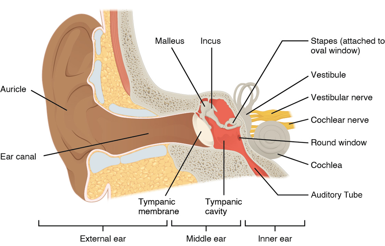
You can see that the cochlea looks like a snail's shell, a spiral moving inwards. On the lateral portion of the cochlea is the oval window, an oval-shaped hole in the bone where the stapes sits. Inside the bony labyrinth of the inner ear is a fluid-filled tube that spirals along with the bone to the center. Just inferior to the oval window is the round window, a round-shaped hole in the cochlea covered by a membrane. As sound waves strike the tympanic membrane and move it inwards, the ossicle bones shift. When the stapes moves, it presses into the oval window, causing a fluid wave within the cochlea. The round window will bulge out to accommodate the wave and pucker in when the wave recedes. The frequency of the fluid waves will match the frequency of the sound waves.
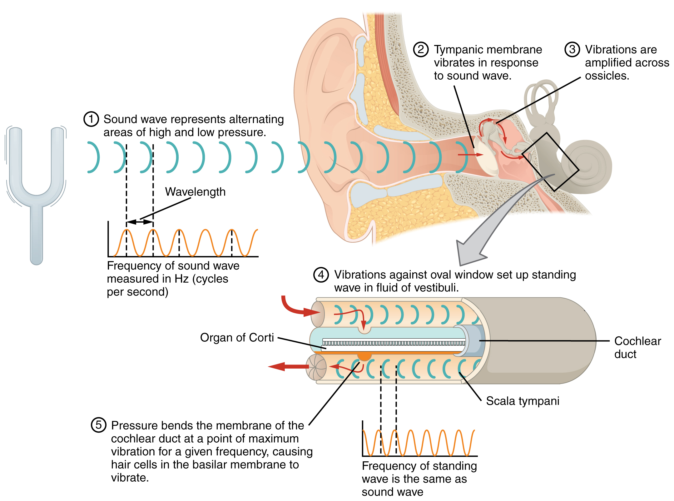
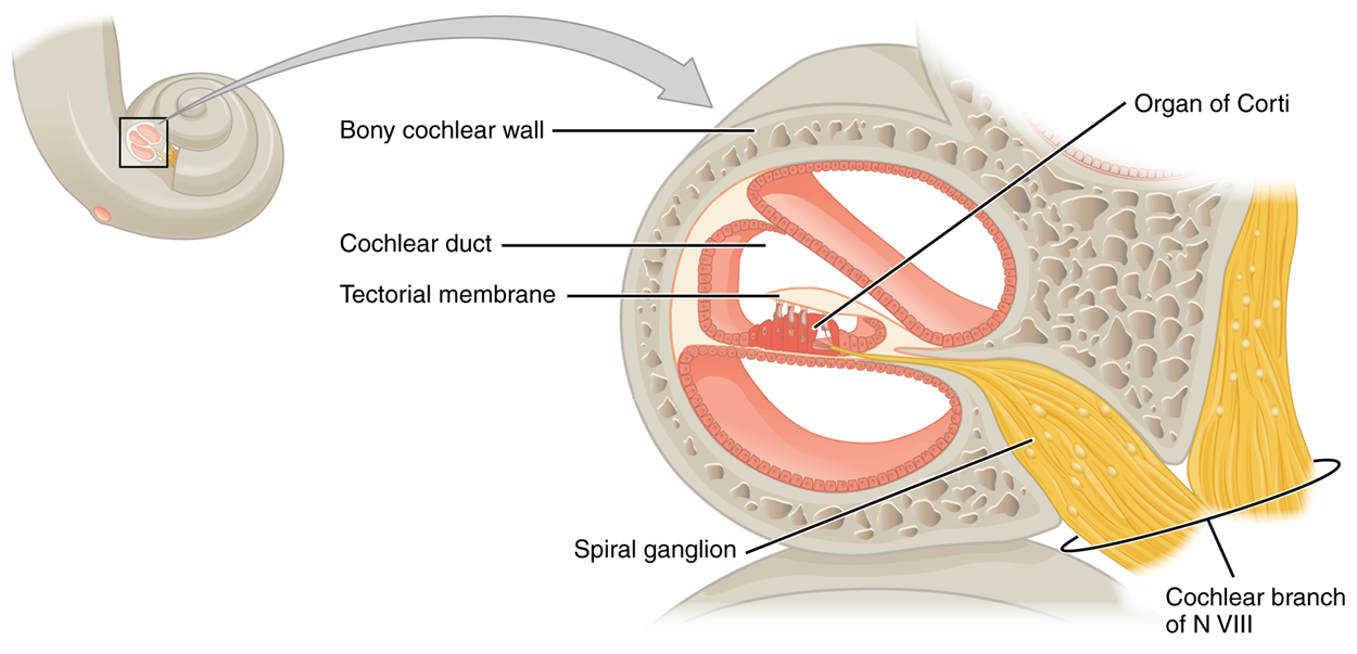
Along the center of the winding cochlea is a cavity called the cochlear duct, which contains an auditory sensory organ called the organ of Corti. As the fluid waves move through the cochlea, the cochlear duct moves at a specific spot, depending on the frequency of the waves. Higher frequency waves move the region of the duct that is close to the base of the cochlea. Lower frequency waves move the region that is near the tip of the cochlea.
The organ of Corti contains sensory receptor cells that are specialized mechanoreceptors called hair cells, which are named for the hair-like stereocilia extending from the cell’s apical surfaces. The stereocilia are an array of microvilli-like structures arranged from tallest to shortest. Protein fibers tether adjacent hairs together within each array, such that the array will bend in response to movements of the underlying tissue.
The stereocilia extend up from the hair cells to the overlying tectorial membrane, which is attached medially to the organ of Corti. When the pressure waves move the organ of Corti, the tectorial membrane slides across the stereocilia. This bends the stereocilia either toward or away from the tallest member of each array. When the stereocilia bend toward the tallest member of their array, tension in the protein tethers opens ion channels in the hair cell membrane. This will depolarize the hair cell membrane. When the stereocilia bend toward the shortest member of their array, the tension on the tethers slackens and the ion channels close.
When no sound is present, and the stereocilia are standing straight, a small amount of tension still exists on the tethers, keeping the membrane potential of the hair cell slightly depolarized. Depolarized hair cells send signals to the auditory sensory neuron. All auditory sensory neurons leave the cochlea as part of the cochlear nerve, which is the auditory portion of the vestibulocochlear nerve (CN VIII).
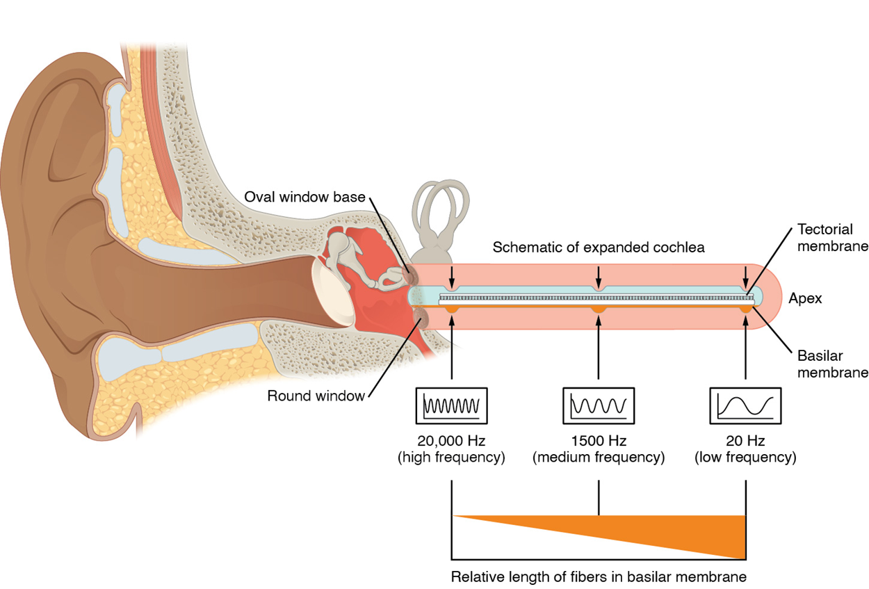
As stated above, a given region of the cochlear duct will only move if the incoming sound is at a specific frequency. Because the tectorial membrane only moves where the cochlear duct moves, the hair cells in this region will also only respond to sounds of this specific frequency. Therefore, as the frequency of a sound changes, different hair cells are activated all along the basilar membrane.
The unit of Hertz measures the frequency of sound waves in terms of cycles produced per second. Frequencies as low as 20 Hz are detected by hair cells at the apex, or tip, of the cochlea. Frequencies in the higher ranges of 20 KHz are encoded by hair cells at the base of the cochlea, close to the round and oval windows. Most auditory stimuli contain a mixture of sounds at a variety of frequencies and intensities (represented by the amplitude of the sound wave). The hair cells along the length of the cochlear duct, which are each sensitive to a particular frequency, allow the cochlea to separate auditory stimuli by frequency just as a prism separates visible light into its component colors.
Along with audition, the inner ear is responsible for encoding information about vestibulation, also known as the sense of equilibrium or balance. Recall that the inner ear is a bony labyrinth composed of the cochlea and vestibule. The cochlea contains auditory structures, whereas the vestibule contains vestibular structures. Within the vestibule are three semicircular canals that detect rotational movement as well as the utricle and saccule that detect linear movement and position.
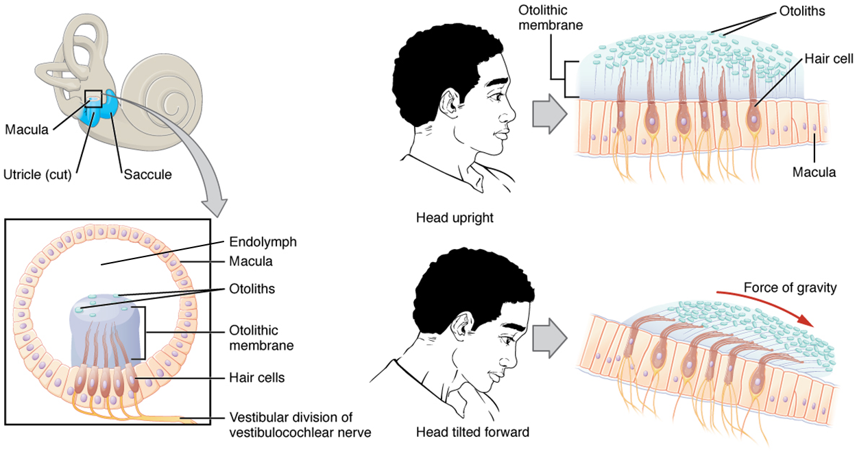
The utricle and saccule both contain hair cells (which are mechanoreceptors) embedded in them. The stereocilia of the hair cells extend into a viscous gel called the otolithic membrane. On top of the otolithic membrane is a layer of calcium carbonate crystals called otoliths, also known as ear stones.
The otoliths essentially make the otolithic membrane top-heavy. The otolithic membrane moves separately from the underlying tissue in response to head movements. Tilting the head causes the otolithic membrane to slide in the direction of gravity. The moving otolithic membrane, in turn, bends the stereocilia, causing some hair cells to depolarize as others hyperpolarize. The exact position of the head is interpreted by the brain based on the pattern of hair-cell depolarization.
The utricle and saccule can also detect changes in linear movement. The utricle is positioned in the horizontal plane and can detect horizontal acceleration or deceleration such as your car picking up speed or slowing down. The saccule is positioned vertically and can detect vertical acceleration or deceleration such as an elevator.
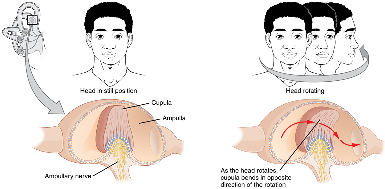
The semicircular canals are three ring-like extensions of the vestibule. One is oriented in the horizontal plane, whereas the other two are oriented in the vertical plane. The base of each semicircular canal, where it meets with the vestibule, connects to an enlarged region known as the ampulla (of the inner ear), which contains hair cells. These hair cells have stereocilia embedded into the cupula, a membrane that attaches to the top of the ampulla.
When the head moves in a rotational movement (i.e., spinning in circles, doing a somersault, or doing a cartwheel), fluid inside one or more of the semicircular canals shifts. As this fluid moves along the ampulla, it bends the cupula and in turn, the stereocilia. This depolarizes the hair cells and sends a signal to a connected ampullary nerve, which extends as the vestibular nerve. This nerve combines with the cochlear nerve to become the vestibulocochlear nerve.
By comparing the signals from all ampullae, the vestibular system can detect the direction of most head movements within three-dimensional (3-D) space.
 Interactive 3-D Model
SOURCE: THIS TUTORIAL HAS BEEN ADAPTED FROM OPENSTAX “ANATOMY AND PHYSIOLOGY 2E”. ACCESS FOR FREE AT OPENSTAX.ORG/BOOKS/ANATOMY-AND-PHYSIOLOGY-2E/PAGES/1-INTRODUCTION. LICENSE: CREATIVE COMMONS ATTRIBUTION 4.0 INTERNATIONAL.