Table of Contents |
As discussed in other lessons, prokaryotes in Kingdom Bacteria can be classified as gram-positive or gram-negative based on their color after Gram staining. Gram-positive cell walls retain the primary crystal violet stain during the decolorization step, whereas gram-negative cell walls are more easily decolorized and therefore show the color of the safranin counterstain.
IN CONTEXT
Knowing whether bacteria are gram-positive or gram-negative can be useful in a variety of ways. For example, it can help in knowing which antibiotics are most likely to be effective against them. However, the terms “gram-positive” and “gram-negative” are not formal taxonomic categories. Although each group has distinctive characteristics that determine how they will stain, these terms describe how bacterial cells respond to staining and should not be confused with taxonomic classification.
The Proteobacteria are the largest phylum of gram-negative bacteria. This phylum includes classes Alphaproteobacteria, Betaproteobacteria, Gammaproteobacteria, Deltaproteobacteria, and Epsilonproteobacteria.
Some of the Proteobacteria are medically important. You will learn more about bacterial diseases in other lessons.
The Alphaprotebacteria include many obligate or facultative intracellular bacteria. Obligate intracellular bacteria can only survive within a cell. Facultative intracellular bacteria have the ability to live within a cell, but do not have to live there. Some alphaproteobacteria are oligotrophs, meaning that they can survive in low-nutrient environments such as deep oceanic sediments, glacial ice, or deep undersurface soil.
EXAMPLE
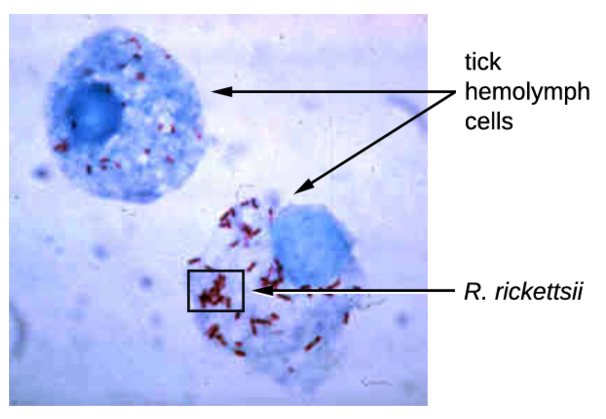
A medically important group of Alphaproteobacteria are the members of genus Rickettsia. These bacteria are considered atypical because they are too small to be evaluated using the Gram staining technique. These bacteria are obligate intracellular parasites. They are metabolically inactive outside the host cell. They cannot synthesize ATP and rely on the host cell to meet their energy needs. This genus includes Rickettsia rickettsii, a tick-borne pathogen that can cause a potentially severe illness called Rocky Mountain spotted fever.The image below shows rod-shaped R. rickettsii inside cells. Other medically important species include R. prowazekii, which is spread by lice and causes epidemic typhus, and R. typhi, which causes murine or endemic typhus.
The Betaprotebacteria are diverse. They live in many different habitats using varied metabolic strategies.
EXAMPLE
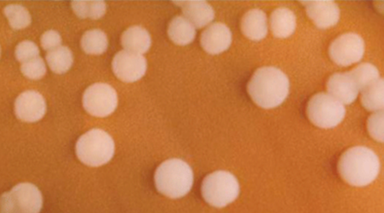
Medically important members of this group include Bordetella pertussis, which causes whooping cough (pertussus), and Neisseria meningitides, which causes bacterial meningitis.The micrograph below shows round, white colonies of N. meningitides on chocolate agar, which is brown.
The most diverse class of gram-negative bacteria is Gammaproteobacteria. This taxon includes a number of human pathogens.
EXAMPLE
Gammaproteobacteria include the human pathogens Pseudomonas aeruginosa (which causes diverse infections), Haemophilis influenzae (which causes upper and lower respiratory tract infections), and Vibrio cholerae (which produces a toxin that causes cholera).Enterobacteriaceae is a large family of enteric (intestinal) bacteria belonging to the Gammaproteobacteria. They are facultative anaerobes and able to ferment carbohydrates. There are two distinct categories of bacteria in this family: coliforms that can ferment lactose completely and noncoliforms that cannot ferment lactose or ferment it incompletely.
EXAMPLE
The prototypical coliform species, Escherichia coli, is exceptionally well-studied and used in research. Many strains of E. coli live in the human intestine as part of the normal microbiota. These strains have mutalistic relationships with humans. However, other strains can cause disease. Some strains produce a deadly toxin called Shiga toxin that is extremely potent and can cause fatal illness.An important noncoliform genus is Salmonella. A number of serotypes (strains or variations also called serovars) cause salmonellosis, characterized by inflammation of the small and large intestine accompanied by fever, vomiting, and diarrhea.
EXAMPLE
Salmonella enterobacterica (serovar typhi), the rod-shaped bacterium shown in the image below, causes typhoid fever, with symptoms including fever, abdominal pain, and skin rashes.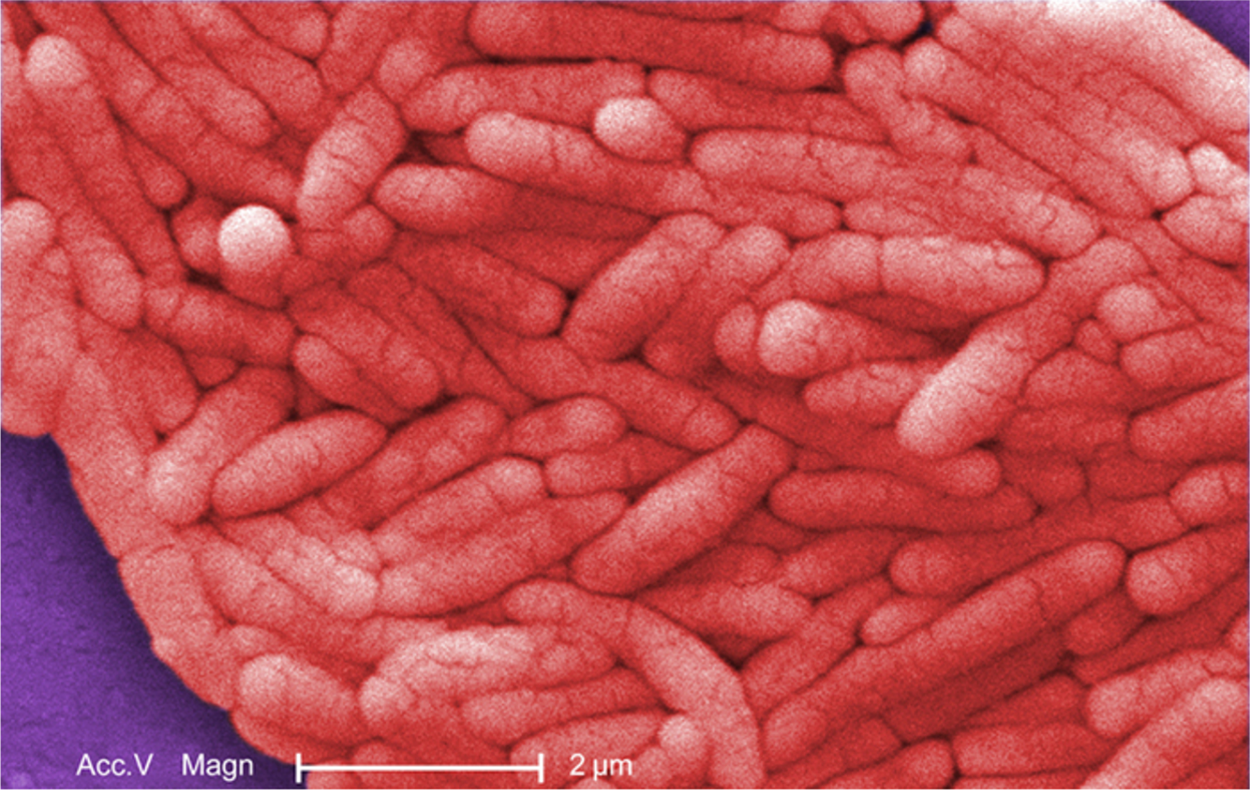
The Deltaprotebacteria is a small class of Proteobacteria. It includes sulfate-reducing bacteria (SRB), which get their name from the way that they use sulfate as an electron acceptor. You will learn more about this type of reaction in the lessons on metabolism.
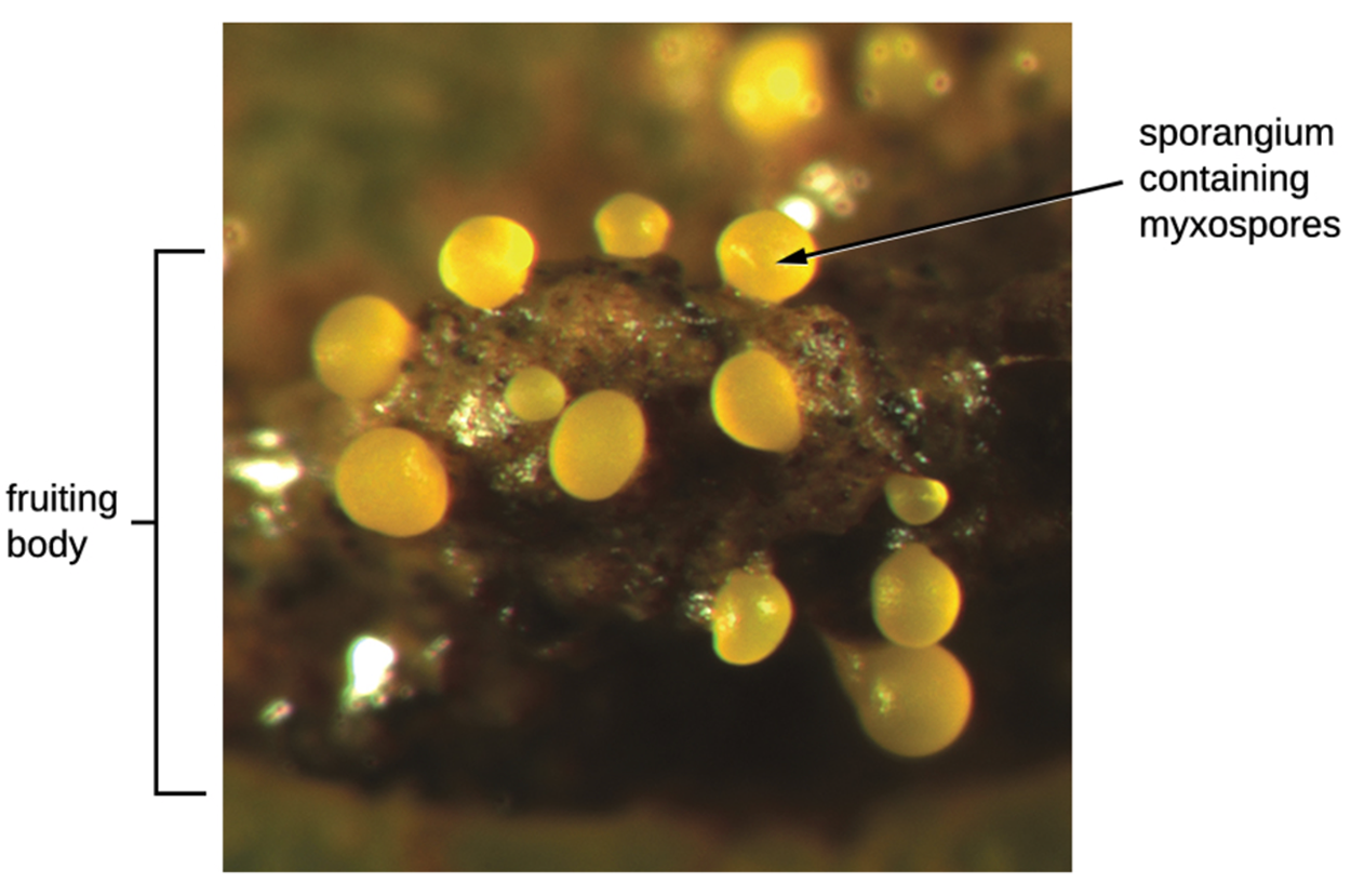
This group also includes the genus Bdellovibrio, species of which are parasites of other gram-negative bacteria, and myxobacteria, which can form multicellular, macroscopic fruiting bodies as shown in the image below.
The smallest class of Proteobacteria is the Epsilonproteobacteria. These bacteria are microaerophilic, meaning that they require lower levels of oxygen than are present in atmospheric air.
EXAMPLE
A medically important species in this class is Helicobacter pylori. This species is helical and flagellated, as shown in the micrograph below of a long, rectangular bacterium with many long flagella attached. It can be a beneficial member of the stomach microbiota, but can also cause stomach inflammation (gastritis) and ulcers in the stomach and duodenum (part of the small intestine). It is also linked to stomach cancer.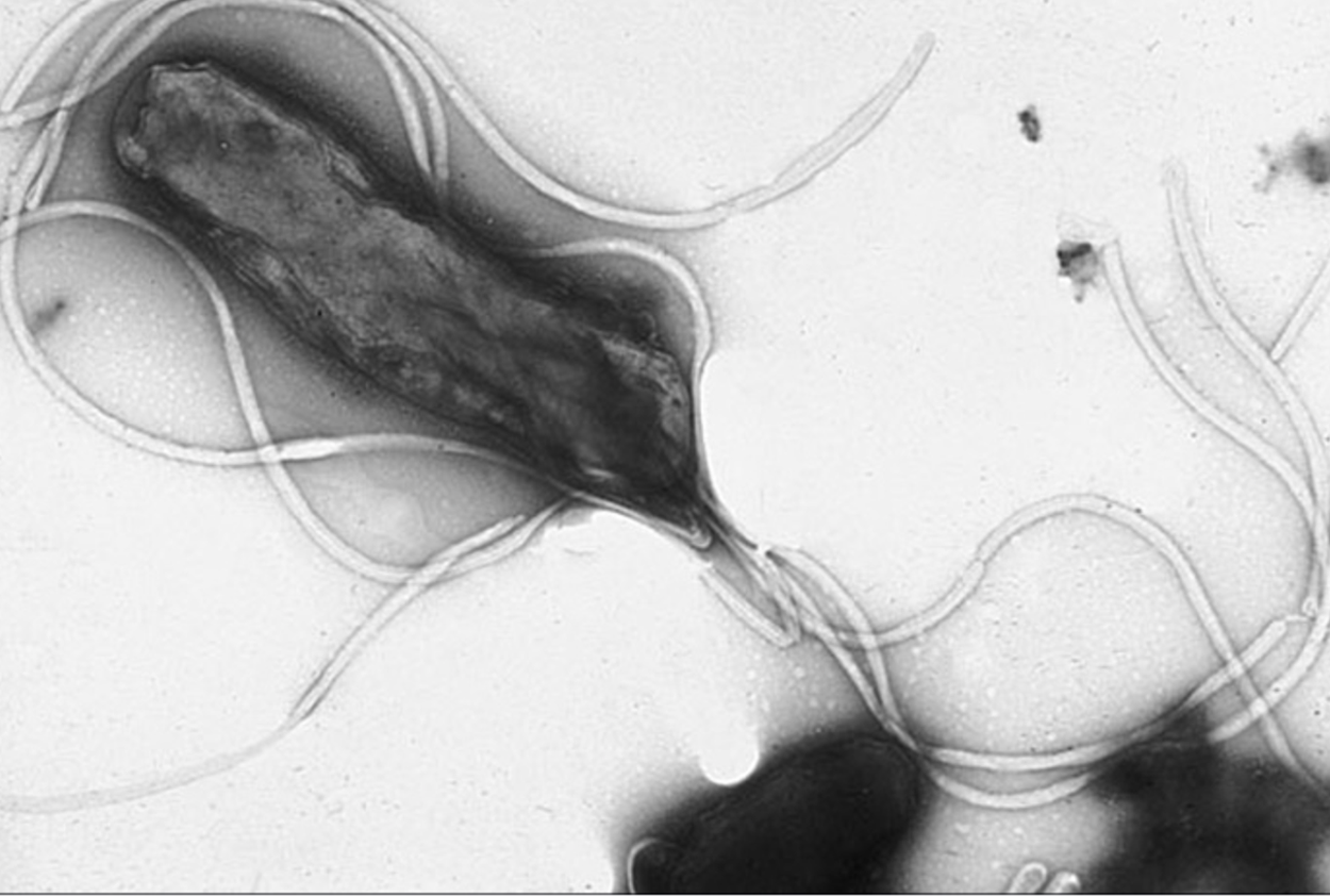
Gram-negative bacteria that are not classified in the Proteobacteria are called the nonproteobacteria. In this lesson we will discuss four classes of gram-negative proteobacteria.
There are other nonproteobacteria, including some that can capture light energy and convert it to chemical energy so that they can use it to make carbon-based compounds such as sugars that they need to grow (phototrophs). You will learn more about types of prototrophs and their metabolic processes in other lessons.
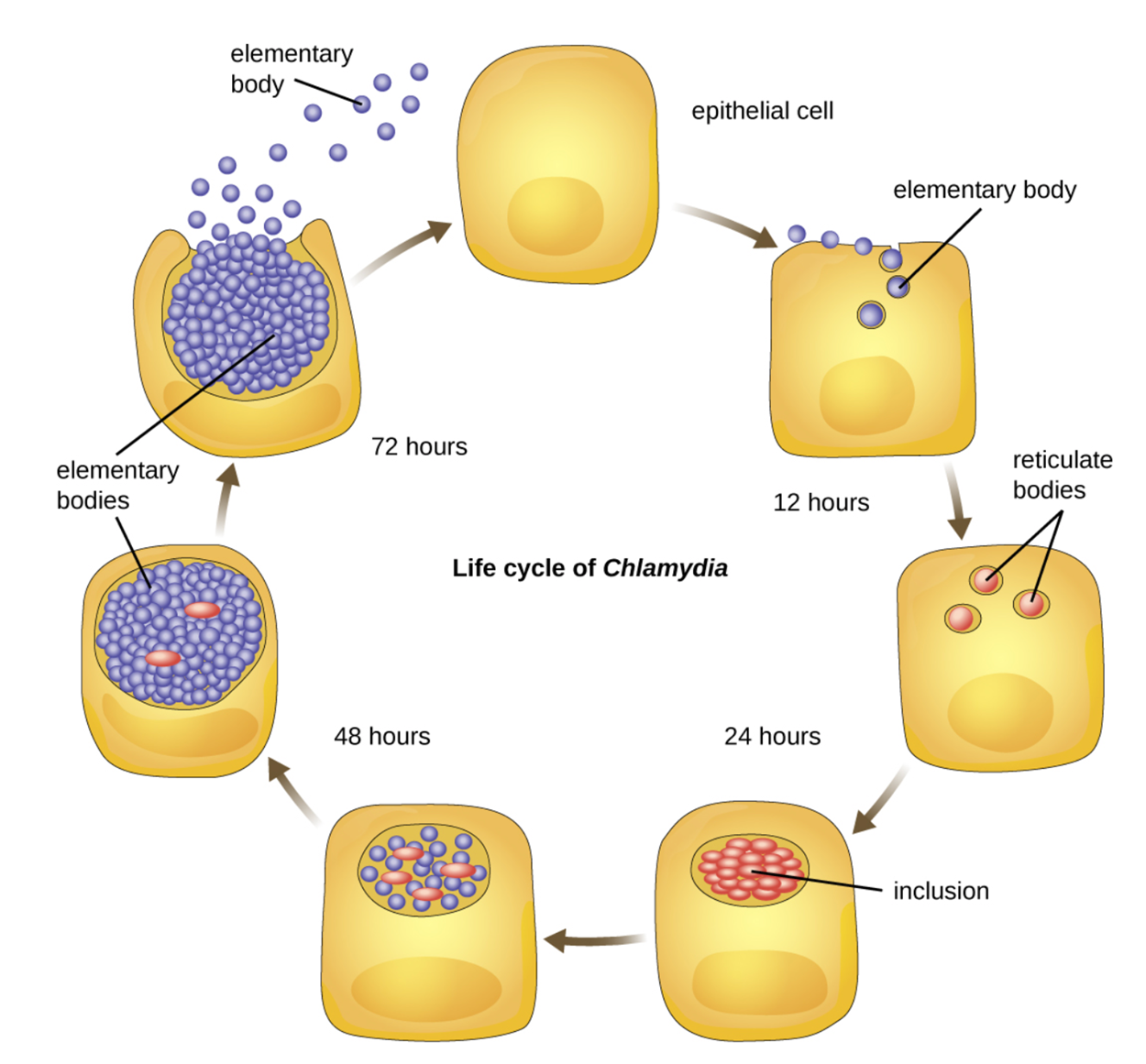
Members of the genus Chlamydia are atypical because they do not stain easily using standard Gram staining procedures. They are obligate intracellular pathogens that are extremely resistant to cellular defenses. They can spread rapidly from host to host via elementary bodies, which are inactive, endospore-like forms of intracellular bacteria. Once they enter an epithelial cell, cells that line body cavities like the stomach or nasal passages, they become active.
Medically important species include Chlamydia trachomatis, which causes a variety of infections such as the sexually transmitted infection chlamydia and types of conjunctivitis.
Spirochetes are characterized by their long (up to 250 μm), spiral-shaped bodies. Most spirochetes are also very thin, which makes it difficult to examine Gram-stained preparations under a conventional brightfield microscope. Darkfield fluorescent microscopy is typically used instead. Spirochetes are also different or even impossible to culture. They are highly motile and use an axial filament to propel themselves. The axial filament is similar to a flagellum, but runs inside the cell body of a spirochete in the periplasmic space between the outer membrane and the plasma membrane as shown in the image below.
EXAMPLE
A medically important spirochete is Treponema pallidum, which causes syphilis.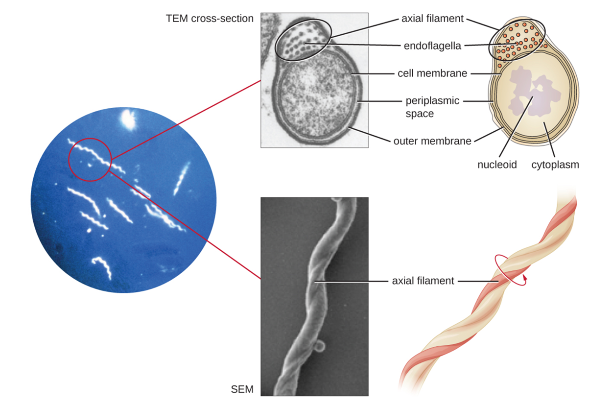
The CFB group includes Cytophaga, Fusobacterium, and Bacteriodes. Members of this group are phylogenetically diverse, but share some similarities in the sequence of nucleotides in their DNA. They are rod-shaped bacteria adapted to anaerobic environments such as the tissue of the gums, gut, and rumen of ruminating animals. They are avid fermenters that process cellulose in the rumen of ruminants, helping in digestion.
EXAMPLE
Cytophaga are motile aquatic bacteria that glide. Fusobacteria inhabit the human mouth and may cause severe infectious diseases. Bacteriodes, the largest of the three groups and shown as rod-shaped cells in the micrograph below, includes many species prevalent in the human large intestine.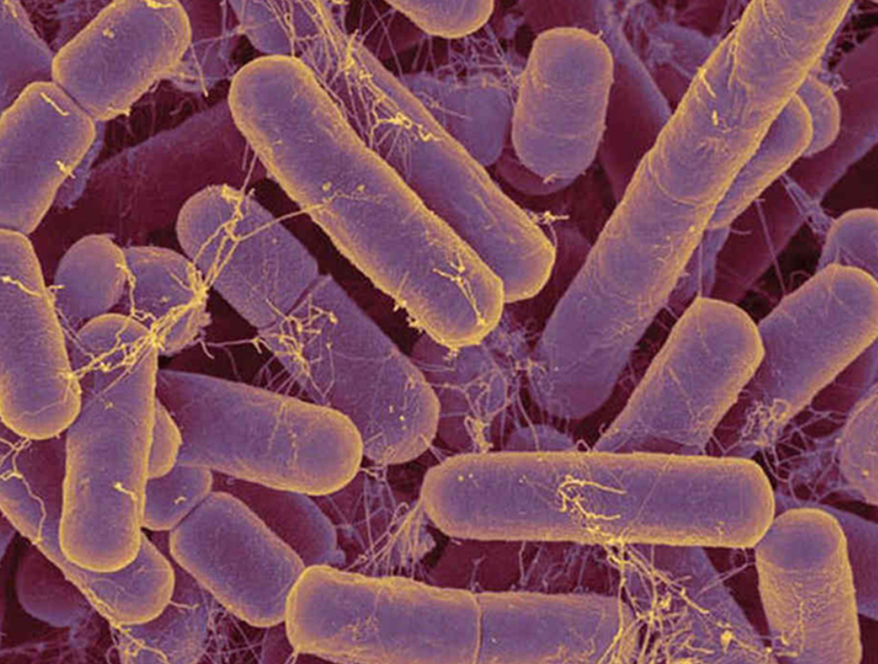
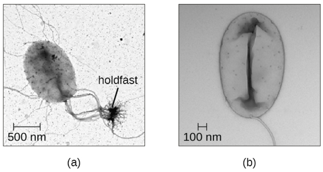
The Planctomycetes are found in aquatic environments, inhabiting freshwater, saltwater, and brackish water. They reproduce by budding. The mother cell produces a bud that detaches and becomes an independent swarmer cell. These swarmer cells are motile and not attached to a substrate. However, they will soon differentiate into cells that have an appendage called a holdfast that attaches them to a surface. They can only reproduce during the part of their life cycle in which they are immobile.
The image below shows an oval cell with thready structures extending to a holdfast in part a as well as a motile cell with no holdfast in part b.
Source: THIS TUTORIAL HAS BEEN ADAPTED FROM OPENSTAX “MICROBIOLOGY.” ACCESS FOR FREE AT openstax.org/details/books/microbiology. LICENSE: CC ATTRIBUTION 4.0 INTERNATIONAL.
REFERENCES
Parker, N., Schneegurt, M., Thi Tu, A.-H., Lister, P., & Forster, B. (2016). Microbiology. OpenStax. Access for free at openstax.org/books/microbiology/pages/1-introduction