Table of Contents |
Genetics is the study of heredity or inheritance of traits. Deoxyribonucleic acid (DNA) carries the genetic blueprint of the cell that is passed from parent to offspring via cell division.
As you have learned, each human cell has 23 pairs of chromosomes: one set of chromosomes is inherited from the female parent and the other set is inherited from the male parent. There is also a mitochondrial genome, inherited exclusively from the female parent, which can be involved in inherited genetic disorders. On each chromosome, there are thousands of genes that are responsible for determining the genotype (genetic makeup) and phenotype (observable characteristics) of the individual. A gene is defined as a sequence of DNA that codes for a functional product.
Genes are composed of DNA and are linearly arranged on chromosomes. Genes specify the sequences of amino acids, which are the building blocks of proteins. In turn, proteins are responsible for orchestrating nearly every function of the cell. Both genes and the proteins they encode are essential to life as we know it.
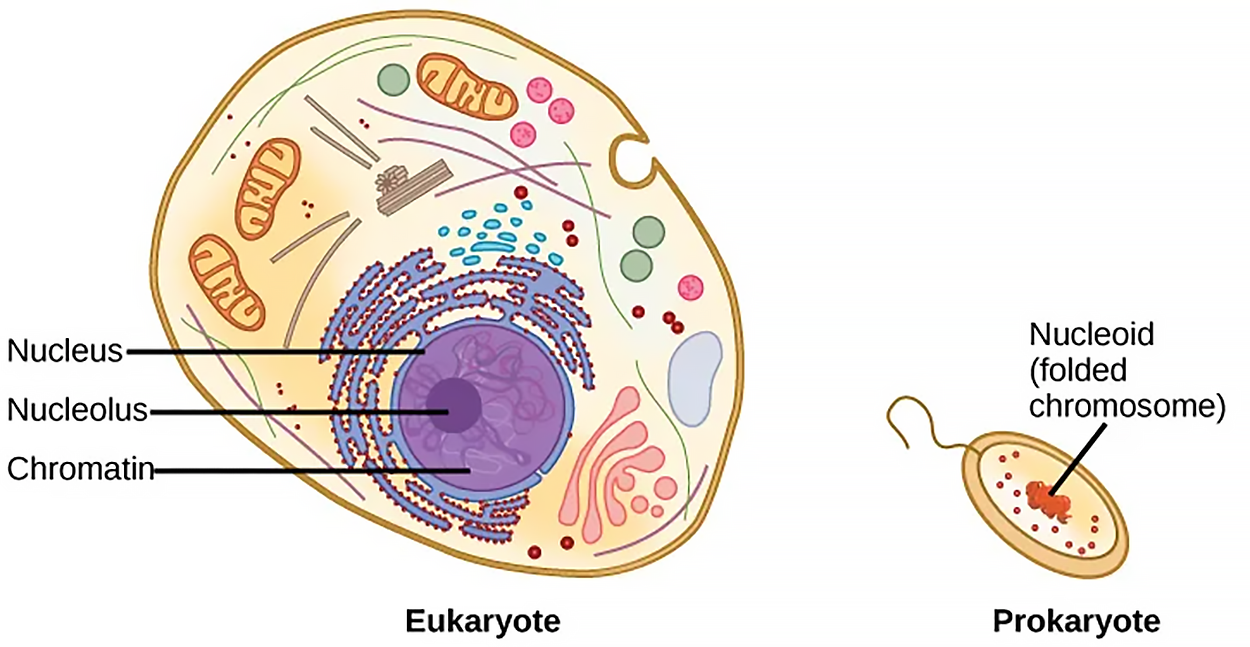
A cell’s complete complement of DNA is called its genome. In prokaryotes like bacteria, the genome is composed of a single, double-stranded DNA molecule in the form of a loop or circle, and the region in the cell containing this genetic material is called a nucleoid. In eukaryotes, such as plants, fungi, some single-celled organisms, and animals (including humans), the genome comprises several double-stranded, linear DNA molecules bound with proteins to form complexes called chromosomes. Each species of eukaryote has a characteristic number of chromosomes in the nuclei of its cells. As you have learned, human body cells (somatic cells) have 46 chromosomes.
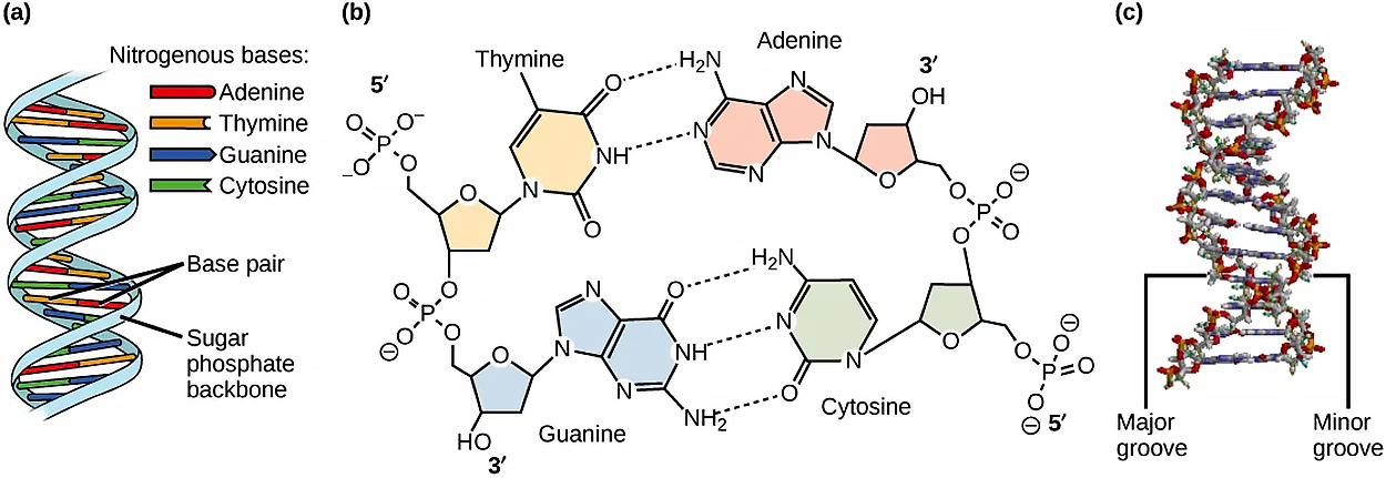
DNA and ribonucleic acid (RNA) are nucleic acids. Nucleic acids are a group of biological macromolecules that contain phosphorus in addition to carbon, hydrogen, oxygen, and nitrogen. The building blocks of nucleic acids are nucleotides. Each nucleotide consists of a pentose sugar (deoxyribose in DNA and ribose in RNA), a nitrogenous base (adenine, cytosine, guanine, and thymine or uracil), and a phosphate group. Each nucleotide is named depending on its nitrogenous base.
DNA is made up of two strands that are twisted around each other to form a double helix. The two strands run in opposite directions (antiparallel), are connected by hydrogen bonds, and are complementary to each other. The purines (adenine and guanine) have a double-ring structure with a six-membered ring fused to a five-membered ring. Pyrimidines are smaller in size; they have a single six-membered ring structure (cytosine, thymine, and uracil). In DNA, purines pair with pyrimidines: adenine pairs with thymine (A-T), and cytosine pairs with guanine (C-G). The term base pair refers to two complementary nucleotides that are paired together.


In RNA, uracil replaces thymine to pair with adenine (U-A). RNA also differs from DNA in that it is single-stranded and has many forms, such as messenger RNA (mRNA), ribosomal RNA (rRNA), and transfer RNA (tRNA) that all participate in the synthesis of proteins. MicroRNAs (miRNAs) regulate the use of mRNA.
IN CONTEXT
Neanderthal Genome: How Are We Related?
The first draft sequence of the Neanderthal genome was published by Richard E. Green et al. in 2010. Neanderthals are the closest ancestors of present-day humans. They were known to have lived in Europe and Western Asia (and now, perhaps, in Northern Africa) before they disappeared from fossil records approximately 30,000 years ago. Green’s team studied almost 40,000-year-old fossil remains that were selected from sites across the world. Extremely sophisticated means of sample preparation and DNA sequencing were employed because of the fragile nature of the bones and heavy microbial contamination. In their study, the scientists were able to sequence some four billion base pairs. The Neanderthal sequence was compared with that of present-day humans from across the world. After comparing the sequences, the researchers found that the Neanderthal genome had 2% to 3% greater similarity to people living outside Africa than to people in Africa. While current theories have suggested that all present-day humans can be traced to a small ancestral population in Africa, the data from the Neanderthal genome suggest some interbreeding between Neanderthals and early modern humans.
Green and his colleagues also discovered DNA segments among people in Europe and Asia that are more similar to Neanderthal sequences than to other contemporary human sequences. Another interesting observation was that Neanderthals are as closely related to people from Papua New Guinea as to those from China or France. This is surprising because Neanderthal fossil remains have been located only in Europe and West Asia. Most likely, genetic exchange took place between Neanderthals and modern humans as modern humans emerged out of Africa, before the divergence of Europeans, East Asians, and Papua New Guineans.
Several genes seem to have undergone changes from Neanderthals during the evolution of present-day humans. These genes are involved in cranial structure, metabolism, skin morphology, and cognitive development. One of the genes that is of particular interest is RUNX2, which is different in modern-day humans and Neanderthals. This gene is responsible for the prominent frontal bone, bell-shaped rib cage, and dental differences seen in Neanderthals. It is speculated that an evolutionary change in the RUNX2 gene was important in the origin of modern-day humans, and this affected the cranium and the upper body.
| Term | Pronunciation | Audio File |
|---|---|---|
| Genome | ge·nome |
|
| Nucleic Acid | nu·cle·ic acid |
|
| Nucleotide | nu·cle·o·tide |
|
Prokaryotes are much simpler than eukaryotes in many of their features, as shown below. Most prokaryotes contain a single, circular chromosome that is found in an area of the cytoplasm called the nucleoid region.

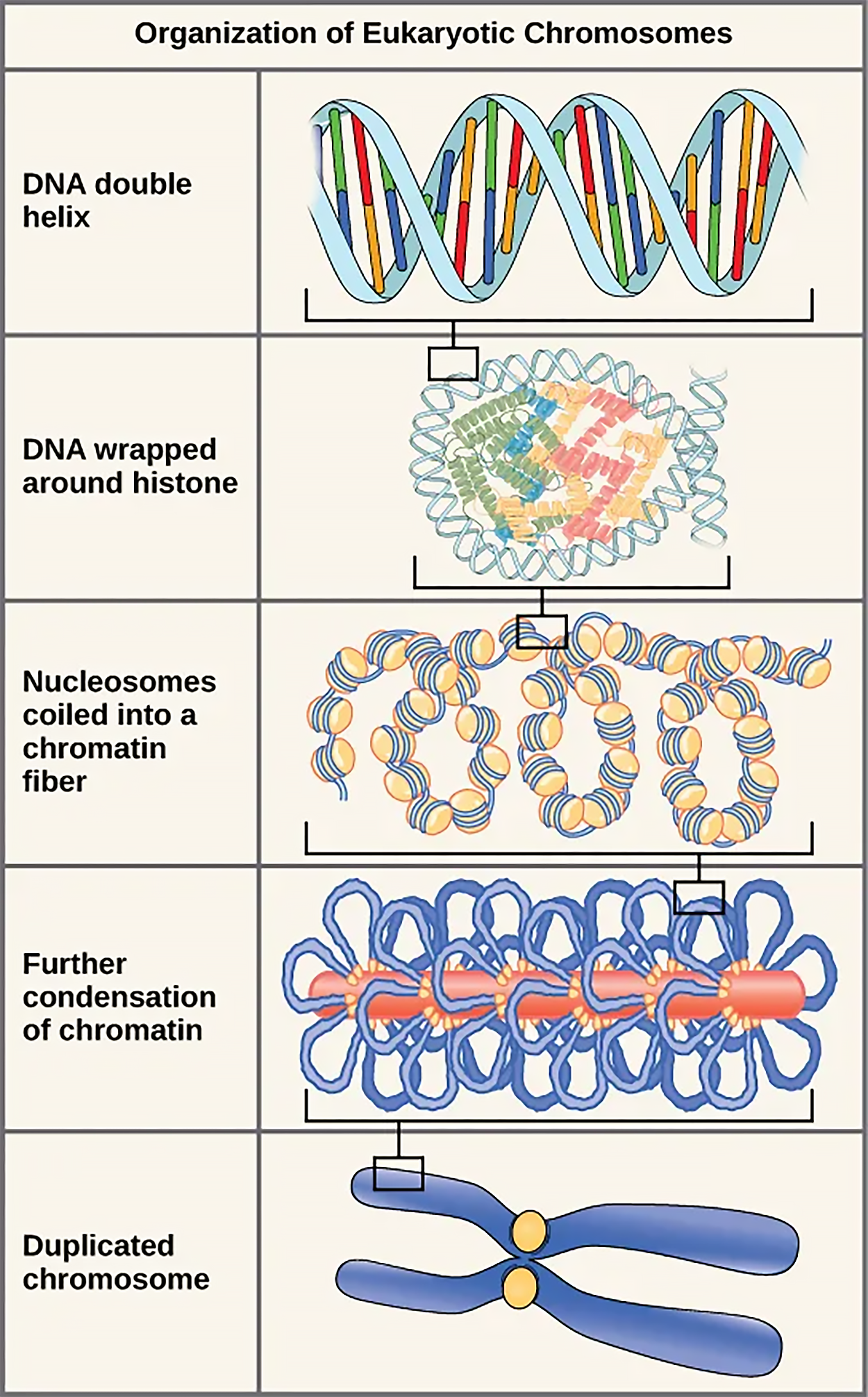
Eukaryotes, whose chromosomes each consist of a linear DNA molecule, employ a different type of packing strategy to fit their DNA inside the nucleus. At the most basic level, DNA is wrapped around proteins known as histones to form structures called nucleosomes. The histones are evolutionarily conserved proteins that are rich in basic amino acids and form an octamer composed of two molecules of each of four different histones. The DNA is wrapped tightly around the histone core. This nucleosome is linked to the next one with the help of a linker DNA. This is also known as the “beads on a string” structure. With the help of a fifth histone, a string of nucleosomes is further compacted into a 30-nm fiber, which is the diameter of the structure. Metaphase chromosomes are even further condensed by association with scaffolding proteins. At the metaphase stage, the chromosomes are at their most compact, approximately 700 nm in width.
In interphase, eukaryotic chromosomes have two distinct regions that can be distinguished by staining. The tightly packaged region is known as heterochromatin, and the less dense region is known as euchromatin. Heterochromatin usually contains genes that are not expressed, and it is found in the regions of the centromere and telomeres. The euchromatin usually contains genes that are transcribed, with DNA packaged around nucleosomes but not further compacted.

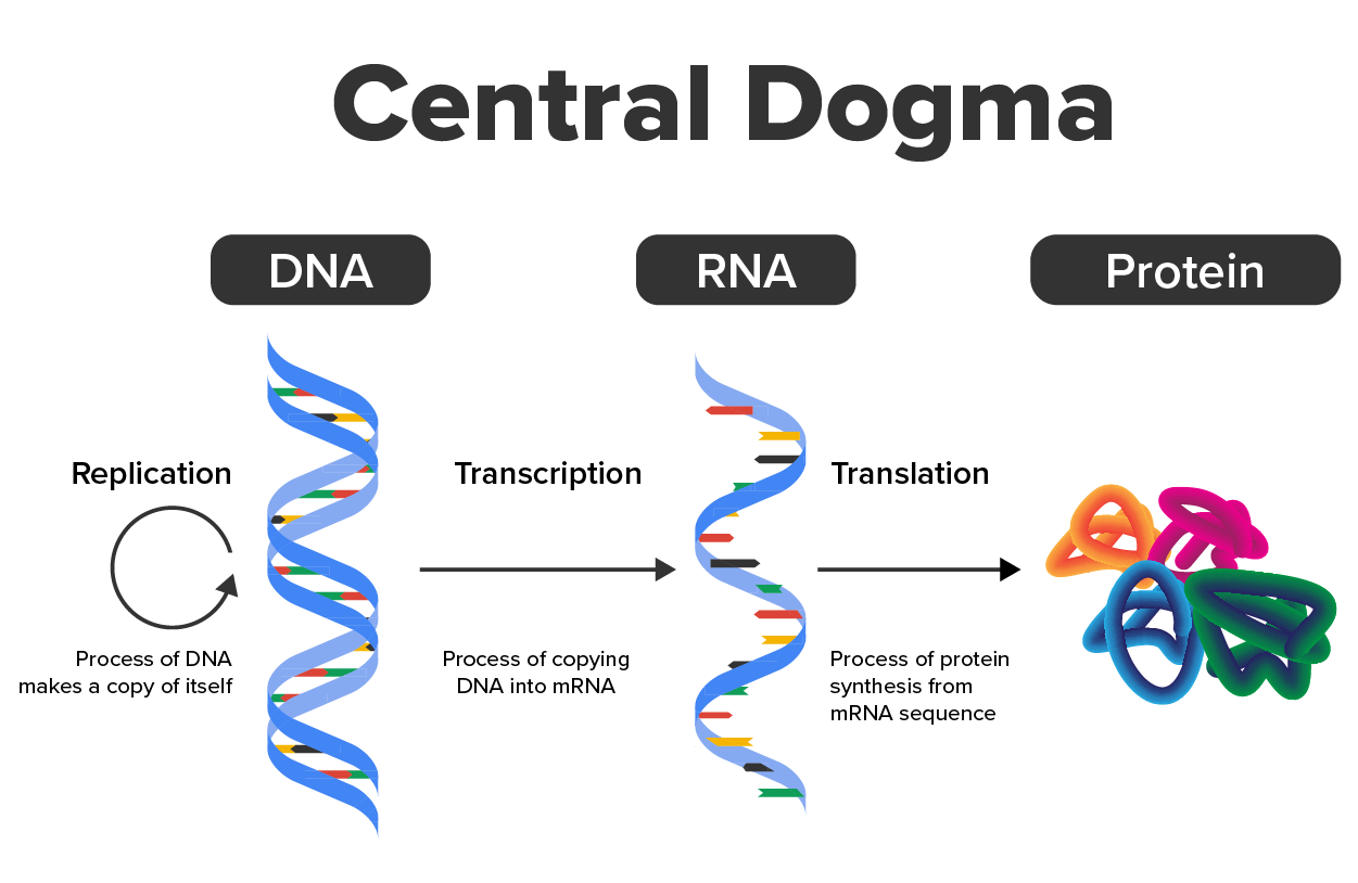
The functions of DNA include replication and providing the information needed to construct the proteins necessary so that the cell can perform all of its functions. Recall from Anatomy & Physiology I that, to do this, the DNA is “read” or transcribed into an mRNA molecule. The mRNA then provides the code to form a protein by a process called translation. Through the processes of transcription and translation, a protein is built with a specific sequence of amino acids that was originally encoded in the DNA. The flow of genetic information is usually DNA → RNA → protein, which is also known as the Central Dogma of Life.
