Table of Contents |
The digestive system has several major functions:
In this lesson, you will learn the anatomy of the digestive system and a little bit about its function. You will learn more about the overall process summarized above later in this course.
This section will provide an overview of the anatomy of the digestive system, including the mouth, tongue, salivary glands, pharynx, esophagus, stomach, small intestine, and large intestine. You will learn more about the accessory organs involved in digestion in other lessons focusing on these organs and on digestive processes.
The mouth, cheeks, tongue, and palate form the oral cavity, which is also called the buccal cavity. The oral cavity includes the teeth.
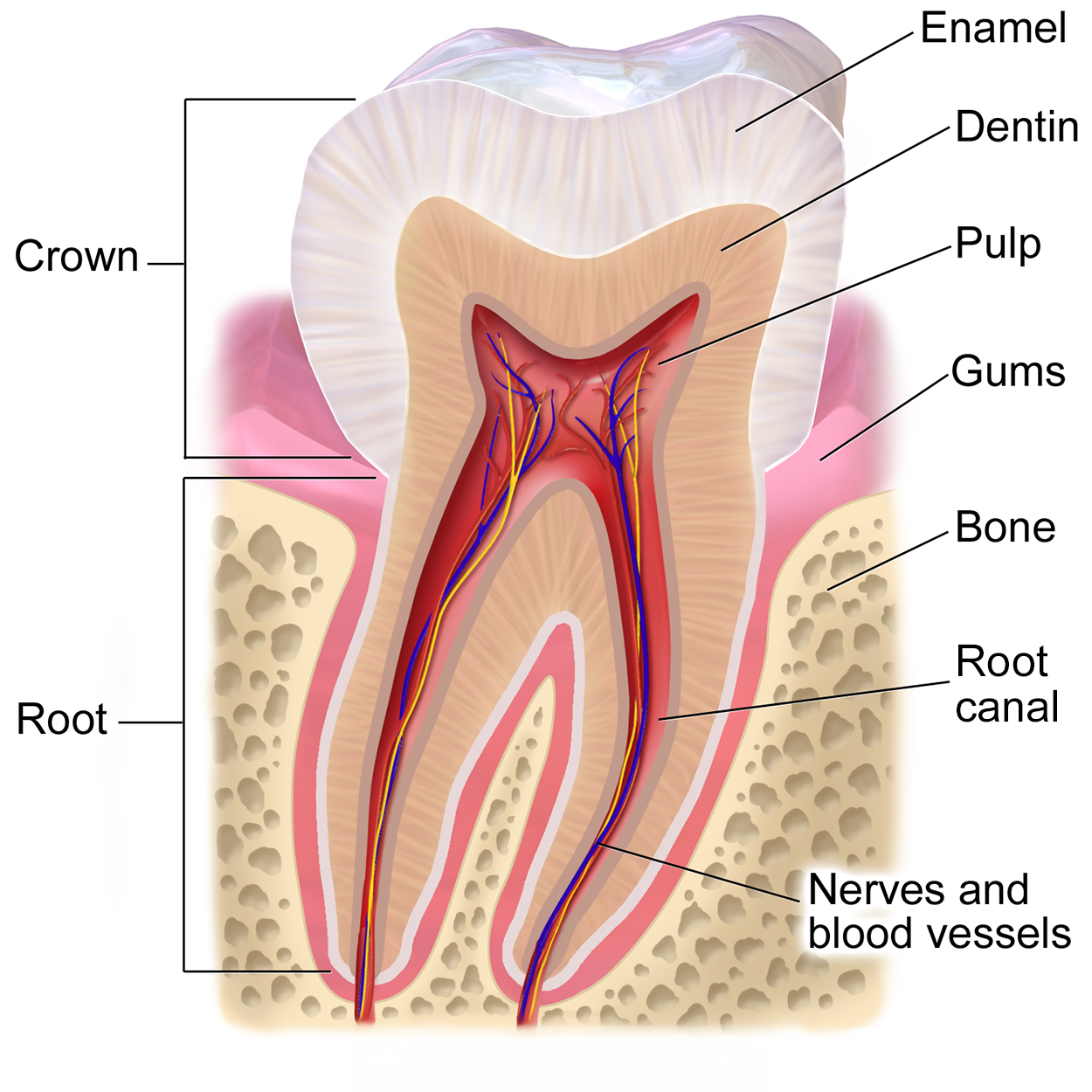
The figure below shows the parts of a human tooth. Note the upper crown that narrows to a root that extends into the fleshy gums (gingiva, or gingivae plural), which are embedded within the bone of the jaw. The root has left and right halves. Note also that the crown has a thick outer covering of enamel, while the enamel is much thinner on the root. Below the enamel is a layer of dentin above the internal pulp. Nerves and blood vessels extend from the base of each half of the root up through each side of the root into the pulp.
If you run your tongue along the roof of your mouth, you’ll notice the roof of the mouth has an arch called a hard palate. The anterior region of the palate serves as a septum (i.e., wall) between the oral and nasal cavities, as well as a rigid shelf against which the tongue can push food when swallowing. The hard palate ends in the posterior oral cavity, where the tissue becomes fleshier. This part of the palate, known as the soft palate, is composed mainly of skeletal muscle that can be manipulated for actions like swallowing or singing.
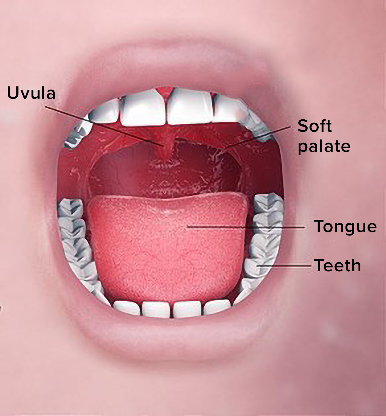
In the figure below, you can see an open mouth with the upper and lower teeth exposed. Note the tongue (a fleshy, muscular structure) lying on the floor of the mouth. The tongue can move and taste food (Merriam-Webster, n.d.a.). Note the soft palate forming the rear roof of the mouth. The uvula is a small, fleshy structure that hangs down from the rear roof of the oral cavity (the center of the soft palate). When swallowing, the soft palate and uvula move upward, helping to keep foods and liquids from entering the nasal cavity and respiratory tract.
Two muscular folds extend downward from the soft palate on either side of the uvula. Between these two folds are the palatine tonsils, clusters of lymphoid tissue involved in the immune system. The lingual tonsils are located at the base of the tongue.
The tongue facilitates ingestion, digestion, sensation (of taste, texture, and temperature of food), swallowing, and vocalization. It has a mucous membrane covering and is composed of skeletal muscles that facilitate eating, swallowing, and speaking. The top and sides of the tongue are studded with papillae (bumpy structures) and taste buds to facilitate sensation (note that papilla is a general term for small bumps or protrusions; Merriam-Webster, n.d.b.). Lingual glands secrete mucus and a watery fluid that contains the enzyme lipase, which becomes active in the acidic environment of the stomach.
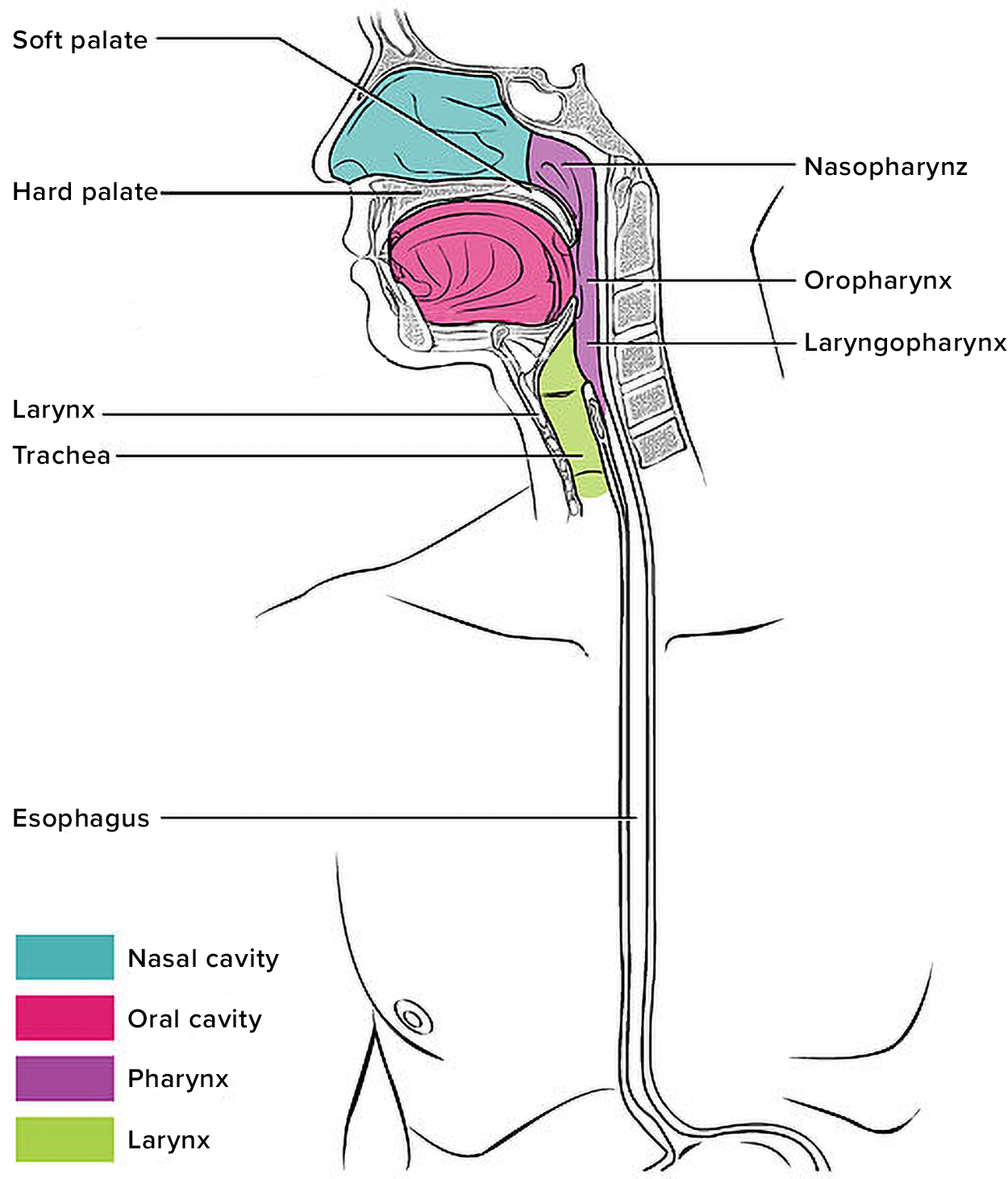
The pharynx is a muscular tube lined with a mucous membrane that runs from the posterior oral and nasal cavities to the esophagus and the larynx.
The pharynx has three subdivisions. The superior section, the nasopharynx, is involved only in breathing and speech. The other two subdivisions, the oropharynx and the laryngopharynx, are used for both functions of breathing and digestion. The oropharynx begins inferior to the nasopharynx and continues to the laryngopharynx. In the laryngopharynx, the inferior border of the laryngopharynx connects to the esophagus, whereas the anterior portion connects to the larynx.
The pharynx is involved in both digestion and breathing. It receives air from the mouth and nasal cavities and food from the mouth. When food enters the pharynx, involuntary muscle contractions close a flap of tissue called the epiglottis to prevent food from entering the trachea and to ensure it enters the esophagus.
The figure above shows the parts of the pharynx described above.
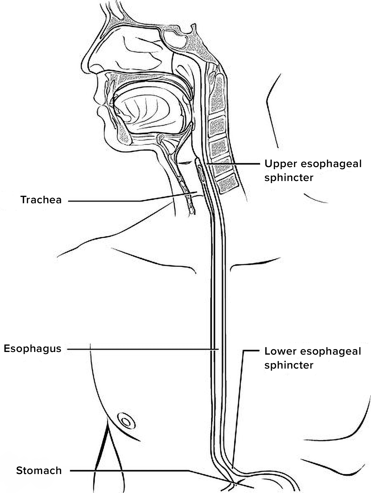
The esophagus is a muscular tube that connects the laryngopharynx to the stomach. It is located posteriorly to the trachea and is approximately ten inches long. The upper esophageal sphincter is a muscular ring that controls the movement of food from the pharynx to the esophagus.
A series of wave-like muscle contractions called peristalsis push food through the esophagus and into the stomach (peristalsis continues through the rest of the digestive tract). Just before the opening to the stomach is another ring-shaped muscle called the lower esophageal sphincter, also known as the cardiac sphincter. Recall that sphincters are muscles that surround tubes and serve as valves, closing the tube when the sphincters contract and opening it when they relax. This sphincter opens to let food pass into the stomach and closes to keep it from backing up into the esophagus.
The figure above shows the esophagus running from the mouth to the stomach, posterior to the trachea. Note the upper esophageal sphincter at the top, where the pharynx meets the esophagus, and the lower esophageal sphincter at the bottom, where the esophagus meets the stomach.
The stomach is a sac-like organ where food is mixed with gastric juice. There are four main regions in the stomach: the cardia, fundus, body, and pylorus. The funnel-shaped pylorus connects the stomach to the duodenum, the first part of the small intestine. The pyloric sphincter is located at this latter point of connection and controls stomach emptying into the small intestine.
The inner lining of the stomach is covered by a mucous membrane that secretes a protective coat of alkaline mucus. The mucus protects the stomach from corrosive stomach acid. Gastric glands lie under the surface of this lining and secrete a complex digestive fluid referred to as gastric juice, which contains hydrochloric acid and other enzymes.
Cells in the stomach lining secrete digestive hormones, such as gastrin and pepsin, as well as hydrochloric acid. The acidity stimulates digestive hormones and also kills most of the bacteria ingested with food. A common digestive disorder called gastroesophageal reflux disease (GERD) is caused by hydrochloric acid that backs up into the lower esophagus.
The muscular walls of the stomach move food through the stomach and vigorously churn it, mechanically breaking it down into smaller particles.
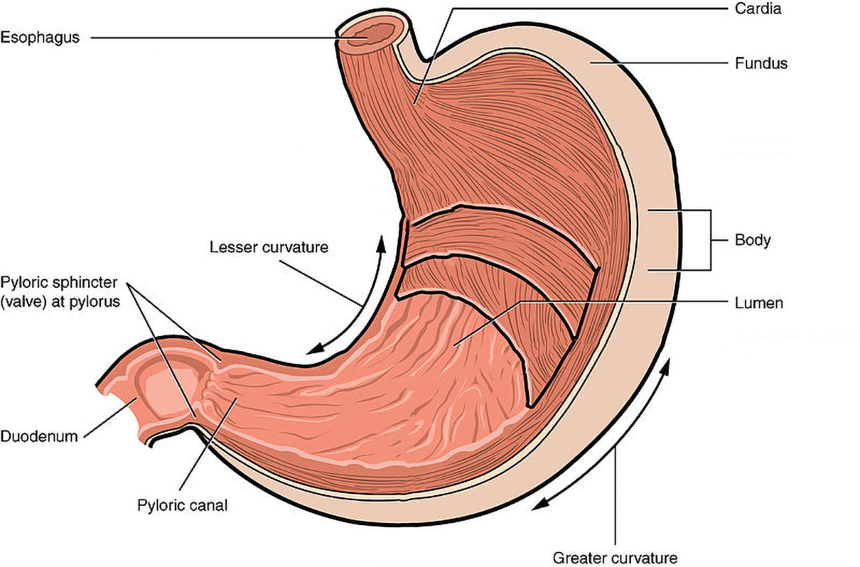
The figure above shows the stomach. Note its curved shape from the esophagus at the upper right to the pyloric canal at the lower right. The lesser curvature is the concave curve along the right side. The greater curvature is the concave opposite side. The lumen is the opening, which is lined by folds in the mucosa (rugae). The cardia is just below the entry to the esophagus, with the fundus extending up to the left. The body is in the middle, and the pylorus is at the lower right. The stomach narrows to form the pyloric canal, which meets the duodenum of the small intestine at the pyloric sphincter.
Partially digested food, called chyme, is released from the stomach via the pyloric sphincter and enters the small intestine, the part of the digestive tract where most of the digestion and absorption into the blood occurs. Approximately 90 percent of nutrients from food are absorbed through villi, small fingerlike extensions on the inner surface of the small intestine. The small intestine is about ten feet long. Its name derives from its small diameter of about one inch, compared to the large intestine’s diameter of about three inches.
The coiled tube of the small intestine is subdivided into three regions called the duodenum, jejunum, and ileum. The ileum joins the cecum of the large intestine at the ileocecal valve. The ileocecal valve controls the flow of chyme from the small intestine to the large intestine.
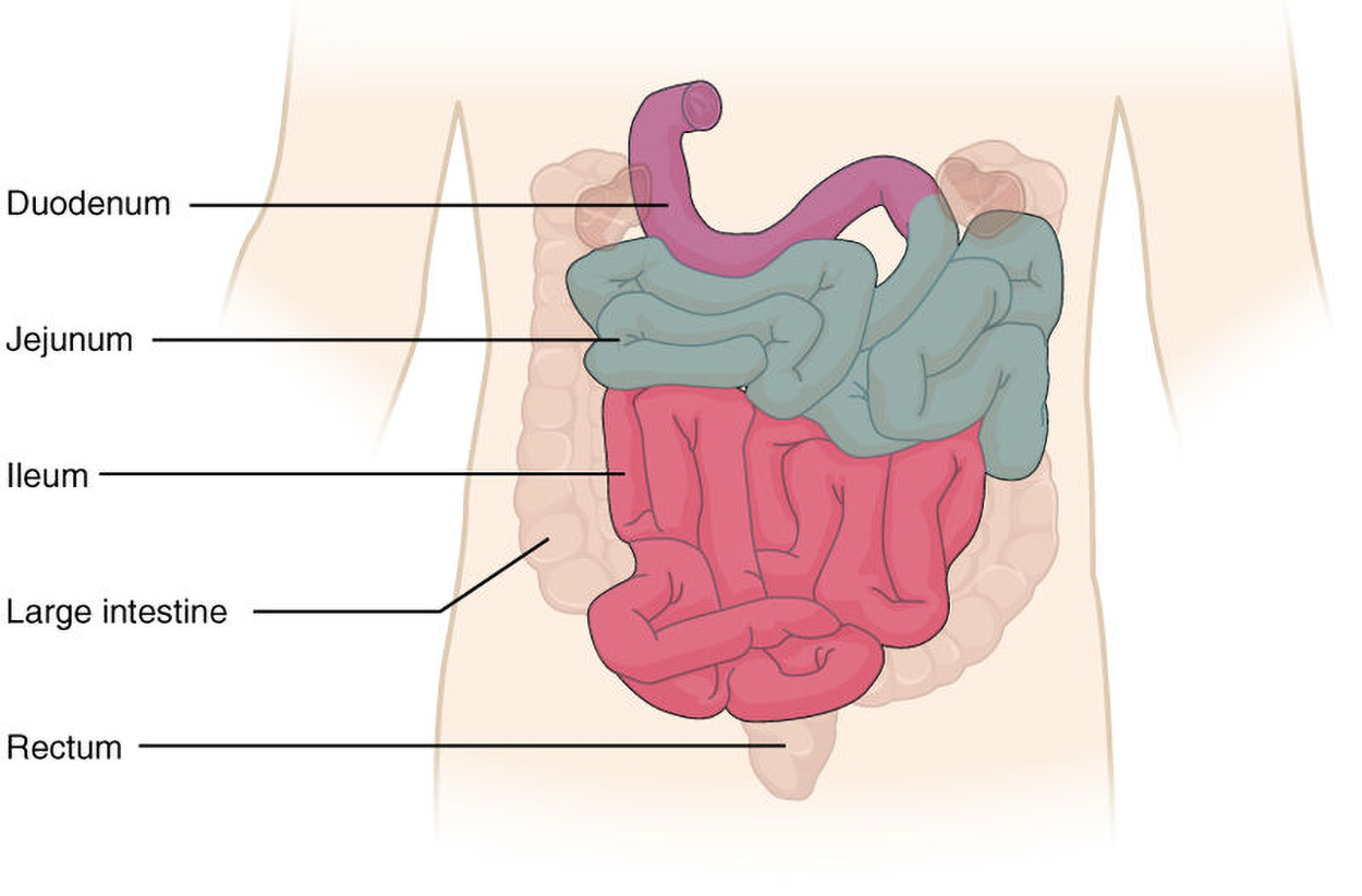
The figure above shows how the coiled small intestine begins with a small piece of duodenum extending from the stomach. The middle region is the ileum, and the jejunum is at the bottom. The large intestine is shown in the background.
The large intestine runs from the small intestine to the anus. The primary functions of the large intestine are to finish absorption of nutrients and water, synthesize certain vitamins, and form and eliminate feces, also called stool. The large intestine is about one-half the length of the small intestine, but it is called large because its diameter is three inches, about twice the diameter of the small intestine. The large intestine is subdivided into four main regions: cecum, colon, rectum, and anal canal.
The figure below shows major features of the large intestine, which are explained in more detail below. Note that the large intestine begins with the small cecum at the lower right, with a small, tubular vermiform appendix (commonly just called the appendix) extending down from it. The cecum joins the ascending colon that runs up the right side of the abdomen, then bends at the right colic (hepatic) flexure. The transverse colon runs across the top, above the small intestine, then bends downward at the left colic (splenic) flexure. The descending colon runs down the left side, then meets the sigmoid colon. The sigmoid colon curves down, then up slightly to the right, before meeting the rectum, which extends down to the anal canal that ends at the anus.
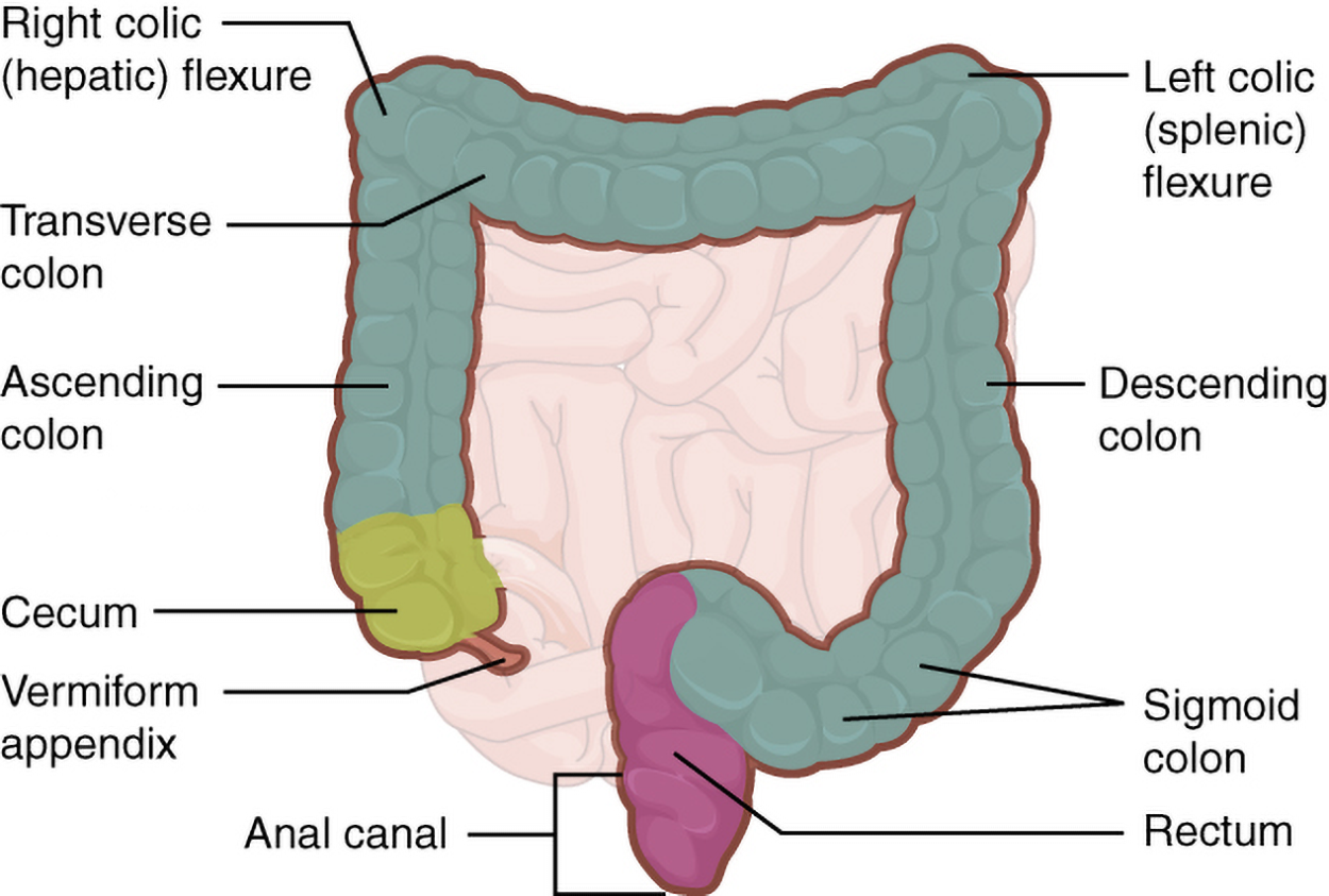
The cecum is the initial part of the large intestine. It receives the contents of the small intestine and continues absorbing water and salts. The appendix is a small pouch attached to the end of the cecum. Its twisted anatomy provides a haven for the accumulation and multiplication of intestinal bacteria that can lead to appendicitis.
Food residue entering the colon travels up the ascending colon on the right side of a person’s abdomen. At the inferior surface of the liver, the colon bends to become the transverse colon, where it travels across to the left side of their abdomen. From there, chyme passes through another bend inferior to the spleen and down the descending colon, which runs down the left side of the posterior abdominal wall and becomes the S-shaped sigmoid colon.
The rectum is the final eight inches of the large intestine. It has three folds called rectal valves that help separate feces from flatus, commonly referred to as gas.
The anal canal is about three inches long and opens to the exterior of the body at the anus, the end of the digestive tract. The anal canal includes two sphincters. The internal anal sphincter is made of smooth muscle, and its contractions are involuntary. The external anal sphincter is made of skeletal muscle, which is under voluntary control. This allows an individual to control the final step in which wastes leave the body. Except when defecating, both sphincters usually remain closed.
Source: THIS TUTORIAL HAS BEEN ADAPTED FROM OPEN RN "MEDICAL TERMINOLOGY 2E". ACCESS FOR FREE AT wtcs.pressbooks.pub/medterm/ LICENSING: CREATIVE COMMONS ATTRIBUTION 4.0 INTERNATIONAL. Accessed by March 2025.
REFERENCES
Merriam-Webster. (n.d.a.). Tongue. Merriam-Webster.com dictionary. Retrieved July 19, 2025, from www.merriam-webster.com/dictionary/tongue
Merriam-Webster. (n.d.b.). Papilla. Merriam-Webster.com dictionary. Retrieved July 19, 2025, from www.merriam-webster.com/dictionary/papilla