Table of Contents |
Sexual reproduction requires the union of two specialized cells, which you learned are called gametes, each of which contains one set of chromosomes. When gametes unite, they form a zygote, or fertilized egg that contains two sets of chromosomes. (Note: Cells that contain one set of chromosomes are called haploid; cells containing two sets of chromosomes are called diploid.) If the reproductive cycle is to continue for any sexually reproducing species, then the diploid cell must somehow reduce its number of chromosome sets to produce haploid gametes; otherwise, the number of chromosome sets would double with every future round of fertilization. Therefore, sexual reproduction requires a nuclear division (division of the nucleus) that reduces the number of chromosome sets by half.
Most animals and plants as well as many unicellular organisms are diploid and therefore have two sets of chromosomes. In each somatic cell of the organism (all cells of a multicellular organism except the gametes or reproductive cells), the nucleus contains two copies of each chromosome, called homologous chromosomes. Homologous chromosomes are matched pairs containing the same genes in identical locations along their lengths. Diploid organisms inherit one copy of each homologous chromosome from each genetic contributor.
Meiosis is cell division that forms haploid cells from diploid cells, and it employs many of the same cellular mechanisms as mitosis, which is cell division that promotes growth, development, and repair of the human body. However, mitosis produces daughter cells whose nuclei are genetically identical to the original parent nucleus. In mitosis, both the parent and the daughter nuclei are at the same “ploidy level”—diploid in the case of most multicellular animals. You will learn more about mitosis in a future challenge.
In meiosis, the starting nucleus is always diploid, and the daughter nuclei that result are haploid. To achieve this reduction in chromosome number, meiosis consists of one round of chromosome replication followed by two rounds of nuclear division. Because many events that occur during each of the division stages are analogous to the events of mitosis, the same stage names are assigned. However, because there are two rounds of division in meiosis, the major process and the stages are designated with a “I” or a “II.” Thus, meiosis I is the first round of meiotic division and consists of prophase I, prometaphase I, and so on. Likewise, meiosis II (during which the second round of meiotic division takes place) includes prophase II, prometaphase II, and so on.
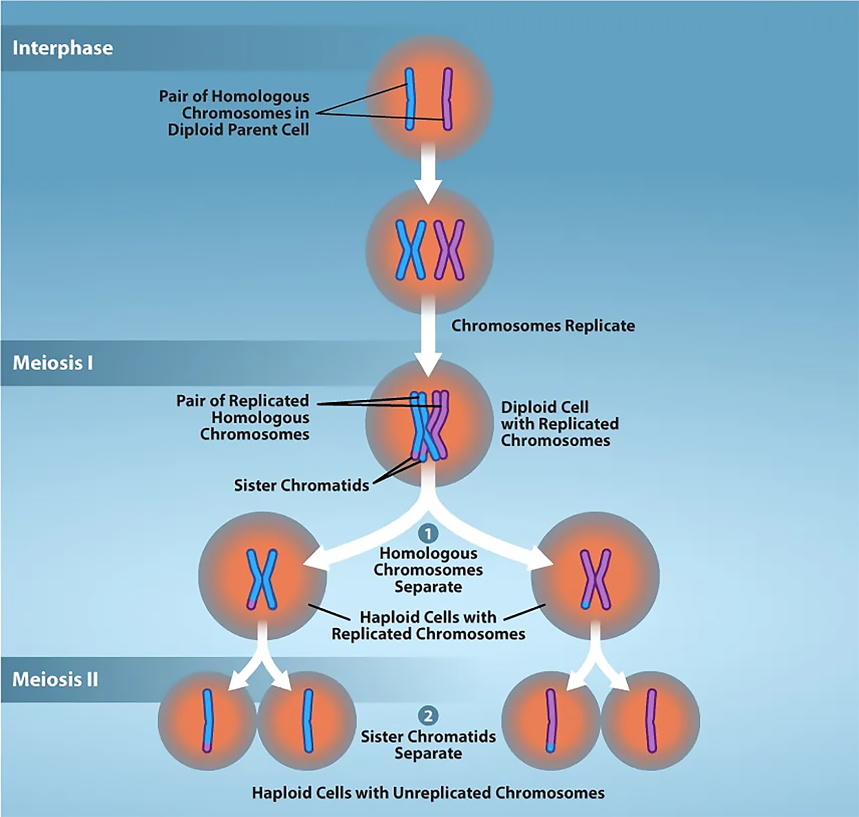
Meiosis is preceded by an interphase consisting of G1, S, and G2 phases, which are nearly identical to the phases preceding mitosis. The G1 phase (the “first gap phase”) is focused on cell growth. During the S phase—the second phase of interphase—the cell copies or replicates the DNA of the chromosomes. Finally, in the G2 phase (the “second gap phase”), the cell undergoes the final preparations for meiosis.
During DNA duplication in the S phase, each chromosome is replicated to produce two identical copies—sister chromatids that are held together at the centromere by cohesin proteins, which hold the chromatids together until anaphase II. (Note: these chromosome copies are called sister chromatids regardless of whether they are in a female gamete or a male gamete.)
As a result of DNA synthesis (replication), the germ cell (the cell that will develop into a gamete) looks a lot like a somatic cell that's about to divide: Its 23 pairs of homologous chromosomes in humans (46 total) now each have two sister chromatids (92 chromatids total).
The phases of meiosis I include:
After telophase I is done, the cells begin meiosis II. During meiosis II, the sister chromatids within the two daughter cells separate, forming four new haploid gametes. The mechanics of meiosis II are similar to mitosis, except that each dividing cell has only one set of homologous chromosomes, each with two chromatids. Therefore, each cell has half the number of sister chromatids to separate out as a diploid cell undergoing mitosis.
The phases of meiosis II include:
EXAMPLE
In humans, both the sperm cell and the egg contain 23 chromosomes which combine to give a total of 46 chromosomes. In you, half of these came from your mother and half from your father.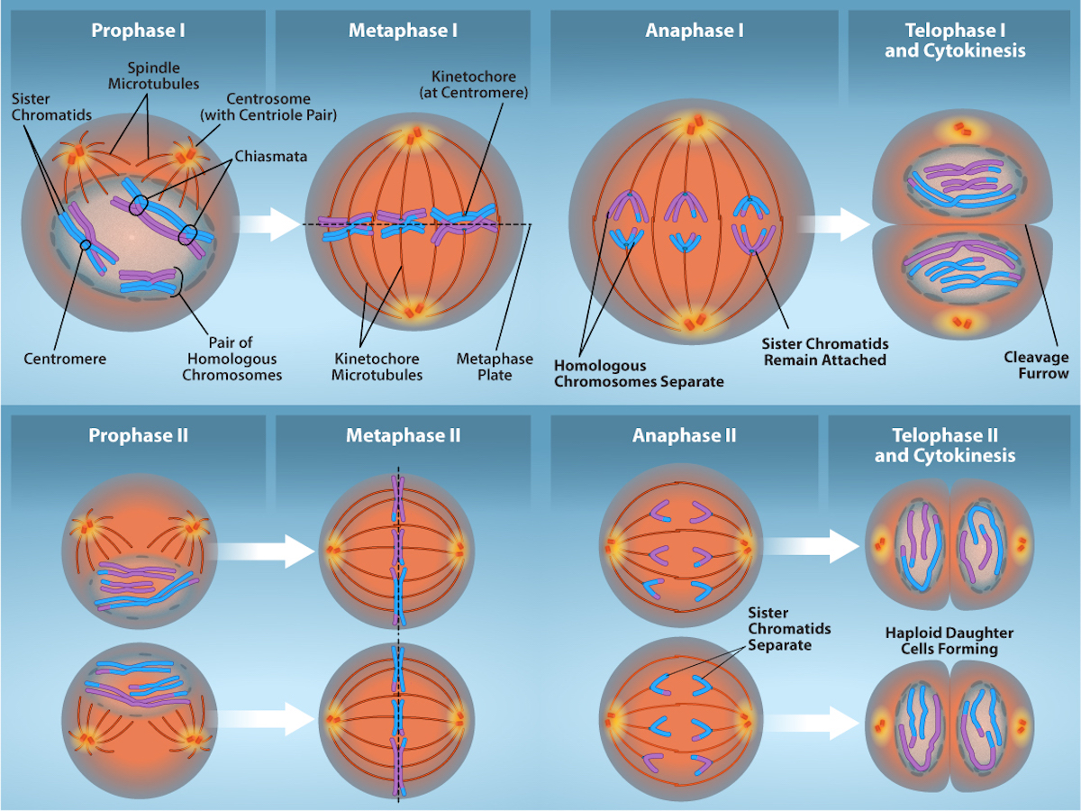
Spermatogenesis occurs in the seminiferous tubules that form the bulk of each testis. The process begins at puberty, after which time sperm are produced constantly throughout a male's life.
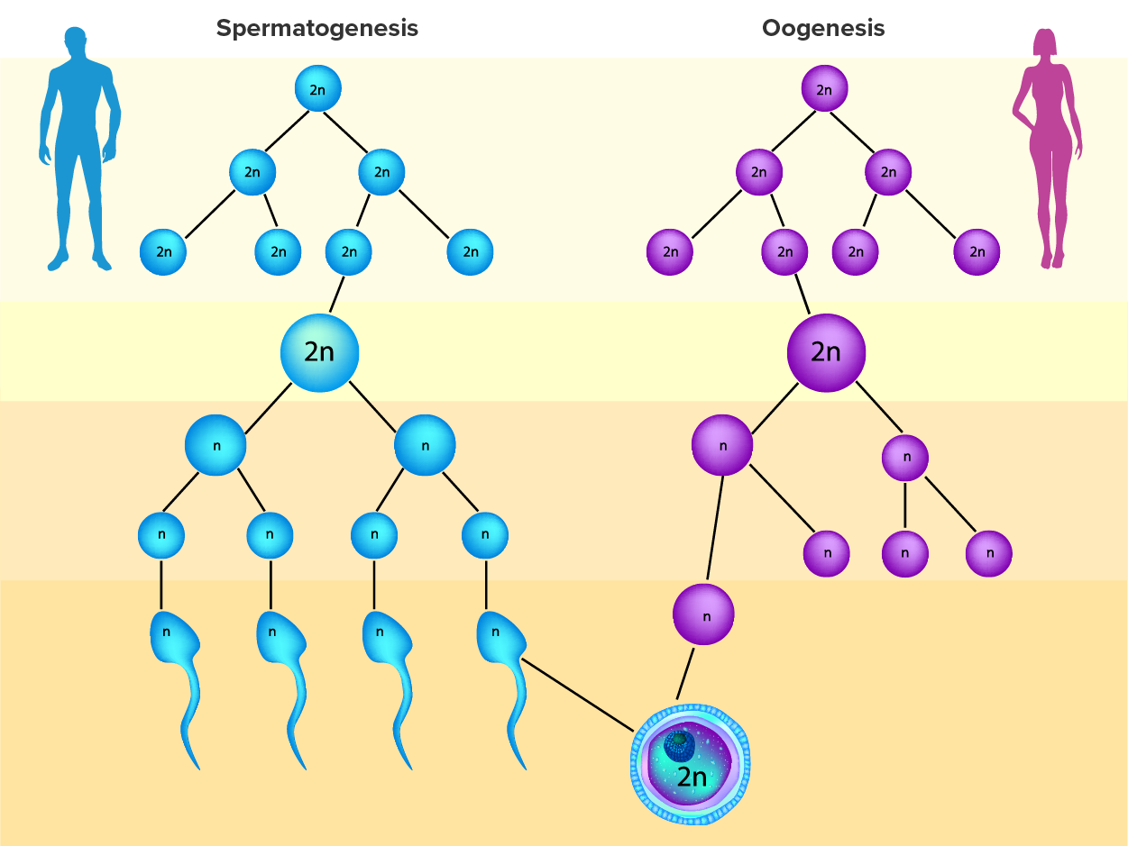
Two identical diploid cells result from spermatogonia mitosis. One of these cells remains a spermatogonium, and the other becomes a primary spermatocyte, the next stage in the process of spermatogenesis. As in mitosis, DNA is replicated in a primary spermatocyte before it undergoes a cell division called meiosis I. During meiosis I, each of the 23 pairs of chromosomes separates. This results in two cells, called secondary spermatocytes, each with only half the number of chromosomes.
Now a second round of cell division (meiosis II) occurs in both of the secondary spermatocytes. As you have learned, during meiosis II, each of the 23 replicated chromosomes divides. Thus, meiosis results in separating the chromosome pairs. This second meiotic division results in a total of four cells with only half of the number of chromosomes. Each of these new cells is a spermatid. Although haploid, early spermatids look very similar to cells in the earlier stages of spermatogenesis, with a round shape, central nucleus, and a large amount of cytoplasm. A process called spermiogenesis transforms these early spermatids, reducing the cytoplasm and beginning the formation of the parts of a true sperm. The fifth stage of germ cell formation—spermatozoa (singular, spermatozoon), or formed sperm—is the result of this process, which occurs in the portion of the tubule nearest the lumen.
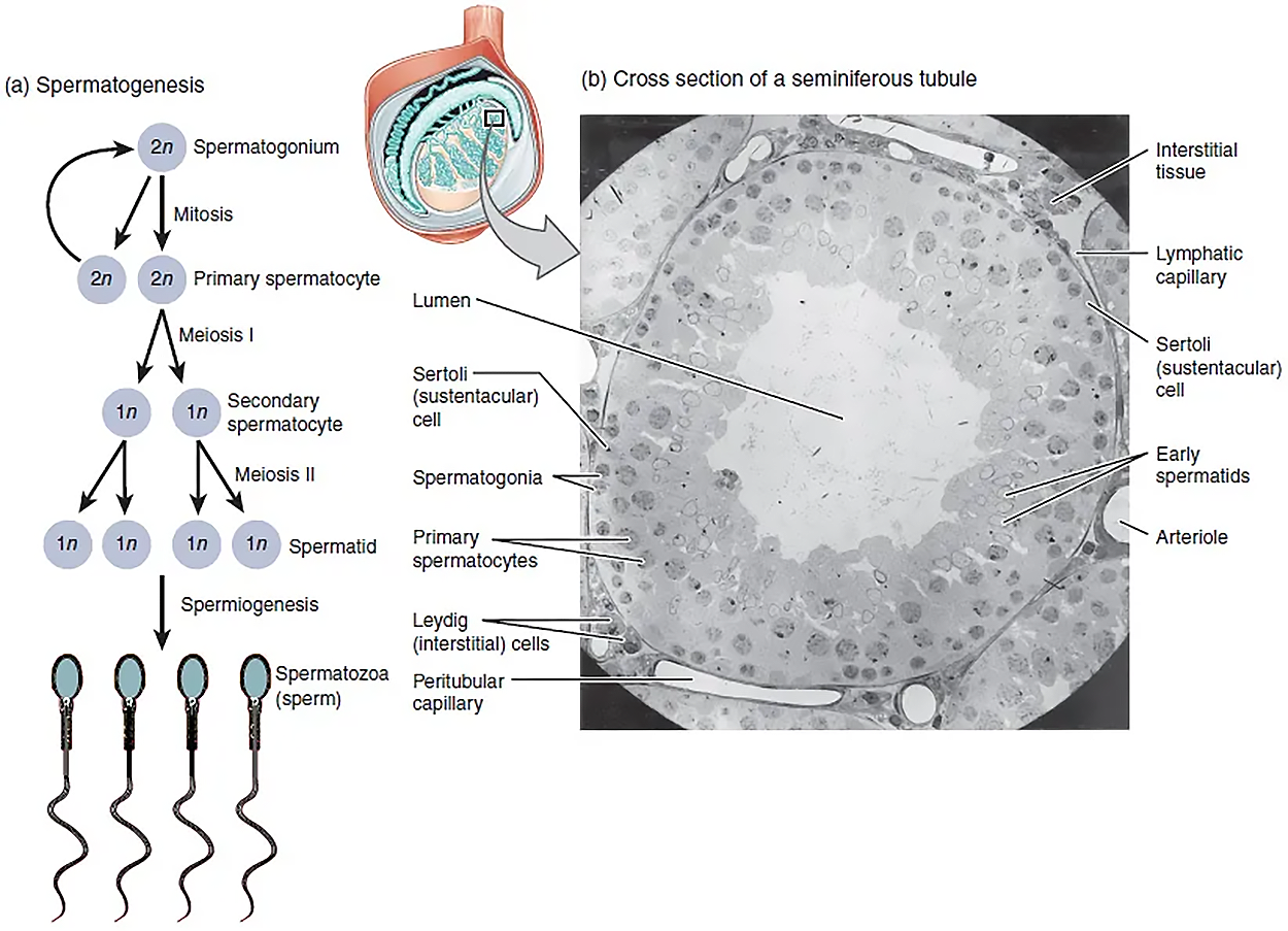
Eventually, the sperm are released into the lumen and are moved along a series of ducts in the testis toward the epididymis for the next step of sperm maturation, in which surface molecules (including proteins and carbohydrates) become coated with seminal plasma proteins. Seminal plasma is the fluid portion of semen that is secreted by both the epididymis and accessory glands.
Gametogenesis in females is called oogenesis. The ovarian cycle governs the preparation of endocrine tissues and the release of eggs, and it includes two interrelated processes: oogenesis (the production of gametes) and folliculogenesis (the growth and development of ovarian follicles). You will learn about oogenesis in this lesson and about folliculogenesis in a future lesson.
Oogenesis involves the process of meiosis, and this process begins with ovarian stem cells called oogonia (singular, oogonium), which are located in the outermost layers of the ovaries. In oogenesis, oogonia form during the embryological development of the individual. Oogonia divide via mitosis, much like spermatogonia in the testis. Unlike spermatogonia, however, oogonia undergo mitosis and form primary oocytes in the fetal ovary prior to birth (approximately one to two million oocytes by the time of birth).
During meiosis, two nuclear divisions separate the paired chromosomes in the nucleus and then separate the chromatids that were made during an earlier stage of the cell’s life cycle. Meiosis and its associated cell divisions produce haploid cells. Recall that haploid cells have half of each pair of chromosomes normally found in diploid cells.
Oocytes are contained within the ovarian follicles. These primary oocytes in primordial follicles are then arrested (stop development) at prophase I of meiosis I. At the time of birth, all future eggs are in prophase I. They again resume development years later, beginning at puberty, and continue until the person is near menopause, which marks the cessation (end) of a female's reproductive functions as the menstrual cycle ends.
Starting in adolescence, anterior pituitary hormones cause the development of a few follicles in an ovary each month. The initiation of ovulation—the release of an oocyte from the ovary—marks the transition from puberty into reproductive maturity. From then on, throughout the reproductive years, ovulation occurs approximately once every 28 days. Just prior to ovulation, a surge of luteinizing hormone (LH) triggers the resumption of meiosis in a primary oocyte. This results in a primary oocyte finishing the first meiotic division.
However, as you can see in the figure below, this cell division does not result in two identical cells. The cell divides unequally, with most of the cytoplasm and organelles going to one cell, called a secondary oocyte, and only one set of chromosomes and a small amount of cytoplasm going to the other cell. The larger cell, the secondary oocyte, eventually leaves the ovary during ovulation. The smaller cell, called the first polar body, remains within the ovary and may or may not complete meiosis and produce second polar bodies; ultimately, these cells will usually die and eventually disintegrate.
Cell division is again arrested in the secondary oocyte, this time at metaphase II. At ovulation, this secondary oocyte is released and travels toward the uterus through the oviduct. If the secondary oocyte is fertilized, the cell continues through meiosis II, producing a second polar body and haploid egg, which fuses with the haploid sperm to form a fertilized egg (zygote) containing all 46 chromosomes. Even though oogenesis produces up to four cells, only one survives.
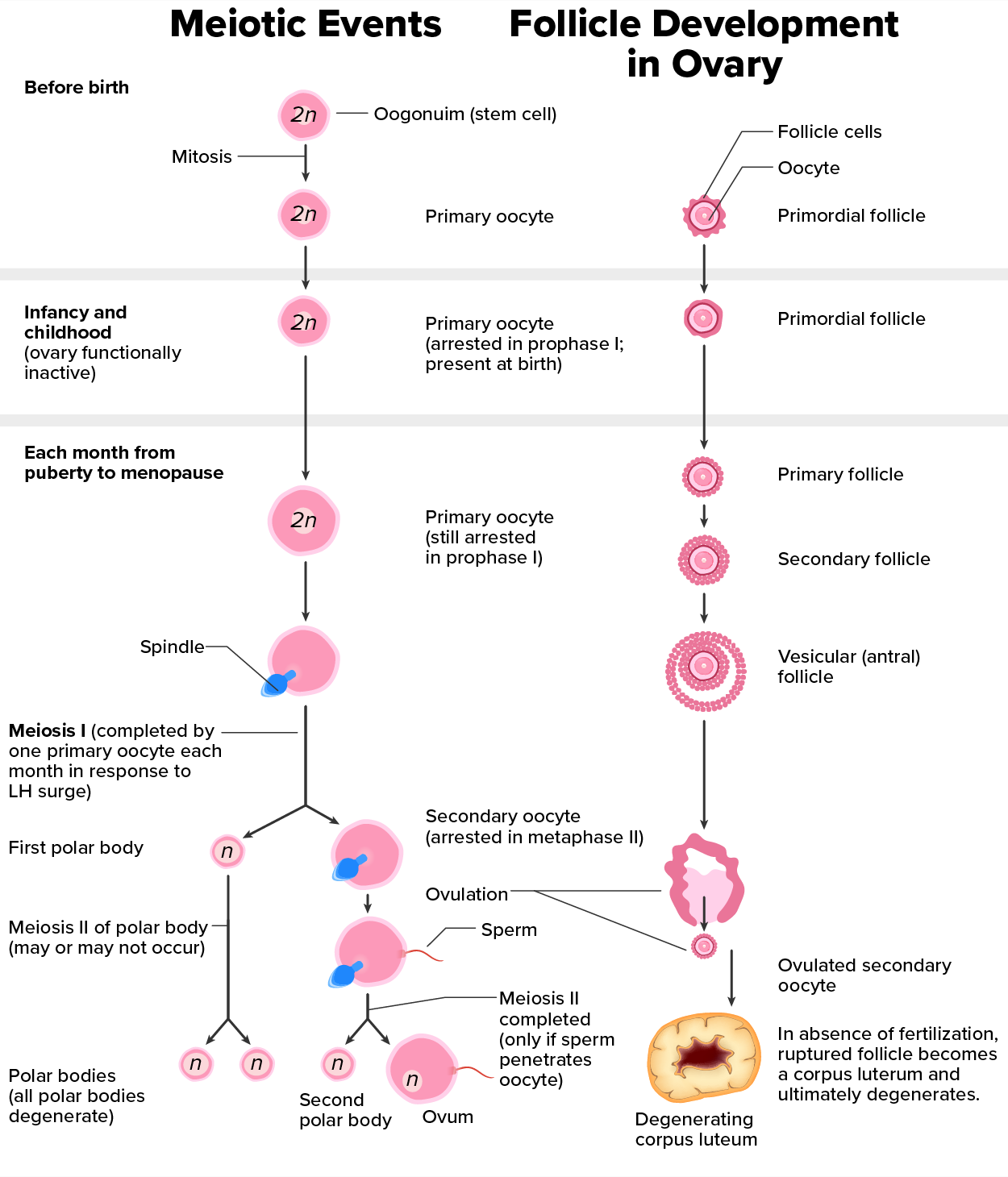
How does the diploid secondary oocyte become an ovum—the haploid female gamete? Meiosis of a secondary oocyte is completed only if a sperm succeeds in penetrating its barriers. Meiosis II then resumes, producing one haploid (“n”) ovum that, at the instant of fertilization by a (haploid) sperm, becomes the first diploid cell of the new offspring (a diploid, or “2n,” zygote). Thus, the ovum can be thought of as a brief, transitional, haploid stage between the diploid oocyte and diploid zygote.
The larger amount of cytoplasm contained in the female gamete is used to supply the developing zygote with nutrients during the period between fertilization and implantation into the uterus. Interestingly, sperm contribute only DNA at fertilization—not cytoplasm. Therefore, the cytoplasm and all of the cytoplasmic organelles in the developing embryo are of egg-derived origin. This includes mitochondria, which contain their own DNA. Scientific research in the 1980s determined that mitochondrial DNA was maternally inherited, meaning that you can trace your mitochondrial DNA directly to your biological mother, her mother, and so on back through your female ancestors.
IN CONTEXT
Mapping Human History With Mitochondrial DNA
When we talk about human DNA, we are usually referring to nuclear DNA; that is, the DNA coiled into chromosomal bundles in the nucleus of our cells. We inherit half of our nuclear DNA from one parent and half from the other parent. However, mitochondrial DNA (mtDNA) comes only from the mitochondria in the cytoplasm of the ovum we inherit from our mother. Our mother received mtDNA from their mother, who got it from their mother, and so on. Each of our cells contains approximately 1,700 mitochondria, with each mitochondrion packed with mtDNA containing approximately 37 genes.
Mutations (changes) in mtDNA occur spontaneously in a somewhat organized pattern at regular intervals in human history. By analyzing these mutational relationships, researchers have been able to determine that we can all trace our ancestry back to one female who lived in Africa about 200,000 years ago. More precisely, that human is our most recent common ancestor through matrilineal (maternal line) descent.
This doesn’t mean that everyone’s mtDNA today looks exactly like that of our common ancestor. Because of the spontaneous mutations in mtDNA that have occurred over the centuries, researchers can map different “branches” off of the “main trunk” of our mtDNA family tree. Your mtDNA might have a pattern of mutations that aligns more closely with one branch, and your neighbor’s may align with another branch. Still, all branches eventually lead back to the common ancestor.
But what happened to the mtDNA of all of the other Homo sapiens females who were living 200,000 years ago? Researchers explain that, over the centuries, their female descendants died childless or with only male children, and thus, their maternal line—and its mtDNA—ended.
During oogenesis, errors in meiosis can result in issues such as abnormal chromosome segregation and oocyte maturation arrest. These meiotic defects can lead to female infertility because of oocyte maturation arrest, increased aneuploidy (an abnormal number of chromosomes in a cell), and poor quality of female gametes. This can result in low rates of success for fertility procedures, such as in vitro fertilization and intracytoplasmic sperm injection, as well as early pregnancy loss.
A striking difference between oogenesis in females and spermatogenesis in males is that about 20% of oocytes have the wrong number of chromosomes, whereas only 3%–4% of sperm have the wrong number of chromosomes.
SOURCE: THIS TUTORIAL HAS BEEN ADAPTED FROM (1) OPENSTAX “BIOLOGY 2E”. ACCESS FOR FREE AT OPENSTAX.ORG/BOOKS/BIOLOGY-2E/PAGES/1-INTRODUCTION (2) OPENSTAX “ANATOMY AND PHYSIOLOGY 2E”. ACCESS FOR FREE AT OPENSTAX.ORG/BOOKS/ANATOMY-AND-PHYSIOLOGY-2E/PAGES/1-INTRODUCTION. LICENSING (1 & 2): CREATIVE COMMONS ATTRIBUTION 4.0 INTERNATIONAL.