Table of Contents |
The external female reproductive structures, referred to collectively as the vulva, include the mons pubis, labia majora, labia minora, vestibule, and perineum.
The mons pubis is a pad of fat that is located anteriorly over the pubic bone. After puberty, it becomes covered in pubic hair.
The labia majora and labia minora are folds located just posterior to the mons pubis. The labia majora are larger outer folds of hair-covered skin. The labia minora are thinner, hairless, and more pigmented folds found medially to the labia majora. The labia minora serve to protect the female urethra and the entrance to the female reproductive tract. They naturally vary in shape and size from woman to woman.
The superior, anterior portions of the labia minora come together to encircle the clitoris. The clitoris is erectile tissue that originates from the same fetal cells as the penis and has abundant nerves that are important in sexual sensation and orgasm.
The vestibule is the area between the labia minora and behind the clitoris that contains the urethral and vaginal openings. It is flanked by outlets to the vestibular glands, also known as Bartholin’s glands, that secrete mucus to keep the vestibular area moist.
The perineum is the region that separates the genitals and anus. In women, it is specifically the area between the vaginal opening and the anus.
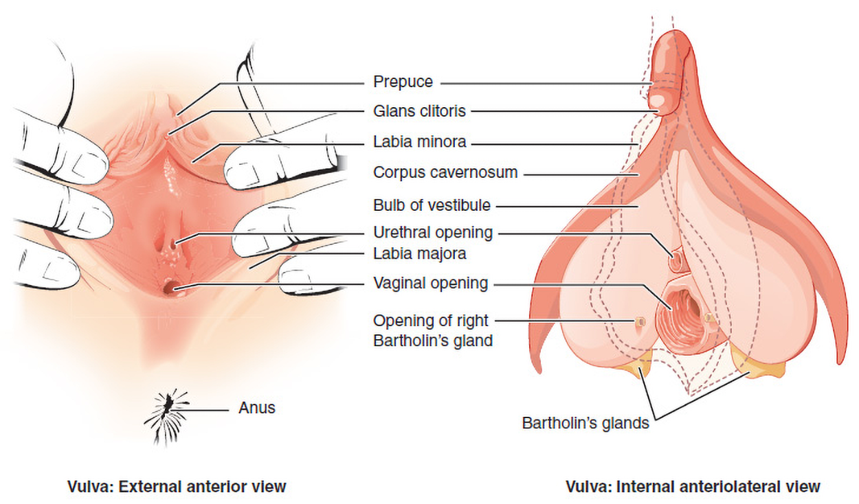
The figure above shows external and internal views of the vulva, both positioned with the anus below. The external view is presented with fingers pulling aside the labia. The prepuce is at the top, with the glans clitoris visible below (this structure is a protrusion in the internal anterolateral view). The labia minora is medial to the labia majora, forming the inner boundary of the tissue being pulled apart. The small urethral opening is above the larger vaginal opening. The inside view shows that the corpus cavernosum is tissue that branches to the left and right sides of the bulb of the vestibule. Bartholin’s glands are visible to the lower left and right of the vaginal opening. The opening of the right Bartholin’s gland is just lateral to the vaginal opening.
Although the breasts are located far from the other reproductive organs, they are considered accessory organs of the female reproductive system. The function of female breasts is to supply milk to an infant in a process called lactation (technically, lactation is the process of producing milk in the milk-producing mammary glands to feed an infant).
The external features of the breast include a nipple surrounded by a pigmented areola. The areolar region is characterized by small, raised areolar glands that secrete lubricating fluid during lactation to protect the nipple from chafing. When a baby nurses (i.e., draws milk from the breast), the entire areolar region is taken into their mouth.
A breast is made up of three main parts: lobules, ducts, and connective tissue. The lobules are the mammary glands composed of alveoli (tiny sacs) that produce milk. The lactiferous ducts are tubes that carry milk to the nipple. The connective tissue (which consists of fibrous and fatty tissue) surrounds and holds everything together.
An infant can draw milk from the lobules and through the ducts and the nipple by suckling. Alveoli are surrounded by fat tissue, which determines the size of the breast. Breast size differs between individuals and does not affect the amount of milk produced.
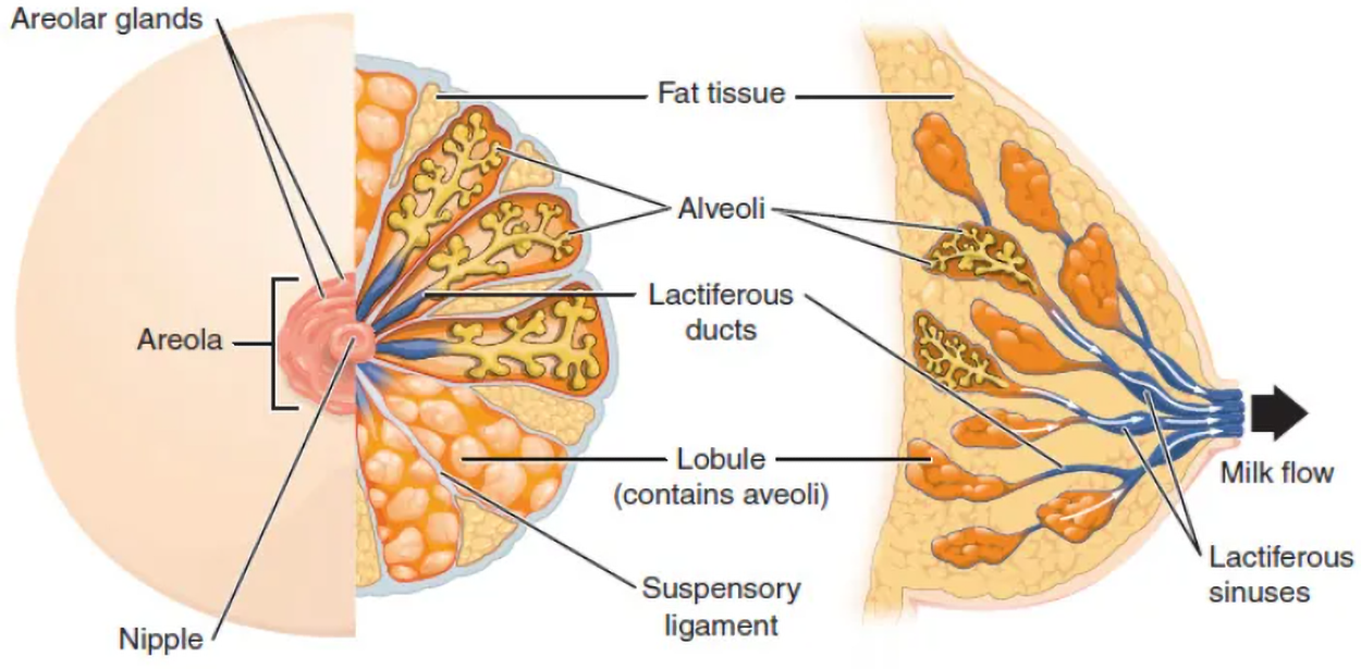
The figure above shows an illustration of a breast and milk flow. The left view shows the central nipple surrounded by the darker areola. The areola has bumpy areolar glands. Half of the breast is presented to show the underlying tissue. There are segmented regions extending from the nipple to the exterior, with suspensory ligaments between them. There are lactiferous sinuses that narrow into lactiferous ducts as they extend away from the areola into lobules that contain alveoli. In some places, the alveoli are shown; fat tissue is visible in others. A side view presented with the nipple to the right shows that much of the breast contains fat tissue. The lactiferous sinus extends from the nipple and narrows into lactiferous ducts that extend to lobules, which contain alveoli. Milk flows from the alveoli into lactiferous ducts that lead into lactiferous sinuses. Milk is released from the nipple.
The internal female reproductive organs include the vagina, uterus, cervix, ovaries, and uterine tubes (fallopian tubes). The figure below shows an illustration of these structures from the side (lateral view) and front (anterior view).
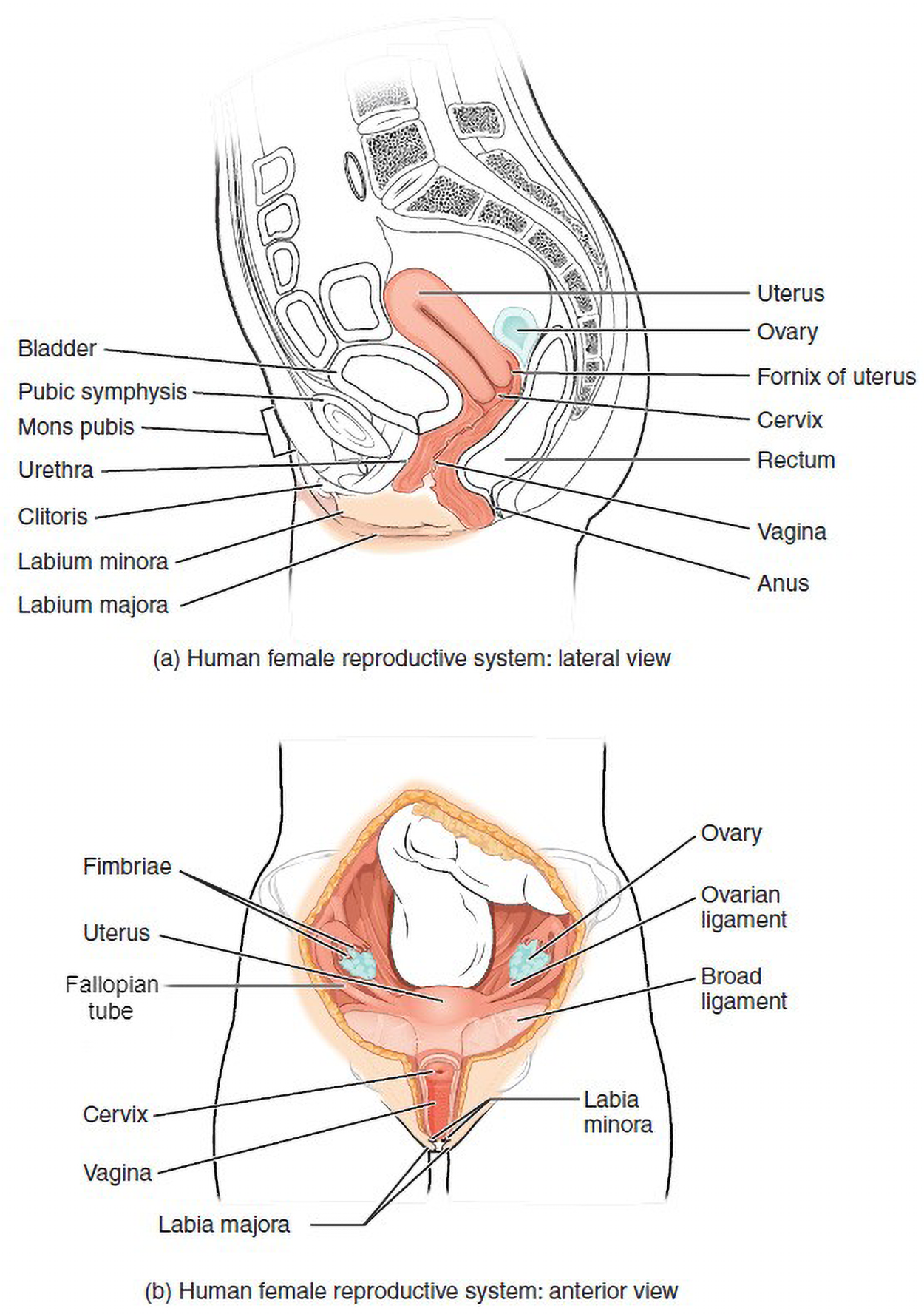
The lateral view above shows the bladder behind the pubic symphysis (joining the pubic bones). The uterus is posterior to the bladder with an ovary nearby. The uterus narrows to the cervix, and the folds of the fornix of the uterus are located here. The uterus is angled from anterior to posterior, and then the vagina below is angled in the opposite direction, descending anteriorly. The mons pubis is anterior to the pubic symphysis. The urethra extends anteriorly and downward, where it ends just below the clitoris. The labia majora and labia minora are shown around the urethra and clitoris. Posterior to the vagina, the rectum angles down to the anus. The anterior view shows the uterus with the ovaries to the left and right. The uterus is supported by broad ligaments. Each ovary is joined by a uterine tube (fallopian tube) to the uterus. Each uterine tube has fimbriae, which align with its corresponding ovary. The ovaries are held in place by ovarian ligaments. The uterus extends down to the cervix, which meets the vagina. The labia majora and labia minora are visible as external anatomy below the vagina.
The vagina is a muscular canal that is approximately 10 centimeters (cm) long. It serves as the entrance to the reproductive tract, as well as the exit from the uterus during menstruation and childbirth.
The vagina is composed of smooth muscle that allows for expansion during intercourse and childbirth. The vagina is lined with mucous membranes that secrete mucus to keep it moist. The superior portion of the vagina meets the cervix (the opening and lower part of the uterus). The inferior portion of the vagina may have a thin, perforated hymen that partially surrounds the opening to the vagina.
The vagina contains a normal population of healthy bacteria called normal flora that help protect against infection. In a healthy woman, the most common type of normal flora is Lactobacillus that secretes lactic acid; normal populations of Lactobacillus help to maintain health and reduce infection risk, while imbalances are associated with infections (Superti and De Seta, 2020). The lactic acid protects the vagina by maintaining an acidic pH (below 4.5). Lactic acid, in combination with other vaginal secretions, makes the vagina a self-cleansing organ. However, douching (a method of rinsing) can disrupt the normal balance of healthy microorganisms and increase a woman’s risk for infections and irritation. It is recommended that women do not douche and that they allow the vagina to maintain its normal healthy population of protective normal flora.
The uterus is a muscular, pear-shaped organ that is approximately five cm wide by seven cm long. It is composed of three sections:
The wall of the uterus is made up of these three layers:
The ovary is the female reproductive gland located in the pelvic cavity. There are two ovaries, one at the entrance to each uterine tube, which are attached to the uterus via the ovarian ligaments. The ovaries create oocytes and hormones. Oocytes are cells that develop into ova. Technically, an oocyte is called an ovum (egg) once it can be fertilized (Duncan et al., 2020).
The uterine tubes (fallopian tubes) transport developing ova from the ovary to the uterus. Each of the uterine tubes is close to, but not directly connected to, the ovary, so the fimbriae catch the ovum like a baseball in a glove. The middle region of the uterine tube (fallopian tube), called the ampulla, is where fertilization often occurs. The fertilized egg then moves from the uterine tube into the uterus, where it implants into the endometrium.
Source: THIS TUTORIAL HAS BEEN ADAPTED FROM OPEN RN "MEDICAL TERMINOLOGY 2E". ACCESS FOR FREE AT wtcs.pressbooks.pub/medterm/ LICENSING: CREATIVE COMMONS ATTRIBUTION 4.0 INTERNATIONAL. Accessed by March 2025.
REFERENCES
Superti, F., & De Seta, F. (2020). Warding Off Recurrent Yeast and Bacterial Vaginal Infections: Lactoferrin and Lactobacilli. Microorganisms, 8(1), 130. doi.org/10.3390/microorganisms8010130
Duncan, F. E., Schindler, K., Schultz, R. M., Blengini, C. S., Stein, P., Stricker, S. A., Wessel, G. M., & Williams, C. J. (2020). Unscrambling the oocyte and the egg: clarifying terminology of the female gamete in mammals. Molecular Human Reproduction, 26(11), 797–800. doi.org/10.1093/molehr/gaaa066