Use Sophia to knock out your gen-ed requirements quickly and affordably. Learn more
×

External Female Reproductive Structures

Author: Sophia

what's covered
In this lesson, you will learn about the external structures of the female reproductive system and their basic functions. Specifically, this lesson will cover:

Table of Contents

before you start
The female, or ovarian, reproductive system functions to produce gametes and reproductive hormones, just like the male reproductive system; however, it also has the additional task of supporting the developing fetus and delivering it to the outside world. Unlike its male counterpart, the female reproductive system is primarily located inside the pelvic cavity. Recall that the ovaries are the female gonads. The gamete they produce is called an oocyte. We will discuss the production of oocytes in detail shortly. First, we will look at some of the structures of the female reproductive system. In this lesson, you will learn about the external structures of the female reproductive system, and you will learn about the internal structures in a future lesson.

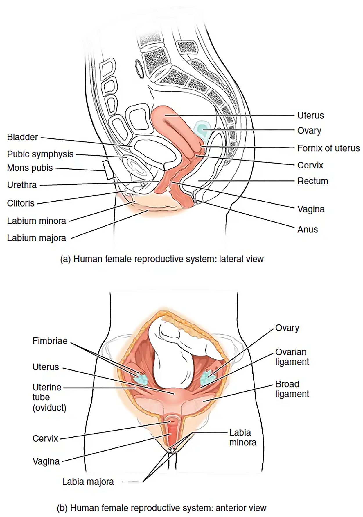
This figure shows the structure and the different organs in the female reproductive system. The top panel shows the lateral view and the bottom panel shows the anterior view.
Female Reproductive System - The major organs of the female reproductive system are located inside the pelvic cavity.

1. External Female Genitals

The external female reproductive structures are referred to collectively as the vulva. The mons pubis is a pad of fat that is located at the anterior, over the pubic bone. After puberty, it becomes covered in pubic hair. The labia majora (labia = “lips”; majora = “larger”) are folds of hair-covered skin that begin just posterior to the mons pubis. The thinner and more pigmented labia minora (labia = “lips”; minora = “smaller”) extend medial to the labia majora. Although they naturally vary in shape and size from person to person, the labia minora serve to protect the urethra and the entrance to the reproductive tract.

The superior, anterior portions of the labia minora come together to encircle the clitoris (or glans clitoris), an organ that originates from the same cells as the glans penis and has abundant nerves that make it important in sexual sensation and orgasm. The clitoris is covered by a layer of skin called the prepuce.

key concept
You learned previously that the prepuce covers the glans penis in males. In females, the prepuce is the hood of the clitoris and is formed by the folds of the vulva. The prepuce in males and females is considered homologous, which means they have a shared origin, and they develop from the same embryonic tissue. You will learn more about embryonic tissue differentiation in a future lesson.

The hymen is a thin membrane that sometimes partially covers the entrance to the vagina.

did you know
An intact hymen cannot be used as an indication of “virginity”. Even at birth, this is only a partial membrane because menstrual fluid and other secretions must be able to exit the body, regardless of penile–vaginal intercourse.

The vaginal opening is located between the opening of the urethra and the anus. It is flanked by outlets to the greater vestibular glands (or Bartholin’s glands). There are also two vestibular bulbs (or bulbs of vestibule), which are formed from corpus spongiosum tissue; these are bulbs of erectile tissue that are close to the body of the clitoris and engorge with blood during sexual arousal.
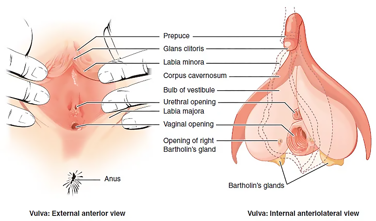
This figure shows the parts of the vulva. The right panel shows the external anterior view and the left panel shows the internal anterolateral view. The major parts are labeled.
The Vulva - The external female genitalia are referred to collectively as the vulva.

Term Pronunciation Table

Term Pronunciation Audio File
Mons Pubis mons pu·bis
Labia Majora la·bia ma·jo·ra
Labia Minora la·bia mi·no·ra
Clitoris cli·to·ris
Hymen hy·men

terms to know
Vulva
The external female genitalia.
Mons Pubis
The mound of fatty tissue located at the front of the vulva.
Labia Majora
The hair-covered folds of skin located behind the mons pubis.
Labia Minora
Thin, pigmented, hairless flaps of skin located medial and deep to the labia majora.
Clitoris
(also, glans clitoris) The nerve-rich area of the vulva that contributes to sexual sensation during intercourse.
Hymen
A piece of tissue that can cover part of the vaginal opening.
Greater Vestibular Glands
(also, Bartholin’s glands) Glands that produce a thick mucus that maintains moisture in the vulva area.
Vestibular Bulb
One of two bulbs of erectile tissue close to the clitoris that become engorged with blood during sexual arousal.


2. The Breasts

The breasts are located far from the other female reproductive organs, and they are therefore considered accessory organs of the female reproductive system. The function of the breasts is to supply milk to an infant in a process called lactation. The external features of the breast include a nipple surrounded by a pigmented areola, whose coloration may deepen during pregnancy. The areola is typically circular and can vary in size from 25 to 100 mm in diameter. The areolar region is characterized by small, raised areolar glands that secrete lubricating fluid during lactation to protect the nipple from chafing. When a baby nurses, or draws milk from the breast, the entire areolar region is taken into the mouth.

Breast milk is produced by the mammary glands, which are modified sweat glands. The milk itself exits the breast through the nipple via 15 to 20 lactiferous ducts that open on the surface of the nipple. These lactiferous ducts each extend to a lactiferous sinus that connects to a glandular lobe within the breast itself that contains groups of milk-secreting cells in clusters called alveoli (of the breast). The clusters can change in size depending on the amount of milk in the alveolar lumen. Once milk is made in the alveoli, stimulated myoepithelial cells that surround the alveoli contract to push the milk to the lactiferous sinuses. From here, the baby can draw milk through the lactiferous ducts by suckling. The lobes themselves are surrounded by fat tissue, which determines the size of the breast; breast size differs between individuals and does not affect the amount of milk produced. Supporting the breasts are multiple bands of connective tissue called suspensory ligaments that connect the breast tissue to the dermis of the overlying skin.
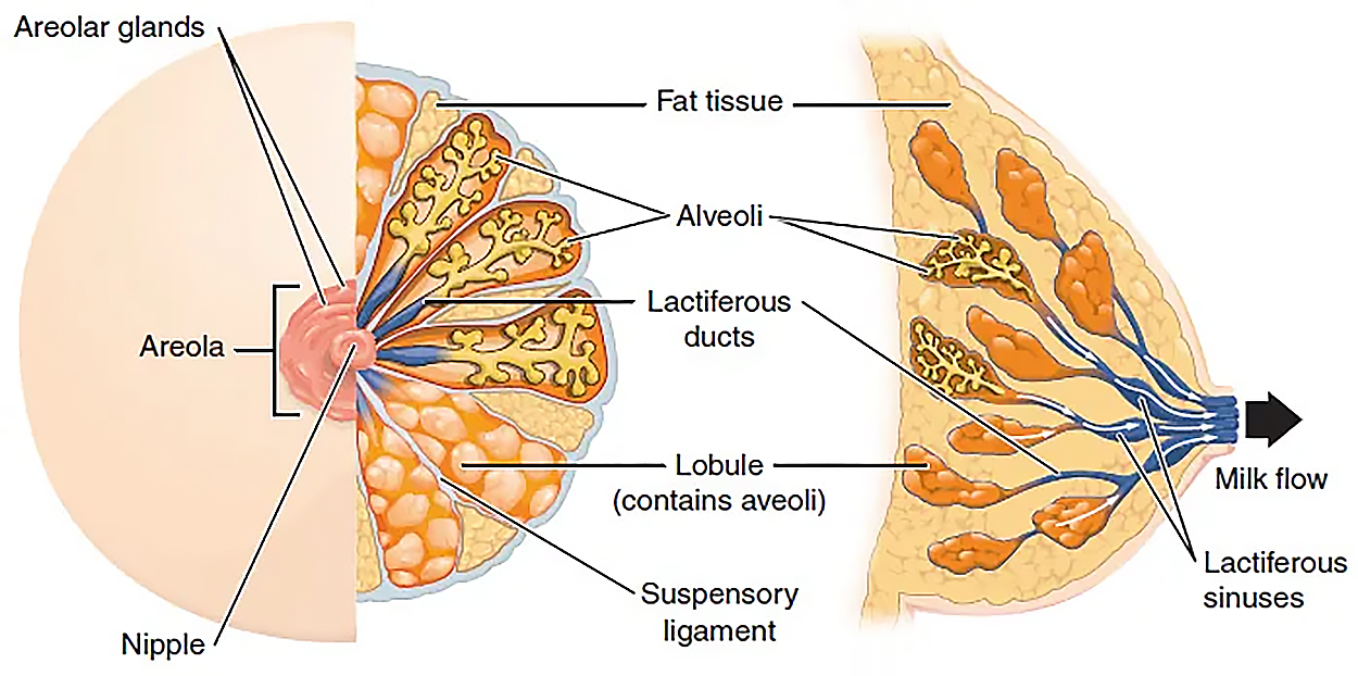

This figure shows the anatomy of the breast. The left panel shows the front view and the right panel shows the side view. The main parts are labeled.
Anatomy of the Breast - During lactation, milk moves from the alveoli through the lactiferous ducts to the nipple.

During the normal hormonal fluctuations in the menstrual cycle, breast tissue responds to changing levels of estrogen and progesterone, which can lead to swelling and breast tenderness in some individuals, especially during the secretory phase. If pregnancy occurs, the increase in hormones leads to further development of the mammary tissue and enlargement of the breasts.

Term Pronunciation Table

Term Pronunciation Audio File
Areola are·o·la
Lactiferous Ducts lac·tif·er·ous duct·s
Alveoli al·ve·o·li

terms to know
Areola
The highly pigmented, circular area surrounding the raised nipple and containing areolar glands that secrete fluid important for lubrication during suckling.
Mammary Glands
Modified sweat glands inside the breast that secrete milk.
Lactiferous Ducts
Ducts that connect the mammary glands to the nipple and allow for the transport of milk.
Lactiferous Sinus
Area of milk collection between alveoli and lactiferous duct.
Alveoli (of the Breast)
Milk-secreting cells in the mammary gland.
Suspensory Ligaments
Bands of connective tissue that suspend the breast onto the chest wall by attachment to the overlying dermis.

summary
In this lesson, you learned about the external structures of the female reproductive system and their functions. You first explored the external female genitals, which help protect the internal genitalia. You then learned about how the breasts are accessory organs of the female reproductive system that can supply milk to an infant.

Source: THIS TUTORIAL HAS BEEN ADAPTED FROM OPENSTAX "ANATOMY AND PHYSIOLOGY 2E". ACCESS FOR FREE AT OPENSTAX.ORG/DETAILS/BOOKS/ANATOMY-AND-PHYSIOLOGY-2E. LICENSE: CREATIVE COMMONS ATTRIBUTION 4.0 INTERNATIONAL. Accessed by August 2023.


Terms to Know
Alveoli (of the Breast)

Milk-secreting cells in the mammary gland.

Areola

The highly pigmented, circular area surrounding the raised nipple and containing areolar glands that secrete fluid important for lubrication during suckling.

Clitoris

(also, glans clitoris) The nerve-rich area of the vulva that contributes to sexual sensation during intercourse.

Greater Vestibular Glands

(also, Bartholin’s glands) Glands that produce a thick mucus that maintains moisture in the vulva area.

Hymen

A piece of tissue that can cover part of the vaginal opening.

Labia Majora

The hair-covered folds of skin located behind the mons pubis.

Labia Minora

Thin, pigmented, hairless flaps of skin located medial and deep to the labia majora.

Lactiferous Ducts

Ducts that connect the mammary glands to the nipple and allow for the transport of milk.

Lactiferous Sinus

Area of milk collection between alveoli and lactiferous duct.

Mammary Glands

Modified sweat glands inside the breast that secrete milk.

Mons Pubis

The mound of fatty tissue located at the front of the vulva.

Suspensory Ligaments

Bands of connective tissue that suspend the breast onto the chest wall by attachment to the overlying dermis.

Vestibular Bulb

One of two bulbs of erectile tissue close to the clitoris that become engorged with blood during sexual arousal.

Vulva

The external female genitalia.