In this lesson, you will explore the structure and function of the first of the four primary tissue categories, epithelial tissue. Specifically, this lesson will cover:
Epithelial tissue refers to cells that cover exterior surfaces of the body, line internal cavities and passageways, and form secreting organs.
Exterior surfaces of the body include not only skin but the passageways of the respiratory, digestive, urinary, and reproductive tracts.
Internal cavities and passageways of the body include the body cavities as well as any hollow structure or organ such as blood vessels and the heart.
Secreting organs, called glands, are made of and located within epithelial tissues, are found throughout the body, and make and release a wide variety of products (i.e., sweat, oil, hormones, enzymes, and more).
An epithelial tissue that covers an external or internal surface is called an epithelium (plural, epithelia). An epithelial tissue that forms a secreting organ is called a gland. This lesson will focus on the structure and function of epithelia. The gland will be covered in a future lesson.
Epithelial tissues provide protection in two main forms:
Shielding: They function as a shield against the external environment, protecting the body from physical, chemical, and biological wear and tear. Some tissues place the epithelial cells in a position to act as direct shields against the external environment. Others are capable of producing and releasing mucus or other compounds that cover the superficial cells.
Regulating: Epithelial tissues also control permeability, regulating the movement of materials into or out of the body such as the air you breathe or food you consume. All substances that enter the body must cross an epithelium. Some epithelia often include structural features that allow the selective transport of molecules and ions across their cell.
All epithelia share some important structural and functional features. This tissue is highly cellular, with densely packed cells and little or no extracellular material present between cells. Each cell has polarity, or directionality, in relation to its surroundings. The apical surface of an epithelial cell is the exposed surface, facing the exterior or interior space. The basal surface (basal, base) of an epithelial cell is the non-exposed surface and faces the underlying material. The lateral surface of an epithelial cell faces neighboring cells. At times, the non-exposed surfaces of the epithelial cell are referred to collectively as the basolateral surface. This polarity within epithelial cells helps to orient the tissue and direct its function in a specific direction. You may also find this polarity helpful in identifying various epithelial tissues.
hint
Here’s a helpful way to remember the names of the surfaces of epithelial cells and tissue.
Apical, or apex, means the highest level. When you find the epithelial surface that touches air or fluid, that is the “top” or apex.
Basal, or base, means the lowest level—think “basement.” When you find the epithelial surface that touches a different, non-epithelial material opposite the apex, you’ve found the “bottom” or base.
Lateral, like from the directional terms, means away from the midline. To the right and left side are other neighboring epithelial cells.
Cross Section of Epithelial Tissue - Epithelial cells and tissues have polarity with an exposed apical surface and a basal surface connected to underlying connective tissues by the basement membrane.
Furthermore, epithelial cells are typically characterized by the polarized distribution of organelles, membrane-bound proteins, and surface structures between their basal and apical surfaces, an adaptation for specific functions. Cilia and microvilli are microscopic extensions of the apical cell membrane supported by microtubules that are found in certain epithelial cells. They both manipulate the movement of material in the external environment, causing it to speed up or slow down.
The basal surface of epithelial tissues is attached to the basement membrane, a combination of extracellular materials secreted by the epithelial tissue and underlying connective tissue. This membrane serves as the attachment point for epithelial tissues as well as a separation from the underlying connective tissue. The basement membrane is formed from materials secreted by both the epithelial and connective tissues. The epithelial tissue secretes a mixture of glycoprotein and collagen, forming the basal lamina. The connective tissue secretes its own extracellular material to form the reticular lamina.
key concept
Epithelial tissues are nearly completely avascular (a, without; vascular, blood vessels), meaning they have no blood vessels, which is what delivers nutrients to the body and takes away wastes. No blood vessels cross the basement membrane. Therefore, nutrients must come by diffusion or absorption from underlying tissues or the surface. Wastes must be diffused or absorbed in the opposite direction.
Many epithelial tissues are capable of rapidly replacing damaged and dead cells. Sloughing off of damaged or dead cells is a characteristic of surface epithelium and allows our airways and digestive tracts to rapidly replace damaged cells with new cells.
terms to know
Epithelium
An epithelial tissue that covers an external or internal surface.
Gland
An epithelial tissue that forms a secreting organ.
Apical Surface
The exposed surface of an epithelial cell or tissue that faces the external or internal space.
Basal Surface
The non-exposed surface of an epithelial cell or tissue that faces the underlying material.
Lateral Surface
The surface of an epithelial cell that neighboring cells.
Basement Membrane
The extracellular material secreted by epithelial and connective tissues serves as the attachment point for epithelial tissues as well as a separation from the underlying connective tissue.
Basal Lamina
An extracellular substance secreted by epithelial tissue that makes up part of the basement membrane.
Reticular Lamina
An extracellular substance secreted by connective tissue that makes up part of the basement membrane.
2. Cell to Cell Junctions
Epithelial cells form special connections between themselves and other cells or their surrounding material called cell junctions. These connections are formed by specific transmembrane proteins, binding to one another or to extracellular material. Three basic types of connections allow varying degrees of interaction between the cells: tight junctions, anchoring junctions, and gap junctions.
Tight junctions form water-tight connections (hence their name) on the lateral surface using multiple membrane proteins to hold the membranes of two adjacent cells together. In this type of cell junction, there is no extracellular space between the two participating cells, and extracellular substances are forced to use membrane transport to travel through the tissue layer instead of diffusing between the cells.
Anchoring junctions are found in the lateral and basal surfaces and stabilize epithelial tissues. The membrane proteins that are involved function like velcro, connecting a cell’s internal cytoskeleton to the cytoskeleton of another cell or to the extracellular matrix. There are three types of anchoring junctions: desmosomes, hemidesmosomes, and adherens.
Desmosomes form between adjacent cells, forming cell-to-cell anchors. Each participating cell forms a collection of membrane proteins together in a patch. Once aligned, the transmembrane proteins interlock. On the internal side, each patch is connected to the cytoskeleton. These cell junctions help to maintain the integrity of an epithelial tissue as it is distorted by external pressure.
Hemidesmosomes (hemi, half), which look like half a desmosome, form a patch of membrane proteins on the basal surface and connect cells to the extracellular matrix (i.e., the basement membrane). These cell junctions keep epithelial tissue attached to the underlying material.
Adherens junctions form between adjacent cells and form cell-to-cell anchors like desmosomes. However, these cell junctions are also connected to contractile proteins inside the cell which can be attached to other adherens junctions at other points in the membrane. Multiple adherens junctions can form a belt-like structure through a tissue referred to as an adhesion belt, which can influence cell or tissue shape.
In contrast with the tight and anchoring junctions, a gap junction forms an intercellular passageway between the membranes of adjacent cells to facilitate the movement of small molecules and ions between the cytoplasm of adjacent cells. Each participating cell forms a complex of transmembrane proteins. When two adjacent cells align their proteins, they interlock and open to form a narrow passageway. These junctions allow electrical and metabolic coupling of adjacent cells, which coordinates function in large groups of cells.
terms to know
Cell Junction
A specialized cell-to-cell connection.
Tight Junction
A cell junction that forms a water-tight seal between adjacent epithelial cells.
Anchoring Junction
A cell junction that stabilizes epithelial tissue.
Desmosome
An anchoring junction that forms a connection on the lateral surface of epithelial cells that helps to hold cells together.
Hemidesmosome
An anchoring junction that links epithelial cells to the basement membrane.
Adherens Junction
An anchoring junction that connects to the cytoskeleton and influences cell shape and folding.
Gap Junction
A cell junction that forms an intercellular passageway between the membranes of adjacent cells to facilitate the movement of small molecules and ions between the cytoplasm of adjacent cells.
3. Classification of Epithelial Tissues
Epithelial tissues are classified based on two characteristics, the number of cell layers and cell shape. Cell shape can be squamous (flattened and thin), cuboidal (cube-shaped, as wide as it is tall), or columnar (column-shaped, taller than it is wide). As an exception, transitional describes a form of specialized stratified epithelium in which the shape of the cells can vary. Similarly, an epithelial tissue can contain just one layer of cells, which is a simple epithelium, or more than one layer of cells, which is a stratified epithelium. As an exception, pseudostratified (pseudo, false) describes tissue with a single layer of irregularly shaped cells that give the appearance of more than one layer.
Cells of Epithelial Tissue - Simple epithelial tissue is organized as a single layer of cells and stratified epithelial tissue is formed by several layers of cells.
3a. Simple Epithelium
Epithelial tissues that consist of only one layer of cells are called simple epithelium. The shape of the cells in simple epithelium reflects the functioning of those cells. The cells in simple squamous epithelium have the appearance of thin scales. Squamous cell nuclei tend to be flat, horizontal, and elliptical, mirroring the form of the cell. Squamous cells look like they’ve been squashed.
Simple squamous epithelium can be split into three categories based on the location in the body. Each of these groups of simple squamous epithelium has its own unique name. Endothelium (endo, inside) is the simple squamous epithelial tissue that lines vessels and organs of the lymphatic and cardiovascular system, and it is made up of a single layer of squamous cells. The mesothelium (meso, middle) is a simple squamous epithelium that forms the surface layer of the serous membrane that lines body cavities and internal organs. Its primary function is to provide a smooth and protective surface. Mesothelial cells are squamous epithelial cells that secrete a fluid that lubricates the mesothelium. Both of these line internal parts of the body. In contrast, a true epithelium (epi, outside) is only located on an exposed surface of the body such as the skin or passageway in the respiratory, digestive, urinary, or reproductive system. Simple squamous epithelium, because of the thinness of the cell, is present where rapid passage of chemical compounds is observed. The alveoli of lungs where gasses diffuse, segments of kidney tubules, and the lining of capillaries are also made of simple squamous epithelial tissue.
Simple Squamous Epithelium - Lung tissue is made of a single layer of squamous cells and allows for the fast diffusion of gasses into and out of the body.
reflect
External surfaces of the body are covered in an epithelium as a protective covering. These surfaces include the skin covering your face, arms, torso, and legs as well as the passageways through the respiratory, digestive, urinary, and reproductive systems. But wait! How can the lining of your stomach (which is covered by an epithelial tissue) be considered an external surface? Isn’t that inside the body?
Recall from above that all substances that enter the body must cross an epithelium. Therefore, if a substance has not crossed an epithelium (the external covering), it is still external to the body. And if it has crossed an epithelium, then it is internal to the body.
Let’s consider the last thing you ate—an omelet, a sandwich, or possibly a delicious roast duck. When that food is on your plate, it is external to the body. As you place the food in your mouth to chew, it has not yet crossed an epithelium, so it is still external. As you swallow the food, it enters your stomach but has not yet crossed an epithelium, so it is still external. When the food exits the stomach, it enters your intestines where it crosses the epithelial tissue lining of that organ and enters the blood. At that point, the material that has been absorbed into the blood is internal to the body. Certain foods (those high in fiber) may travel through your digestive system but never be absorbed and therefore remain external to the body the entire time.
This helps us understand how the term “external surface” is defined
In simple cuboidal epithelium, the nucleus of the box-like cells appears round and is generally located near the center of the cell. These epithelia are active in the secretion and absorption of molecules into or out of the cell. Simple cuboidal epithelia are observed in the lining of the kidney tubules and in the ducts (connecting tubes) of glands.
Simple Cuboidal Epithelium - Shown in high power, the kidney tubules are made of a single layer of cuboidal cells that function to absorb and secrete molecules into and out of the center of the tube. This fluid will eventually become urine.
In simple columnar epithelium, the nucleus of the tall column-like cells tends to be elongated and located in the basal end of the cells. Like the cuboidal epithelia, this epithelium is active in the absorption and secretion of molecules. Simple columnar epithelium forms the lining of some sections of the digestive system and parts of the female reproductive tract. Ciliated columnar epithelium is composed of simple columnar epithelial cells with cilia on their apical surfaces. These epithelial cells are found in the lining of the fallopian tubes and parts of the respiratory system, where the cilia help to remove particulate matter.
Simple Columnar Epithelium -Shown in high power, the lining of the small intestine is comprised of a single layer of columnar cells that function to absorb nutrients and secrete enzymes and buffers.
Pseudostratified columnar epithelium is a type of epithelium that appears to be stratified but instead consists of a single layer of irregularly shaped and differently sized columnar cells. In pseudostratified epithelium, nuclei of neighboring cells appear at different levels rather than clustered in the basal end. The arrangement gives the appearance of stratification. Pseudostratified columnar epithelium is found in the respiratory tract, where some of these cells have cilia.
Pseudostratified Columnar Epithelium - As shown in high power, the lining of the trachea (the tube leading to the lungs) contains a single layer of epithelial cells that look like they are stratified but are actually a single layer with cilia on their apical surface.
Both simple and pseudostratified columnar epithelia are heterogeneous epithelia because they include additional types of cells interspersed among the epithelial cells. For example, a goblet cell (image below) is a mucous-secreting unicellular “gland” interspersed between the columnar epithelial cells of mucous membranes.
A stratified epithelium consists of multiple stacked layers of cells. This epithelium protects against physical and chemical wear and tear. The stratified epithelium is named by the shape of the most apical layer of cells, closest to the free space. Stratified squamous epithelium is the most common type of stratified epithelium in the human body. The apical cells are squamous, whereas the basal layer contains either columnar or cuboidal cells. This tissue can be found with or without being filled with a water resistant protein called keratin (more on this protein in a future lesson). Mammalian skin with resists abrasion and dehydration is an example of a tissue with keratin and is called keratinized, stratified squamous epithelium. The lining of the mouth cavity which resists abrasion is an example of a nonkeratinized, stratified squamous epithelium.
Stratified Squamous Epithelium - Shown in high power, the skin (left) contains many layers of squamous cells with many very thin layers on the apical end full of keratin. The lining of the mouth (right) contains many layers of squamous cells without keratin. Both tissues function to resist abrasion, but only the skin can resist dehydration.
Stratified cuboidal epithelium and stratified columnar epithelium are tissues containing multiple layers of cells where the apical layer is cube or column-like, respectively. These tissues can be found in certain glands and ducts but are rare in the human body.
The last stratified epithelium is transitional epithelium, located only in the urinary system, primarily the urinary bladder. This tissue is named ‘transitional’ because the apical layer of cells transitions between two states. When the urinary bladder is empty, there is very little pressure and transitional epithelium is convoluted and has bulbous apical cells with convex, umbrella-shaped, apical surfaces. As the bladder fills with urine, the tissue sustains more pressure and compresses to become squamous. This tissue is able to sustain compression without distortion to allow the bladder to fill and empty as needed.
Transitional Epithelium - The urinary bladder and attached tubing are lined with an epithelium that is found in two different shapes depending on the pressure being applied. An empty bladder (right) applies limited pressure and the apical cells form bulbous shapes with convex surfaces. A full bladder (left) applies pressure to the apical cells forming more squamous-like cells.
Below is a table to summarize the simple and stratified epithelium.
Summary of Simple and Stratified Epithelial Tissue Cells
terms to know
Simple Squamous Epithelium
A simple epithelial tissue with flat apical cells which covers and allow for fast diffusion of molecules.
Endothelium
The simple squamous epithelium that lines vessels and organs of the lymphatic and cardiovascular system.
Mesothelium
The simple squamous epithelium that forms the surface layer of the serous membrane that lines body cavities and internal organs.
Simple Cuboidal Epithelium
A simple epithelial tissue with cube-shaped apical cells which regulates absorption and secretion.
Simple Columnar epithelium
A simple epithelial tissue with column-shaped apical cells which regulates absorption and secretion.
Pseudostratified Epithelium
A simple epithelial tissue that looks stratified and secretes mucus.
Goblet Cell
A mucous-secreting unicellular “gland” interspersed between the columnar epithelial cells of mucous membranes.
Stratified Squamous Epithelium
A stratified epithelial tissue with flat apical cells which resists abrasion.
Keratinized Stratified Squamous Epithelium
A stratified epithelial tissue with flat, keratin-filled apical cells which resists abrasion and dehydration
Non-Keratinized Stratified Squamous Epithelium
A stratified epithelial tissue with flat apical cells which resists abrasion.
Stratified Cuboidal Epithelium
A stratified epithelial tissue with cube-shaped apical cells, located in certain glands and ducts, and functions to regulate secretion and absorption.
Stratified Columnar Epithelium
A stratified epithelial tissue with column-shaped apical cells, located in certain glands and ducts, and functions to regulate secretion and absorption.
Transitional Epithelium
A stratified epithelial tissue with bulbous or squamous-like apical cells depending on pressure, located in the urinary bladder and attached tubes.
summary
In this lesson, you learned to identify the common characteristics of epithelial tissue. You also learned to identify the types of cell-to-cell junctions found in epithelial tissues. Finally, you learned to classify epithelial tissues as to whether they are simple epithelium or stratified epithelium as well as their cell shape.
An anchoring junction that connects to the cytoskeleton and influences cell shape and folding.
Anchoring Junction
A cell junction that stabilizes epithelial tissue.
Apical Surface
The exposed surface of an epithelial cell or tissue that faces the external or internal space.
Basal Lamina
An extracellular substance secreted by epithelial tissue that makes up part of the basement membrane.
Basal Surface
The non-exposed surface of an epithelial cell or tissue that faces the underlying material.
Basement Membrane
The extracellular material secreted by epithelial and connective tissues serves as the attachment point for epithelial tissues as well as a separation from the underlying connective tissue.
Cell Junction
A specialized cell-to-cell connection.
Desmosome
An anchoring junction that forms a connection on the lateral surface of epithelial cells that helps to hold cells together.
Endothelium
The simple squamous epithelium that lines vessels and organs of the lymphatic and cardiovascular system.
Epithelium
An epithelial tissue that covers an external or internal surface.
Gap Junction
A cell junction that forms an intercellular passageway between the membranes of adjacent cells to facilitate the movement of small molecules and ions between the cytoplasm of adjacent cells.
Gland
An epithelial tissue that forms a secreting organ.
Goblet Cell
A mucous-secreting unicellular “gland” interspersed between the columnar epithelial cells of mucous membranes.
Hemidesmosome
An anchoring junction that links epithelial cells to the basement membrane.
Keratinized Stratified Squamous Epithelium
A stratified epithelial tissue with flat, keratin-filled apical cells which resists abrasion and dehydration.
Lateral Surface
The surface of an epithelial cell that neighboring cells.
Mesothelium
The simple squamous epithelium that forms the surface layer of the serous membrane that lines body cavities and internal organs.
Non-Keratinized Stratified Squamous Epithelium
A stratified epithelial tissue with flat apical cells which resists abrasion.
Pseudostratified Epithelium
A simple epithelial tissue that looks stratified and secretes mucus.
Reticular Lamina
An extracellular substance secreted by connective tissue that makes up part of the basement membrane.
Simple Columnar Epithelium
A simple epithelial tissue with column-shaped apical cells which regulates absorption and secretion.
Simple Cuboidal Epithelium
A simple epithelial tissue with cube-shaped apical cells which regulates absorption and secretion.
Simple Squamous Epithelium
A simple epithelial tissue with flat apical cells which covers and allow for fast diffusion of molecules.
Stratified Columnar Epithelium
A stratified epithelial tissue with column-shaped apical cells, located in certain glands and ducts, and functions to regulate secretion and absorption.
Stratified Cuboidal Epithelium
A stratified epithelial tissue with cube-shaped apical cells, located in certain glands and ducts, and functions to regulate secretion and absorption.
Stratified Squamous Epithelium
A stratified epithelial tissue with flat apical cells which resists abrasion.
Tight Junction
A cell junction that forms a water-tight seal between adjacent epithelial cells.
Transitional Epithelium
A stratified epithelial tissue with bulbous or squamous-like apical cells depending on pressure, located in the urinary bladder and attached tubes.

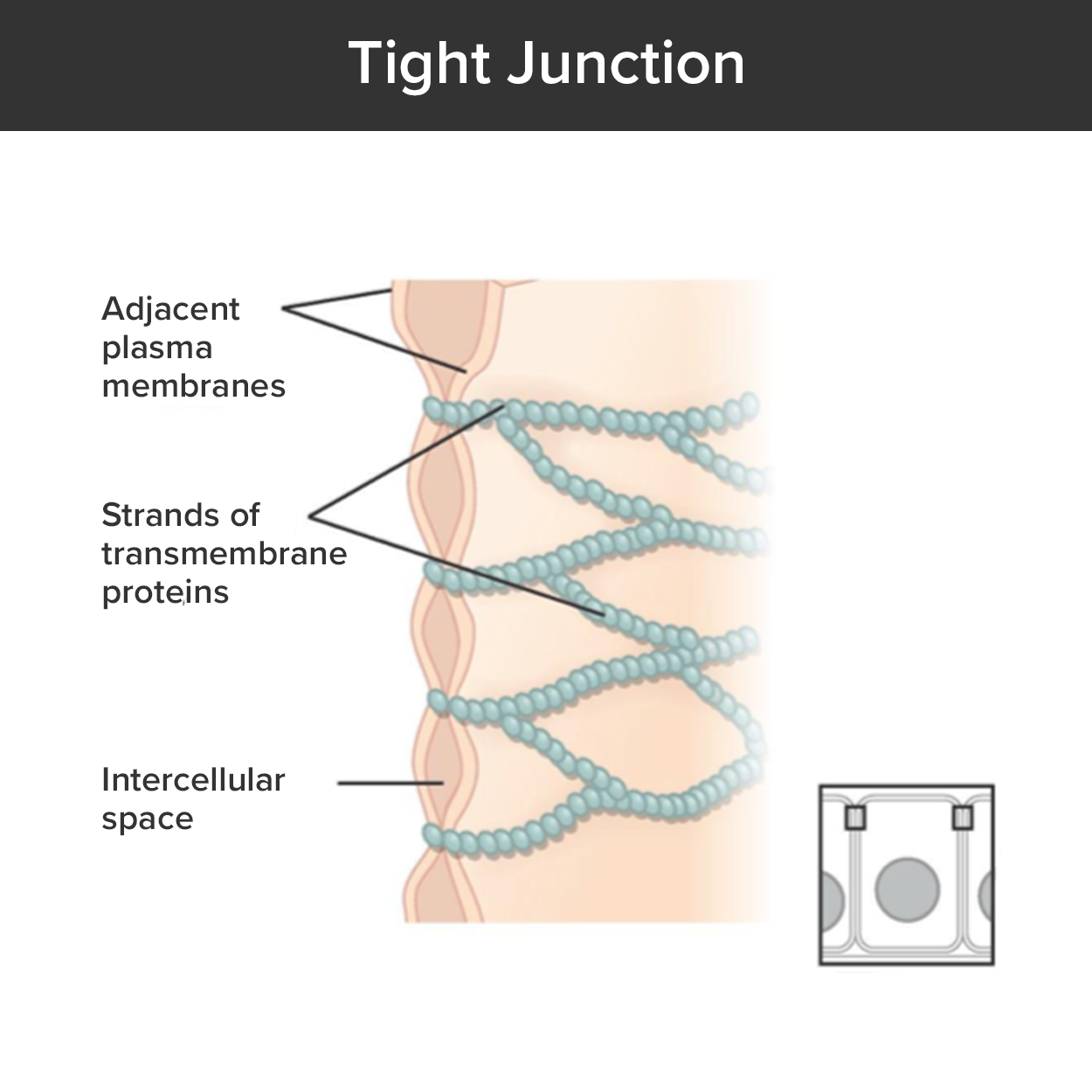 Tight junctions form water-tight connections (hence their name) on the lateral surface using multiple membrane proteins to hold the membranes of two adjacent cells together. In this type of cell junction, there is no extracellular space between the two participating cells, and extracellular substances are forced to use membrane transport to travel through the tissue layer instead of diffusing between the cells.
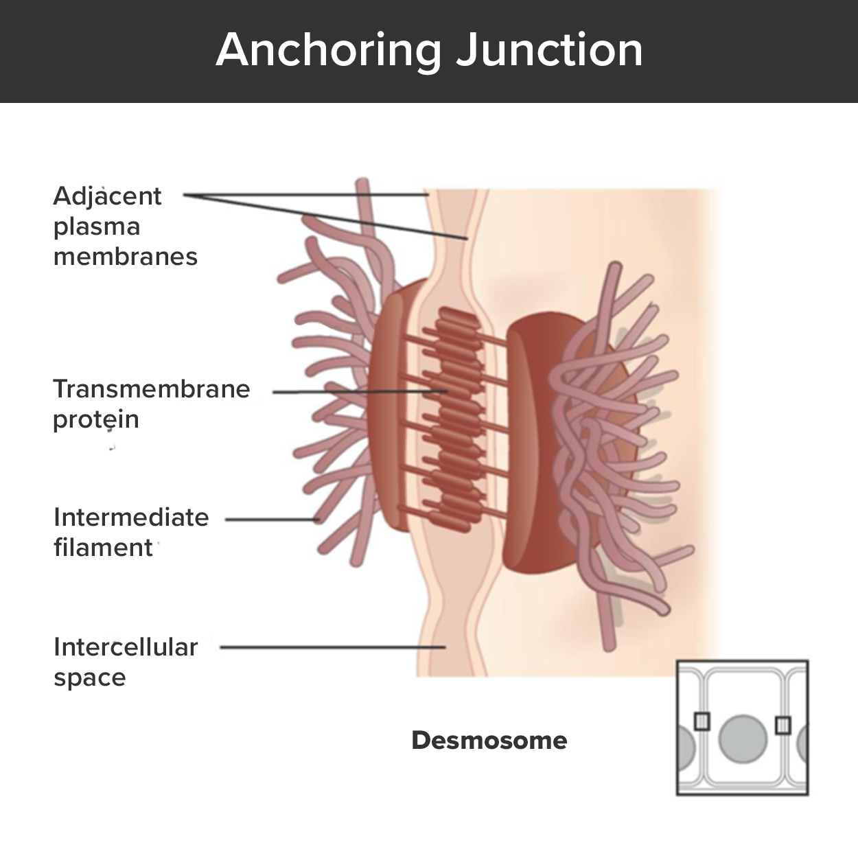 Desmosomes form between adjacent cells, forming cell-to-cell anchors. Each participating cell forms a collection of membrane proteins together in a patch. Once aligned, the transmembrane proteins interlock. On the internal side, each patch is connected to the cytoskeleton. These cell junctions help to maintain the integrity of an epithelial tissue as it is distorted by external pressure.
 Hemidesmosomes (hemi, half), which look like half a desmosome, form a patch of membrane proteins on the basal surface and connect cells to the extracellular matrix (i.e., the basement membrane). These cell junctions keep epithelial tissue attached to the underlying material.
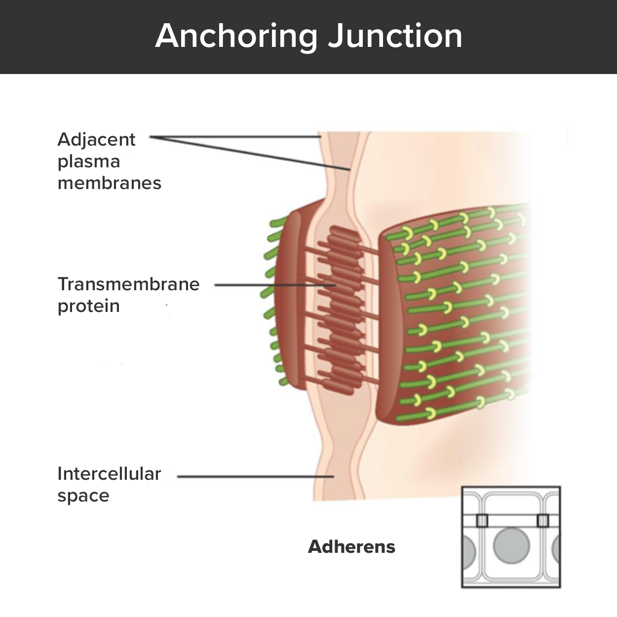 Adherens junctions form between adjacent cells and form cell-to-cell anchors like desmosomes. However, these cell junctions are also connected to contractile proteins inside the cell which can be attached to other adherens junctions at other points in the membrane. Multiple adherens junctions can form a belt-like structure through a tissue referred to as an adhesion belt, which can influence cell or tissue shape.
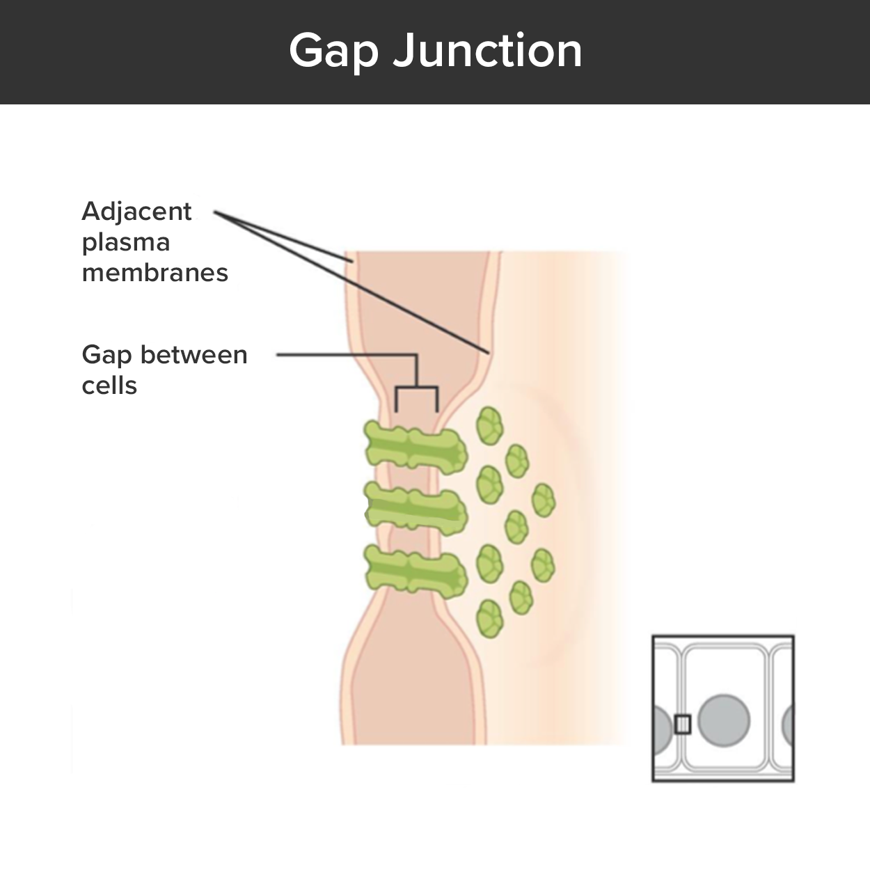 In contrast with the tight and anchoring junctions, a gap junction forms an intercellular passageway between the membranes of adjacent cells to facilitate the movement of small molecules and ions between the cytoplasm of adjacent cells. Each participating cell forms a complex of transmembrane proteins. When two adjacent cells align their proteins, they interlock and open to form a narrow passageway. These junctions allow electrical and metabolic coupling of adjacent cells, which coordinates function in large groups of cells.


 contains a single layer of epithelial cells that look like they are stratified but are actually a single layer with cilia on their apical surface.")
 In the lining of the small intestine, columnar epithelium cells are interspersed with goblet cells. (b) The arrows in this micrograph point to the mucous-secreting goblet cells. LM × 1600. Micrograph provided by the Regents of University of Michigan Medical School © 2012")
 contains many layers of squamous cells with many very thin layers on the apical end full of keratin. The lining of the mouth (right) contains many layers of squamous cells without keratin. Both tissues function to resist abrasion, but only the skin can resist dehydration.")
 applies limited pressure and the apical cells form bulbous shapes with convex surfaces. A full bladder (left) applies pressure to the apical cells forming more squamous-like cells.")
