A full-term pregnancy lasts approximately 270 days (approximately 38.5 weeks) from conception to birth. Because it is easier to remember the first day of the last menstrual period (LMP) than to estimate the date of conception, obstetricians (doctors who specialize in caring for pregnant people before, during, and after childbirth) set the due date as 284 days (approximately 40.5 weeks) from the LMP. This assumes that conception occurred on day 14 of the menstrual cycle, which is usually a good approximation.
The 40 weeks of an average pregnancy are usually discussed in terms of three trimesters, each approximately 13 weeks. The first 2 weeks are characterized by fertilization, and the other 38 weeks are characterized by embryonic and fetal development.
Chart of Pregnancy Months, Weeks, and Trimesters With Stages of Embryo Development
During the second and third trimesters, the pre-pregnancy uterus—about the size of a fist—grows dramatically to contain the fetus, causing a number of anatomical changes in the pregnant person.
Size of Uterus Throughout Pregnancy—The uterus grows throughout pregnancy to accommodate the fetus.
term to know
Trimester
The division of the duration of a pregnancy into three 3-month terms.
2. First Trimester (Weeks 1–13)
The first trimester of pregnancy refers to the first 13 weeks of pregnancy. It is a critical period of embryonic development that includes rapid growth and the formation of major organ systems.
term to know
First Trimester
Marks the start of the pregnancy through the first 13 weeks, where the fertilized egg implants in the uterus and develops into an embryo.
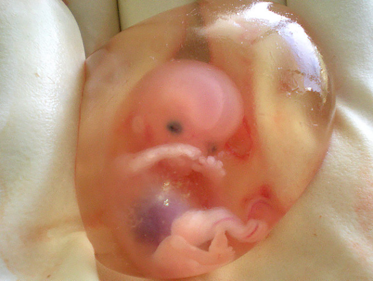
2a. Embryonic Development (Weeks 1–8)
"Weeks of pregnancy" refers to the number of weeks after the first day of the mother's last menstruation. Thus, in the first 2 "weeks of pregnancy", the mother's oocyte hasn't even ovulated, let alone been fertilized.
Recall that when a sperm cell fertilizes an oocyte, the resulting cell is called a zygote. This cell will cleave several times to form many smaller cells. When the zygote has cleaved into 16 cells, it is a ball of cells called a morula (3 to 4 days after fertilization/conception). The morula fills with fluid contained within a thin layer of cells called the trophoblast (this layer of cells will become the placenta); within the fluid is the inner cell mass, which becomes the embryo. It does so by first undergoing gastrulation—becoming the three germ layers (endoderm, mesoderm, and ectoderm). It is around this time (9–12 days after fertilization, or the fourth "week of pregnancy") that the embryo implants in the endometrium.
The following table shows embryonic developmental milestones and risks during weeks 4 through 8 of pregnancy:
Developmental Milestones and Risks
Description
The basic body plan of an embryo will begin to develop.
During the process of gastrulation (when the three germ layers differentiate), an embryonic disc will form. This disc doesn't look much like a person, but it is essentially the first stage of setting up a body plan of the embryo. From there, the neural tube will form within the embryonic disc and will actually become the brain and spinal cord. The cells that form into the neural tube originate from within the ectoderm (the inner layer of germ cells). Following this, other germ cell layers will also begin to specialize and give rise to certain body features, organs, and organ systems. By the end of week 8, the embryo will actually begin to resemble a human being.
The reproductive structures will also begin to develop, depending on the inheritance of the sex chromosomes.
Sex chromosomes are the chromosomes that determine the sex of the individual. If the individual inherited X and Y chromosomes, male reproductive structures will form. If the individual inherited X and X chromosomes, female reproductive structures will form. However, the baby isn't large enough and the sexual features not developed enough to see on an ultrasound until about week 20 of pregnancy. At the end of week 8, the embryo is then called a fetus.
There is always a chance a miscarriage can occur.
Miscarriages can also occur later in development, although it's much less common. Recall that a miscarriage is when an embryo or fetus will be spontaneously expelled from the uterus. The cause or causes of miscarriage can be many factors, but often it is a genetic abnormality. As the body plan is being laid out and as the embryo is starting to develop into a fetus, genetic abnormalities can cause improper development. The body, oftentimes, will expel the abnormal fetus from the uterus because of these genetic abnormalities.
2b. Fetal Development (Weeks 9–13)
When the organism is about 9 weeks old, the fetus is about the size of a kidney bean and begins to take on the recognizable form of a human being, as the “tail” begins to disappear. From this point onward, a fetus undergoes immense growth as it develops organs and dramatically increases in length and weight.
During this time in fetal development, the brain continues to expand, the body elongates, and ossification continues. Fetal movements are frequent during this period but are jerky and not well-controlled. The bone marrow begins to take over the process of erythrocyte production—a task that the liver performs during the embryonic period. The liver now secretes bile. The fetus circulates amniotic fluid by swallowing it and producing urine. The eyes are well-developed by this stage, but the eyelids are fused shut. The fingers and toes begin to develop nails.
Between this time, the sex organs begin to differentiate. The reproductive tissues of male and female humans develop similarly in utero until, in some cases, a low level of the hormone testosterone is released from the gonads. Embryonic males and females, though genetically distinguishable, are morphologically identical. Bipotential gonads, or gonads that can develop into male or female sexual organs, are connected to a central cavity called the cloaca via Müllerian ducts and Wolffian ducts. (The cloaca is an extension of the primitive gut.) Several events lead to sexual differentiation during this period.
Mammalian sex determination is generally determined by X and Y chromosomes. Individuals homozygous for X (XX) are female in sex, and heterozygous individuals (XY) are male. The presence of a Y chromosome triggers the development of a certain set of male characteristics, and its absence results in female characteristics. Nondisjunction during meiosis can produce other combinations such as XXY, XYY, and XO, which are called chromosomal intersex, and you will learn more about these in a future lesson.
key concept
Without much chemical prompting, all fertilized eggs would develop into females. To become a male, an individual must be exposed to the cascade of factors initiated by a single gene on the male Y chromosome. This is called the sex-determining region of the Y chromosome (SRY). Because females do not have a Y chromosome, they do not have the SRY gene. Without a functional SRY gene, an individual will be female.
In both male and female embryos, the same group of cells has the potential to develop into either the male or female gonads; this tissue is considered bipotential. The SRY gene actively recruits other genes that begin to develop the testes and suppresses genes that are important in female development. As part of this SRY-prompted cascade, germ cells in the bipotential gonads differentiate into spermatogonia. Without SRY, different genes are expressed, oogonia form, and primordial follicles develop in the primitive ovary.
Soon after the formation of the testis, the Leydig cells begin to secrete testosterone. Testosterone can influence tissues that are bipotential to become male reproductive structures. Testosterone causes the primitive gonads to differentiate into sexual organs, such as the scrotum and penis. When testosterone is absent, the primitive gonads develop into ovaries. Tissues that produce a penis in males produce a clitoris in females. The tissue that will become the scrotum in a male becomes the labia in a female. Thus, the male and female anatomies arise from a divergence in the development of what were once common embryonic structures.
think about it
The internal reproductive structures (for example, the uterus, uterine tubes, and part of the vagina in females; and the epididymis, ductus deferens, and seminal vesicles in males) form from one of two rudimentary duct systems in the embryo. For typical reproductive function in the adult, one set of these ducts must develop properly, and the other must degrade. In males, secretions from sustentacular cells trigger a degradation of the female duct, called the Müllerian duct. At the same time, testosterone secretion stimulates the growth of the male tract, the Wolffian duct. Without such sustentacular cell secretion, the Müllerian duct will develop; without testosterone, the Wolffian duct will degrade. Thus, the developing offspring will be female.
During male fetal development, upon masculinization, the bipotential gonads become the testes and associated epididymis. The Müllerian ducts degenerate. The Wolffian ducts become the epididymis and ductus deferens, and the cloaca becomes the urethra and rectum.
During female fetal development, upon feminization, the bipotential gonads develop into ovaries. The Wolffian ducts degenerate. The Müllerian ducts become the uterine tubes and uterus, and the cloaca divides and develops into a vagina, a urethra, and a rectum.
Sexual Differentiation—Differentiation of the male and female reproductive systems does not occur until the fetal period of development.
By the 12th week, the fetus has all its body parts including external genitalia. In the following weeks, the fetus will develop hair, nails, teeth, and the excretory and digestive systems will continue to develop. At the end of the 12th week, the fetus is about 3 inches long and weighs about 28 grams.
3. Second Trimester (Weeks 14–28)
The second trimester of pregnancy starts at the fourth month and lasts until the end of the sixth month. During this time of fetal development, organs and organ systems are maturing, and fetal movement can be felt.
Weeks 13–16 are marked by sensory organ development. The eyes move closer together; blinking motions begin, although the eyes remain sealed shut. The lips exhibit sucking motions. The ears move upward and lie flatter against the head. The scalp begins to grow hair. The excretory system is also developing: The kidneys are well-formed, and meconium, or fetal feces, begins to accumulate in the intestines. Meconium consists of ingested amniotic fluid, cellular debris, mucus, and bile.
At about 16 weeks, the fetus is approximately 4.5 inches long. Fingers and toes are fully developed, and fingerprints are visible. At this point, the fetus can kick, urinate, and swallow, and the taste buds are developing.
During approximately weeks 16–20, as the fetus grows and limb movements become more powerful, the pregnant person may begin to feel quickening, or fetal movements. However, space restrictions limit these movements and typically force the growing fetus into the “fetal position,” with the arms crossed and the legs bent at the knees. Sebaceous glands coat the skin with a waxy, protective substance called vernix caseosa that protects and moisturizes the skin and may provide lubrication during childbirth. A silky hair called lanugo also covers the skin during weeks 17–20, but it is shed as the fetus continues to grow. Extremely premature infants sometimes exhibit residual lanugo.
Residual Lanugo on a Newborn’s Ear
The following weeks are characterized by rapid weight gain, which is important for maintaining a stable body temperature after birth. The bone marrow completely takes over erythrocyte synthesis, and the axons of the spinal cord begin to be myelinated, or coated in the electrically insulating glial cell sheaths that are necessary for efficient nervous system functioning. (The process of myelination is not completed until adolescence.) During this period, the fetus grows eyelashes. The eyelids are no longer fused and can be opened and closed. The lungs begin producing surfactant, a substance that reduces surface tension in the lungs and assists in proper lung expansion after birth.
did you know
What happens if not enough surfactant is produced?
Inadequate surfactant production in premature newborns may result in respiratory distress syndrome, and as a result, the newborn may require surfactant replacement therapy, supplemental oxygen, or maintenance in a continuous positive airway pressure (CPAP) chamber during their first days or weeks of life.
In male fetuses, the testes descend into the scrotum near the end of this period.
terms to know
Second Trimester
Marks the start of the fourth month until the end of the sixth month of pregnancy in which the fetus continues to develop as organs and organ systems mature.
Meconium
Fetal wastes consisting of ingested amniotic fluid, cellular debris, mucus, and bile.
Quickening
Fetal movements that are strong enough to be felt by the mother.
Vernix Caseosa
A waxy, cheese-like substance that protects the delicate fetal skin until birth.
Lanugo
Silk-like hairs that coat the fetus; shed later in fetal development.
4. Third Trimester (Weeks 29–40)
The third trimester is marked as the seventh month until birth, which on average is within the ninth month. During this time, organs and organ systems are continuing to mature, and the fetus is preparing for birth. Babies who are born before the third trimester will generally have a low survival rate because their organ systems are too underdeveloped. The time within the mother is very important for organs and organ systems to properly develop. Essentially, if the baby is born before the seventh month, the baby's organ systems aren't well enough developed to allow for survival outside of the uterus.
The fetus at 30 weeks measures 28 cm (11 in) from crown to rump and exhibits the approximate body proportions of a full-term newborn, but still is much leaner. The fetus continues to lay down subcutaneous fat from week 31 until birth. The added fat fills out the hypodermis, and the skin transitions from red and wrinkled to soft and pink. Lanugo is shed, and the nails grow to the tips of the fingers and toes. Immediately before birth, the average crown-to-rump length is 35.5–40.5 cm (14–16 in), and the fetus weighs approximately 2.5–4 kg (5.5–8.8 lbs). Once born, the newborn is no longer confined to the fetal position, so subsequent measurements are made from head to toe instead of from crown to rump. At birth, the average length is approximately 51 cm (20 in).
IN CONTEXT
Meconium-Related Complications
Throughout the second half of gestation, the fetal intestines accumulate a tarry, greenish-black meconium. The newborn’s first stools consist almost entirely of meconium; they later transition to seedy yellow stools or slightly formed tan stools as meconium is cleared and replaced with digested breast milk or formula, respectively. Unlike these later stools, meconium is sterile; it is devoid of bacteria because the fetus is in a sterile environment and has not consumed any breast milk or formula. Typically, an infant does not pass meconium until after birth. However, in 5%–20% of births, the fetus has a bowel movement in utero, which can cause major complications in the newborn.
The passage of meconium in the uterus signals fetal distress, particularly fetal hypoxia (i.e., oxygen deprivation). This may be caused by maternal drug abuse (especially tobacco or cocaine), maternal hypertension, depletion of amniotic fluid, long labor or difficult birth, or a defect in the placenta that prevents it from delivering adequate oxygen to the fetus.
Meconium passage is typically a complication of full-term or post-term newborns because it is rarely passed before 34 weeks of gestation when the gastrointestinal system has matured and is appropriately controlled by nervous system stimuli. Fetal distress can stimulate the vagus nerve to trigger gastrointestinal peristalsis and relaxation of the anal sphincter. Notably, fetal hypoxic stress also induces a gasping reflex, increasing the likelihood that meconium will be inhaled into the fetal lungs.
Although meconium is a sterile substance, it interferes with the antibiotic properties of the amniotic fluid and makes the newborn and pregnant person more vulnerable to bacterial infections at birth and during the perinatal period. Specifically, inflammation of the fetal membranes, inflammation of the uterine lining, or neonatal sepsis (infection in the newborn) may occur. Meconium also irritates delicate fetal skin and can cause a rash.
The first sign that a fetus has passed meconium usually does not come until childbirth, when the amniotic sac ruptures. Normal amniotic fluid is clear and watery, but amniotic fluid in which meconium has been passed is stained greenish or yellowish.
Normal Amniotic Fluid vs. Meconium-Stained Amniotic Fluid
Aspiration of meconium with the first breath can result in labored breathing, a barrel-shaped chest, or a low Apgar score (which is an assessment used to estimate how well a newborn is doing based on five criteria: “appearance,” based on skin color; “pulse,” based on heart rate; “grimace,” based on reflex; “activity,” based on muscle tone; and “respiration”). An obstetrician can identify meconium aspiration by listening to the lungs with a stethoscope for a coarse rattling sound. Blood gas tests and chest X-rays of the infant can confirm meconium aspiration. Inhaled meconium after birth could obstruct a newborn’s airways, leading to alveolar collapse, interfere with surfactant function by stripping it from the lungs, or cause pulmonary inflammation or hypertension. Any of these complications will make the newborn much more vulnerable to pulmonary infection, including pneumonia.
Meconium Aspiration—Meconium aspiration occurs when a newborn breathes meconium with their first breath.
term to know
Third Trimester
Marks the seventh month of pregnancy until birth and is characterized by the continued development of the fetus as it prepares for birth.
Interactive 3-D Model Investigate the fetal development in three dimensions using augmented reality (AR)!
If you’re on a laptop or desktop computer:
Scan the QR code using the camera on your smartphone or tablet.
In this lesson, you learned about the development of an embryo and fetus throughout pregnancy. Specifically, you first learned that stages of embryonic and fetal development are described in terms of trimesters. Then, you learned that the first trimester (weeks 1–13) includes both embryonic development (weeks 1–8) and fetal development (weeks 9–13), and that it is a period of pregnancy with formation, reproductive structure development, and the possibilities of a miscarriage. Then, you learned that the second trimester (weeks 14–28) is a time when organs and organ systems are maturing, and fetal movement can be felt. Finally, you explored the third trimester (weeks 29–40), which is a time when organ and organ systems are continuing to mature, and the fetus is preparing for birth.
Marks the start of the pregnancy through the first 13 weeks, where the fertilized egg implants in the uterus and develops into an embryo.
Lanugo
Silk-like hairs that coat the fetus; shed later in fetal development.
Meconium
Fetal wastes consisting of ingested amniotic fluid, cellular debris, mucus, and bile.
Quickening
Fetal movements that are strong enough to be felt by the mother.
Second Trimester
Marks the start of the fourth month until the end of the sixth month of pregnancy in which the fetus continues to develop as organs and organ systems mature.
Third Trimester
Marks the seventh month of pregnancy until birth and is characterized by the continued development of the fetus as it prepares for birth.
Trimester
The division of the duration of a pregnancy into three 3-month terms.
Vernix Caseosa
A waxy, cheese-like substance that protects the delicate fetal skin until birth.


 Interactive 3-D Model
