Use Sophia to knock out your gen-ed requirements quickly and affordably. Learn more
×

EIA, FEIA, and ELISA Testing

Author: Sophia

what's covered
In this lesson, you will learn about types of enzyme immunoassays and immunostaining. These include many widely used tests, including home pregnancy tests. Understanding these tests is helpful as they are frequently encountered in clinical and laboratory settings as well as everyday life. Specifically, this lesson will cover the following:

Table of Contents

1. Introduction

In this lesson, you will learn about different types of tests that use antibodies to detect the presence of antigens. These tests, called enzyme immunoassays (EIAs), are carried out in microtiter plates or in vivo and share a common characteristic: they use an antibody molecule with a variable region that binds to a specific antigen and a constant region that binds to an enzyme. The enzyme is important because it can undergo a reaction to show the presence of the antibody, allowing rapid visualization and/or quantification of test results.

Chromogens are colorless molecules that can be converted into a colored end product. EIAs often use enzymes that react with chromogens. Alkaline phosphatase and horseradish peroxidase are commonly used.

In some cases, a fluorogen is the substrate for the enzyme. Fluorogens are nonfluorescent molecules that can be converted by the enzyme into a fluorescent form that can be detected using a spectrophotometer or a fluorescence microscope. When EIAs use fluorogens, they are called fluorescent enzyme immunoassays (FEIAs).

terms to know
Enzyme Immunoassay (EIA)
An assay that uses antibodies to detect the presence of antigens.
Fluorogen
A nonfluorescent molecule that can be converted by an enzyme into a fluorescent form that can be detected using a spectrophotometer or a fluorescence microscope.
Fluorescent Enzyme Immunoassay (FEIA)
An enzyme immunoassay that uses a fluorogen.

2. Immunostaining

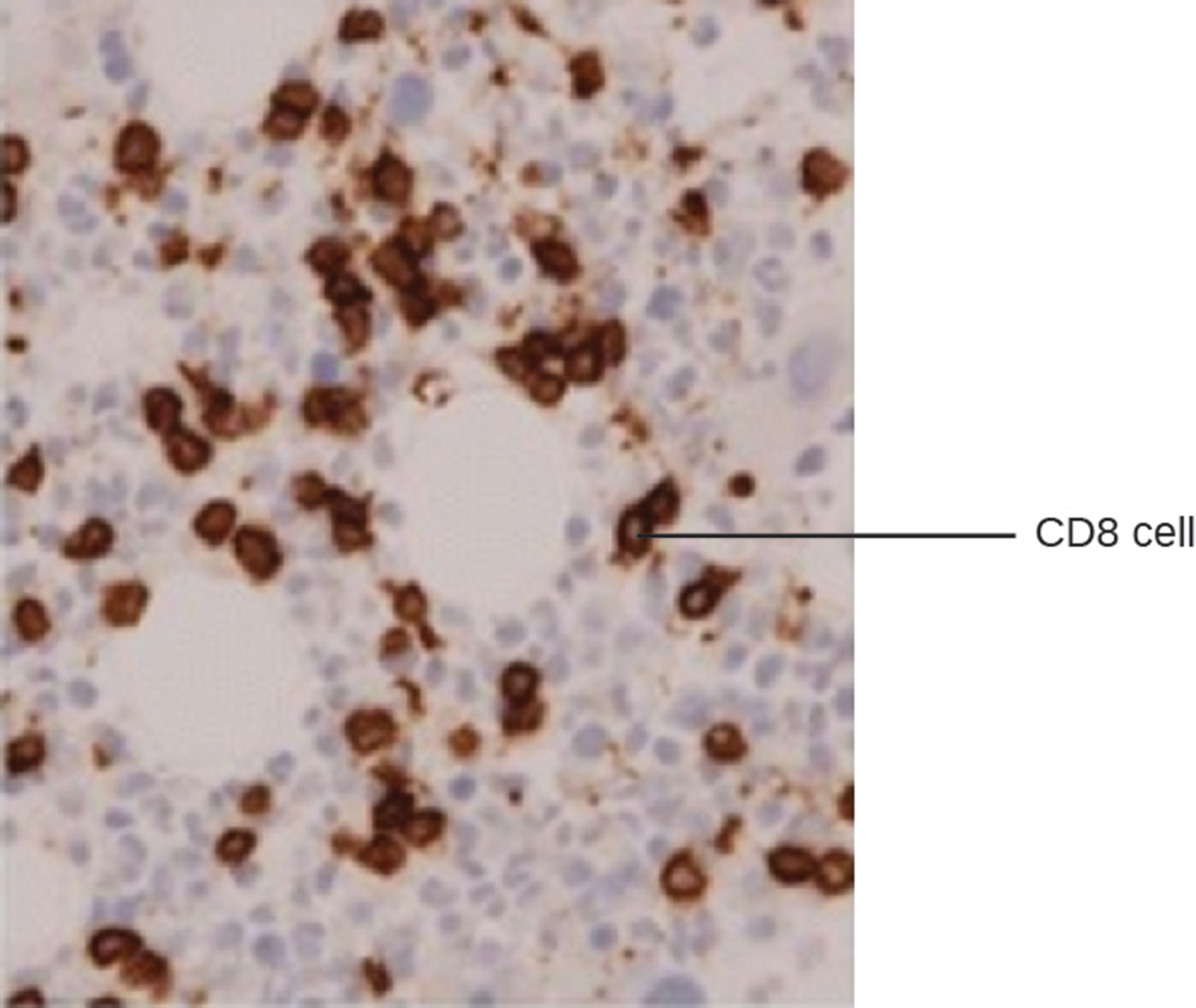
EIA can be used for immunostaining. The antibody–enzyme conjugate enhances microscopy by making antigens of interest more visible. The micrograph below shows an example of immunohistochemistry (IHC), which is similar to immunostaining but used to visualize different cell types. In the micrograph, an mAb against CD8 was used to stain CD8+ cells in a section of tissue. This allows the number of CD8 cells to be counted, located, and compared with the number of CD8 cells in other tissues.

Immunocytochemistry (ICC) is another type of immunostaining that is similar to IHC but is performed after the extracellular matrix has been stripped away and alcohol has been added to make the cell membrane permeable to antibodies. Antibodies can therefore enter the cell to bind to specific targets such as organelles. Some ICC techniques use EIA and others use fluorescent molecules in place of enzymes (a fluorescent immunoassay).

terms to know
Immunostaining
The antibody–enzyme conjugate enhances microscopy by making antigens of interest more visible.
Immunohistochemistry (IHC)
A similar process to immunostaining but used to visualize different cell types.
Immunocytochemistry (ICC)
A type of immunostaining similar to IHC but performed after the cell membrane has been made permeable to allow antibodies to enter and bind to intracellular structures.

3. Enzyme-Linked Immunosorbent Assays

Enzyme-linked immunosorbent assays (ELISAs) are very commonly used examples of EIAs. There are three major types of ELISAs:

  • Direct ELISAs involve antigens immobilized in wells of microtiter plates that are exposed to antibodies bound to enzymes.
  • Sandwich ELISAs are used to quantify specific antigen present in a solution.
  • Indirect ELISAs quantify antigen-specific antibody rather than antigen.
These techniques are summarized in the steps and images below.

Direct ELISA

step by step
Step 1. Antigens are immobilized in the wells of microtiter plates.
Step 2. An antibody specific to the antigen of interest is conjugated to an enzyme and added to each well. If the antigen is present, the antibody will bind.
Washing is performed to remove unbound antibodies.
Step 3. A colorless chromogen is added. If the enzyme is present, the chromogen will be converted to its colored form. If the enzyme is absent, no color will appear.

The image below shows an example of a direct ELISA test that produces a color change, indicating a positive result (i.e., presence of the antigen of interest).

Viral antigens (drawn as diamonds) are attached to a surface. Antibodies (drawn as Y’s) with an enzyme conjugate (purple circle) attached to them bind to the viral antigens. A substrate (drawn as a blue circle) interacts with the enzyme on the antibody and changes color for detection.

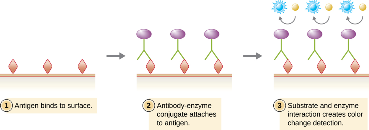
Direct ELISA tests are rapid and relatively simple, but other types of ELISA tests are sometimes preferred for various reasons (such as higher sensitivity).

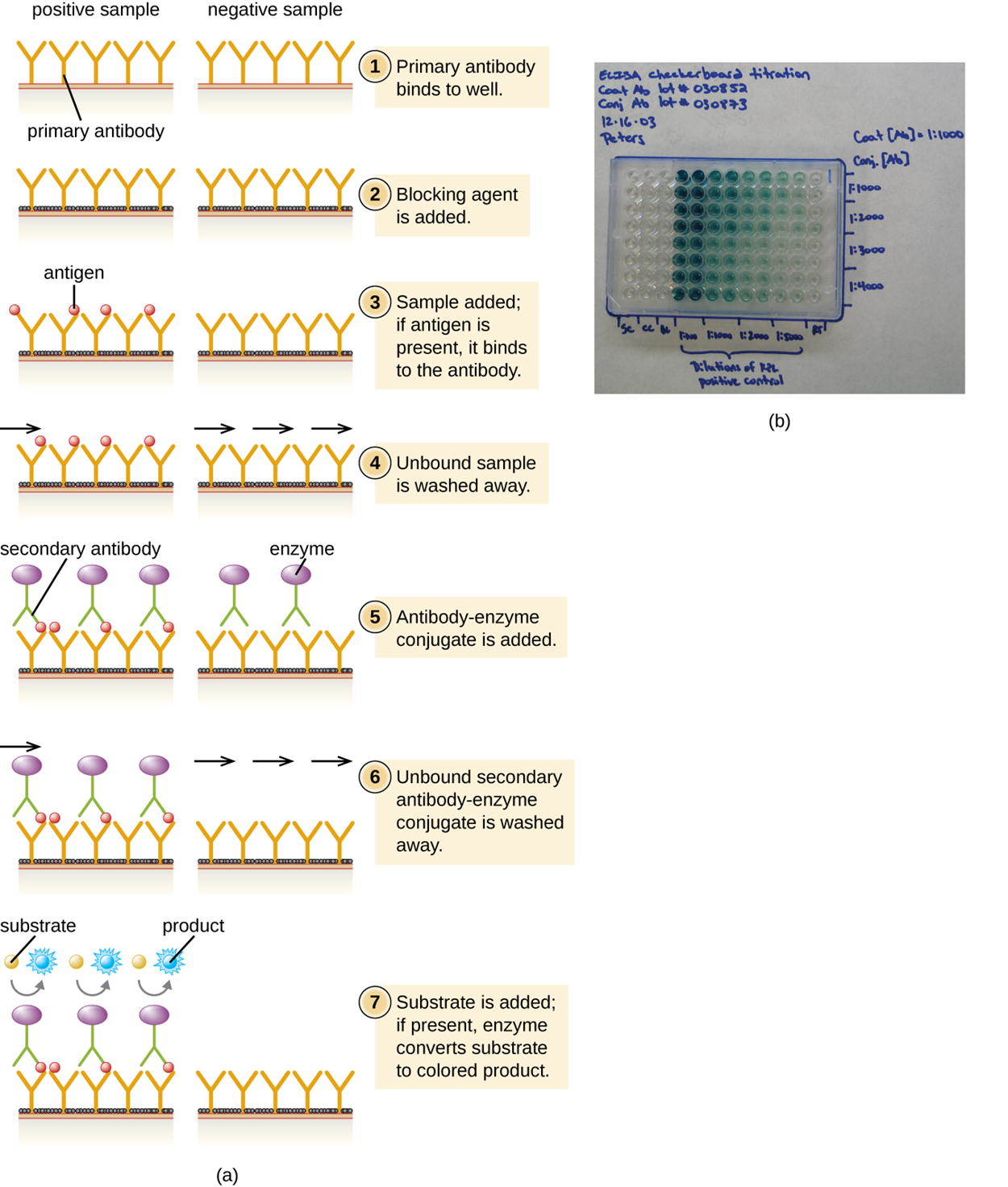
The sandwich ELISA test uses antibodies to quantify the exact amount of an antigen present in a sample. The test is summarized in the steps and image below.

step by step
Step 1. The primary antibody is added to all of the wells of a microtiter plate, to which it sticks through hydrophobic interactions.
Step 2. The microtiter plate is incubated.
Step 3. Unbound antibody is washed away. Washing is repeated between subsequent steps to remove unbound molecules.
Step 4. A blocking protein is added to bind to remaining nonspecific protein-binding sites in the well.
Step 5. Some wells receive known amounts of antigen to allow a standard curve to be produced, whereas other wells receive unknown antigen solutions.
Step 6. The primary antibody captures the antigen.
Step 7. After another wash, the secondary antibody is added (a polyclonal antibody conjugated to an enzyme).
Step 8. After another wash, a colorless chromogen substrate is added.
Step 9. The enzyme converts the chromogen to its colored form.
Step 10. The colored intensity of each sample is measured with a spectrophotometer. The amount of color produced is directly proportional to the amount of enzyme, which is proportional to the amount of captured antigen.
Step 11. The unknown samples can be compared to the standard curve produced using known amounts of antigen to quantify the amount of antigen present in them.

The figure below presents a simplified version of the steps provided above. Note that the figure does not include preparation of a standardized curve, which adds steps above.

(a) Diagram of a sandwich ELISA showing what happens in both a positive and a negative sample. First, the primary antibody binds to the well. This is shown as Ys bound to a surface. Next, a blocking agent is added. This is shown as a black covering on the surface between the antibodies. Next, the sample is added; if the correct antigen is present it binds to the antibody. In the positive well, a circle binds to the antibodies; in the negative well, nothing binds to the antibodies. Next, any unbound sample is washed away. Next, the antibody–enzyme conjugate is added. This is shown in the positive well as another Y shape that binds to the circles. This new Y shape has a purple circle at the end. These antibodies are also in the negative sample but they are not attached to anything. Next, the unbound antibody–enzyme conjugate is washed away; these remain in the positive sample (because they are attached to the antigen) but are washed away in the negative sample. Finally, a substrate is added to both the positive and negative samples. The enzyme in the positive sample turns this substrate blue. (b) A plastic plate with many wells. Some are clear, some are blue.

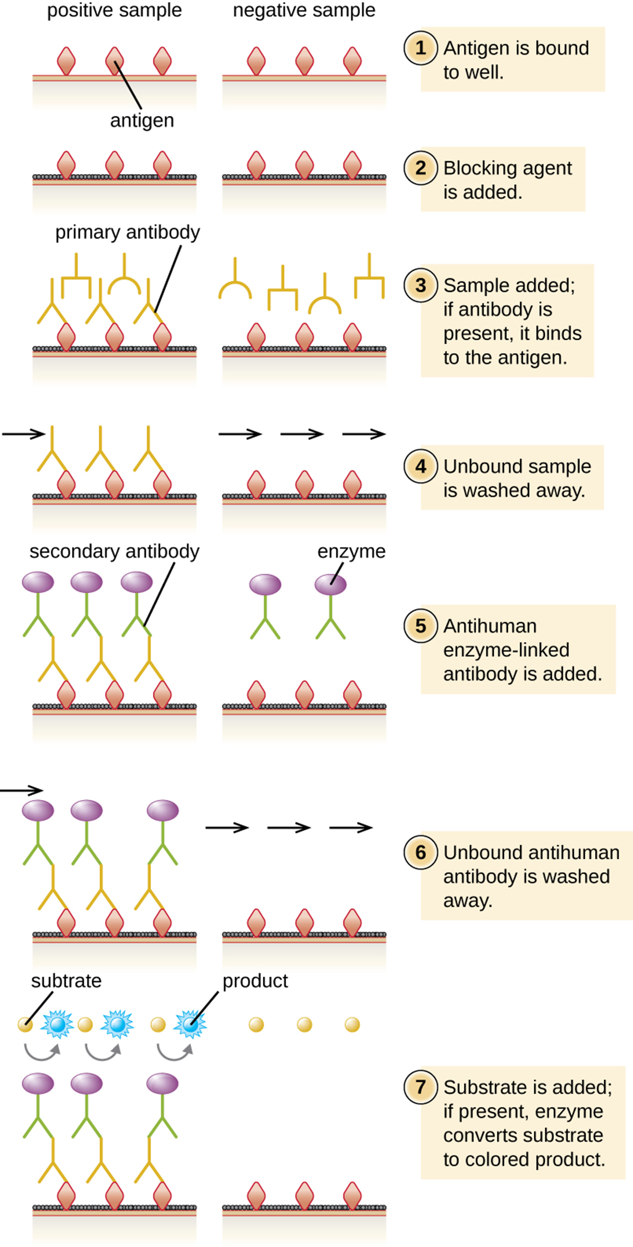
Indirect ELISA is used to quantify antigen-specific antibody instead of antigen. Therefore, it is useful to detect antibodies against a variety of pathogens. However, it has some important differences from direct ELISA. The first step is to attach a known antigen (not antibody) to the bottom of the microtiter plate wells. After unbound sites are blocked, patient serum is added and any primary antibodies present bind to antigen. After washing away unbound proteins, the secondary antibody with its conjugated enzyme is added. This antibody targets the first antibody. The intensity of color produced by the conjugated enzyme–chromogen reaction is used to quantify how much antigen-specific antibody is present in the patient’s serum.

Positive tests must be confirmed, especially as cross-reactivity may occur. This is often accomplished using immunoblots to identify the presence of specific peptides or tests to identify nucleic acids. The steps of indirect ELISA are summarized in the steps and image below.

step by step
Step 1. Primary antibody binds to wells.
Step 2. Blocking agent is added.
Step 3. Sample is added; if antigen is present, it binds to the antibody.
Step 4. Unbound sample is washed away.
Step 5. Secondary antibody–enzyme conjugate is added.
Step 6. Unbound secondary antibody–enzyme conjugate is washed away.
Step 7. Substrate is added. If the enzyme is present, it converts the substrate to a colored product. If the enzyme is absent, no color is produced.

A Diagram of indirect ELISA showing what happens in both a positive and a negative sample. First, the antigens are bound to the well. This is shown as diamonds on the surface of both the positive and negative wells. Next, a blocking agent is added. This is shown as a black covering on the surface between the antigens. Next, the sample is added. If the correct antibody is present it binds to the antigen. This is shown as Ys in both the positive and negative wells. Otherwise, nothing binds to the antigen. Next, any unbound sample is washed away. In the positive well, there is an antibody bound to the antigen, in the negative well, there is nothing bound to the antigen. Next, an anti-human enzyme-linked antibody is added. This is shown as a Ys with a purple circle in both wells. Next, unbound antigen is washed away. In the positive sample, this Y remains bound to the old antibody. In the negative well, it is no longer present. Finally, a substrate is added to both wells. In the positive well, the enzyme changes the substrate to a blue color.

terms to know
Enzyme-Linked Immunosorbent Assays (ELISA)
Commonly used EIAs that detect the presence of antigens or antigen-specific antibodies or quantify antigen in a solution.
Direct ELISA
Uses antigens immobilized in wells of microtiter plates that are exposed to antibodies bound to enzymes.
Sandwich ELISA
Used to quantify specific antigen present in solution.
Indirect ELISA
Used to quantify antigen-specific antibody rather than antigen.

4. Immunofiltration and Immunochromatographic Assays

Immunofiltration is used to detect or quantify antigens or antibodies that are present in very low quantities. In this process, fluid is passed through a porous membrane into an absorbent pad. The pad has either antigens or antibodies attached to capture antibodies or antigens, respectively.

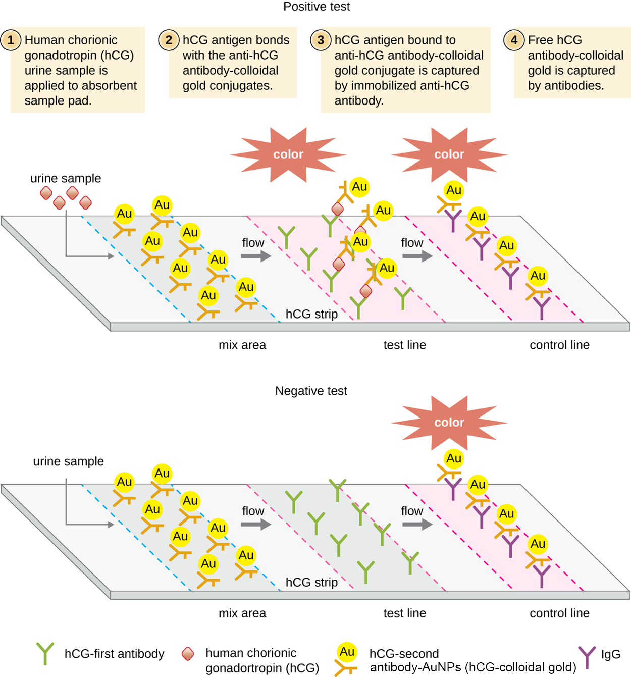
Immunochromatographic assays (also called lateral flow tests) have been developed using an adaptation of immunofiltration. In these tests, fluids are applied to an absorbent pad on a test strip. Fluid flows through capillary action and moves through a strip of beads with antibodies attached to their surfaces. The fluid in the sample hydrates the reagents in the strip. Antibody-coated beads made of latex or tiny gold particles bind to antigens in the test fluid as it passes across them. The antibody–antigen complexes then flow over a second strip that has immobilized antibody against the antigen; this strip retains beads that have bound antigen. A third control strip binds any beads regardless of the presence or absence of antigen. Color develops at the test lines if the test is positive. If the test was performed correctly but the antigen is absent, then color only develops at the control line.

These tests are commonly used for in-home pregnancy tests as well as the TORCH test, which is used to screen pregnant women or newborns for a range of infections called the TORCH complex, which includes toxoplasmosis, others (syphilis, hepatitis B), rubella, cytomegalovirus, and herpes simplex.

These tests take advantage of antibody sandwiches, which provide sensitivity and specificity. They are fast and inexpensive, even though they are not as quantitative as ELISA.

The photo below shows a positive pregnancy test, which is an example of a lateral flow test. Two lines have formed through the process described above.

A pregnancy test stick with 2 red lines; one is labeled test line and the other is labeled control line. A key on the stick states that 2 lines mean pregnant and 1 line means not pregnant.

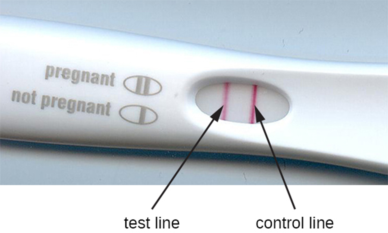
As an example of how immunochromatographic assays (lateral flow tests) work, the steps and image below describe a human pregnancy test.

step by step
Step 1. A urine sample is applied to the absorbent sample pad. In pregnant individuals, levels of human chorionic gonadotropin (hCG) increase and are present in urine. The fluid moves across the strip through capillary action.
Step 2. If present, hCG antigen binds with the anti-hCG antibody-colloidal gold conjugates in a band on the strip.
Step 3. If hCG antigen is present, then hCG antigen bound to anti-hCG-colloidal gold conjugate is captured by immobilized anti-hCG antibody. This produces the test line.
Step 4. Free hCG antibody-colloidal gold is captured by antibodies to produce the control lines.
A diagram showing the lateral flow test of a urine sample. The top panel shows a positive test and the bottom shows a negative test. In both a urine sample is added; in the positive sample, there are antigens present. The label reads hCG urine sample is applied to the absorbent sample pad. In both cases, the urine sample flows across the test area. The first region is a mix area that contains hCG-second antibody-AuNPs (hCG-colloidal gold). These then flow across the hCG strip in both samples. Next, we reach the test line. Here there are antibodies that bind the antigens in the positive sample but bind nothing in the negative sample. In the positive sample, the hCG-colloidal gold also binds to the antigen, forming an antibody sandwich around the antigen. The presence of the hCG-colloidal gold causes a color change here in the positive sample but not in the negative sample. Finally, the control line contains antibodies that bind the hCG-colloidal gold directly; so these bind in both the positive and negative samples. The control line turns color in both.

think about it
Have you taken a rapid antigen test for COVID-19? These are available at medical clinics and as home tests. After swabbing your nose, you add your sample to liquid and then put several drops into a rectangular test strip. The liquid moves across the strip and produces a line labeled C, or control, if the test was carried out correctly. If COVID antigens are present, a second line appears (often labeled T for test). The test device resembles the pregnancy test shown in this lesson because it is also a lateral flow test (Ng et al., 2022; Healthline, 2022).

The table below summarizes the mechanisms and procedures of the assays discussed in this lesson including examples.

Immunoblots and Enzyme Immunoassays
Type of Assay Mechanism Specific Procedures Examples
Immunoblots Uses enzyme–antibody conjugates to identify specific proteins that have been transferred to an absorbent membrane Western blot: Detects the presence of a particular protein Detecting the presence of HIV peptides (or peptides from other infectious agents) in patient sera
Immunostaining Uses enzyme–antibody conjugates to stain specific molecules on or in cells Immunohistochemistry: Used to stain specific cells in a tissue Stain for presence of CD8+ cells in host tissue
Enzyme-linked immunosorbent assay (ELISA) Uses enzyme–antibody conjugates to quantify target molecules Direct ELISA: Uses a single antibody to detect the presence of an antigen Detection of HIV antigen p24 up to one month after being infected
Indirect ELISA: Measures the amount of antibody produced against an antigen Detection of HIV antibodies in serum
Immunochromatographic (lateral flow) assays Techniques use the capture of flowing, color-labeled antigen–antibody complexes by fixed antibody for disease diagnosis Lateral flow assays for pregnancy, COVID-19 antigens, and others Detection of antibodies for various pathogens in patient sera (e.g., rapid strep, malaria dipstick)
Pregnancy test detecting human chorionic gonadotrophin in urine
Detection of COVID-19 antigens


terms to know
Immunofiltration
Used to detect or quantify antigens or antibodies that are present in very low quantities by passing fluid through a porous membrane into an absorbent pad that has either antigens or antibodies attached.
Immunochromatographic Assay (Lateral Flow Test)
A test in which fluids are applied to an absorbent pad on a test strip and flow through capillary action to interact with strips of beads with attached antibodies. One strip detects antibody bound to antigen (producing the test line if these are present) and the other strip binds to any beads regardless of whether they are bound to antigens (producing the control line).

summary
In this lesson, you learned about a variety of powerful testing techniques that include or are based on enzyme immunoassays. After an introduction that explained the basic process involved in enzyme immunoassay testing, you learned about immunostaining and how it can be used to identify structures and even for histological analysis of cells in tissue. Next, you learned about different types of enzyme-linked immunosorbent assays and how each type is used. Finally, you learned about immunofiltration and immunochromatographic assays. Immunochromatographic assays include many common tests such as the current rapid tests for COVID-19 detection and home pregnancy tests.

Source: THIS CONTENT HAS BEEN ADAPTED FROM OPENSTAX’s “MICROBIOLOGY”. ACCESS FOR FREE AT openstax.org/details/books/microbiology

REFERENCES

Jillian, K. MS, RD. (2022, December 16). Rapid COVID-19 Tests: When to Use Them and How They Work. Healthline. www.healthline.com/health-news/rapid-covid-19-tests-when-to-use-them-and-how-they-work

Ng, Q. X., Lim, Y. L., Han, M. X., Teoh, S. E., Thumboo, J., & Tan, B. H. (2022). The Performance of Lateral Flow Tests in the Age of the Omicron: A Rapid Systematic Review. Life (Basel, Switzerland), 12(11), 1941. doi.org/10.3390/life12111941

Terms to Know
Direct ELISA

Uses antigens immobilized in wells of microtiter plates that are exposed to antibodies bound to enzymes.

Enzyme Immunoassay (EIA)

An assay that uses antibodies to detect the presence of antigens.

Enzyme-Linked Immunosorbent Assays (ELISA)

Commonly used EIAs that detect the presence of antigens or antigen-specific antibodies or quantify antigen in a solution.

Fluorescent Enzyme Immunoassays (FEIA)

An enzyme immunoassay that uses a fluorogen.

Fluorogen

A nonfluorescent molecule that can be converted by an enzyme into a fluorescent form that can be detected using a spectrophotometer or a fluorescence microscope.

Immunochromatographic Assay (Lateral Flow Test)

A test in which fluids are applied to an absorbent pad on a test strip and flow through capillary action to interact with strips of beads with attached antibodies. One strip detects antibody bound to antigen (producing the test line if these are present) and the other strip binds to any beads regardless of whether they are bound to antigens (producing the control line).

Immunocytochemistry (ICC)

A type of immunostaining similar to IHC but performed after the cell membrane has been made permeable to allow antibodies to enter and bind to intracellular structures.

Immunofiltration

Used to detect or quantify antigens or antibodies that are present in very low quantities by passing fluid through a porous membrane into an absorbent pad that has either antigens or antibodies attached.

Immunohistochemistry (IHC)

A similar process to immunostaining but used to visualize different cell types.

Immunostaining

The antibody–enzyme conjugate enhances microscopy by making antigens of interest more visible.

Indirect ELISA

Used to quantify antigen-specific antibody rather than antigen.

Sandwich ELISA

Used to quantify specific antigen present in solution.