Table of Contents |
Before viruses can be cultivated, they must be isolated and transferred to an appropriate environment for growth. Because they cannot reproduce without a host, viruses cannot be grown in isolation on artificial nutrient media. Instead, they are allowed to infect host cells that can be cultured and grown. When needed, the viruses can be isolated from liquid growth medium (separated from the cells) by centrifugation or filtration.
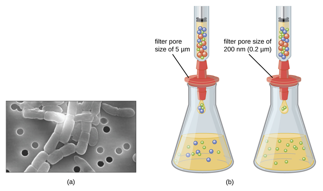
The image below shows filtration. As discussed in other lessons, porcelain Chamberland filters were used to demonstrate the existence of pathogens smaller than bacteria (i.e., viruses). At present, membrane filters are commonly used. Part (a) of the image below shows a scanning electron micrograph of rod-shaped bacteria captured on a membrane filter. The bacteria are too large to fit through the pores in the filter. Image (b) shows filtration of medium through a membrane filter into a flask. The left-hand filter has a pore size of 5 μm and allows bacterial (blue) and animal (red) cells to pass through as well as viruses (green). The right-hand filter has a pore size of 0.2 μm and traps the cells while allowing viruses to pass through.
Although viruses require cells to replicate, this still leaves many options for cultivating them. These methods can be broadly described as in vivo (within a whole living organism) or in vitro (in an artificial environment).
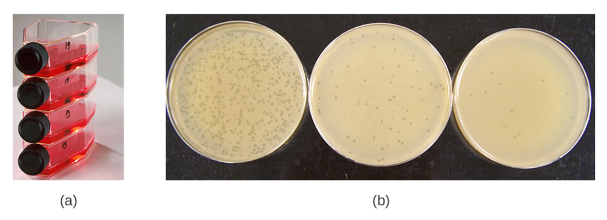
The image below shows examples of each type of cultivation approach. Part (a) shows flat cell culture flasks, which represent an in vitro method. These flasks contain cells, but not entire organisms. Part (b) shows an in vivo approach involving bacteriophages replicating on dense lawns of bacteria growing across the surface of an agar-based culture medium. Clear spots (plaques) appear in areas where bacteriophages have killed the cells.
Microbiologists often need to grow animal viruses for multiple reasons, including the following:
Because viruses often have tissue tropism, it is important to consider the location in which the viruses will be cultivated. The correct tissue must be selected either for culture or by exposing the viruses to the correct location within the animal or embryo.
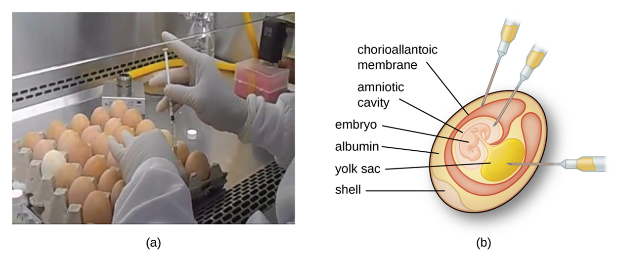
The image below shows a researcher working with chicken eggs (a) and the structure of an embryonated egg (b). Syringe needles pointing into the egg in part b indicate locations where viruses can be replicated. The specific location is chosen based on the requirements of the virus to be cultivated.
When viruses are grown in host animals, embryos, or tissue cultures, they may cause damage to the cells. When grown in embryos, viruses may produce lesions, disrupt embryonic development, or kill the embryo.
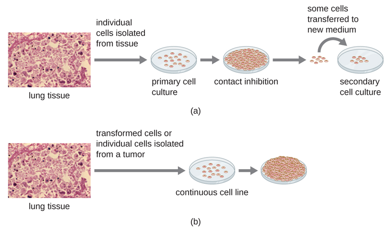
For in vitro studies, various types of cells can be used to support the growth of viruses. A primary cell culture is freshly prepared from animal organs or tissues. To prepare the culture, it is necessary to extract cells from tissues. Mechanical scraping or mincing is one approach. Alternatively, enzymes such as trypsin or collagenase can be used to break up tissue and release individual cells.
Many cells are anchorage dependent, meaning that they only grow when they can attach to a surface. Therefore, primary cell cultures require a liquid culture medium in a petri dish or tissue-culture flask. This gives the cells a glass or plastic surface for attachment and growth.
Most cells in a culture have a limited lifespan and have density-dependent growth, so steps must be taken to maintain appropriate conditions for growth. When these cells come into contact with too many other cells, mitosis stops. This process is called contact inhibition. To prevent contact inhibition, cells from the primary culture must be transferred to another vessel with fresh culture medium. This is called a secondary cell culture. Cell density can be reduced by pouring off some cells and adding fresh medium to provide space and nutrients for continued growth.
Another approach is to use immortal (continuous) cell lines, which are usually derived from transformed cells or tumors. Transformation means that the cells have taken up new genes and you will learn more about this process in other lessons. These cells can sometimes be subcultured repeatedly or even indefinitely. One reason tumors grow inappropriately is that they lack normal controls on cell division, such as contact inhibition. These cells may also lack anchorage dependency, meaning that they can be grown in suspension. The resulting immortal cell lines grow in piles or lumps that resemble small tumors because of these shared characteristics.
The illustration below shows the steps involved in both types of cell culture. Part (a) shows the production of primary and secondary cell cultures. Part (b) shows the production of an immortal cell line.
IN CONTEXT
An important and well-known immortal cell line is the HeLa cell line. These cells were obtained from Henrietta Lacks, a black woman who died of cervical cancer in 1951. HeLa cells were the first immortal tissue cell line and are still being used in research today.
However, the story raises important questions about ethics and patient consent. Henrietta Lacks attended a clinic at Johns Hopkins Hospital that served people who lacked financial resources. She received medical care, but was highly vulnerable. As was common at the time, her family was not asked to consent for researchers to harvest and use her cells for research purposes. Her family did not realize that her cells had been used in this manner and her husband said that the family had not given permission (Cramer, 2021; NPR, 2021).
Henrietta Lacks’ grave is unmarked, and her family lacked financial resources, but others made considerable amounts of money off of her cells. When her family found out, some were angry and even filed a federal lawsuit against a biotechnology company in 2021 with suggestions of additional lawsuits to follow (Cramer, 2021). One of her grandsons emphasized that the lawsuit showed how they wanted the family to reassert control of Lacks’ tremendous legacy (NPR, 2021).
However, even though Lacks’ family is beginning to gain recognition for Henrietta Lacks’ legacy and the way that her cells were used without consent (“stolen”, in the words of her family),, benefits from medical advances made possible by her cells are still unevenly available. For example, her cells were used in the development of COVID-19 vaccinations that are much more accessible in wealthy countries compared with other parts of the world (Cramer, 2021; NPR, 2021).
Today, patient data is regularly used to make important medical discoveries. There are regulations regarding appropriate consent and use of patient data and samples. With some exceptions, Institutional Review Boards must approve studies that involve patients.
Viruses in cultures or in whole organisms can be detected in a variety of ways. Because viral infections such as common colds are familiar to people, many symptoms of viral illness may come to mind. These symptoms will be discussed in lessons on diseases. However, many of these symptoms are common to multiple viruses and it is often important to use laboratory methods to accurately identify specific viruses. The first step is often preparing a sample for further analysis.
Viral infections often cause cytopathic effects (CPEs) at a cellular level. CPEs are distinct, observable cell abnormalities. Some CPEs can be detected using a microscope. Some examples of CPEs include the following:
| Cytopathic Effects of Specific Viruses | ||
|---|---|---|
| Virus | Cytopathic Effect | Example |
| Paramyxovirus | Syncytium and faint basophilic cytoplasmic inclusion bodies (arrows) |
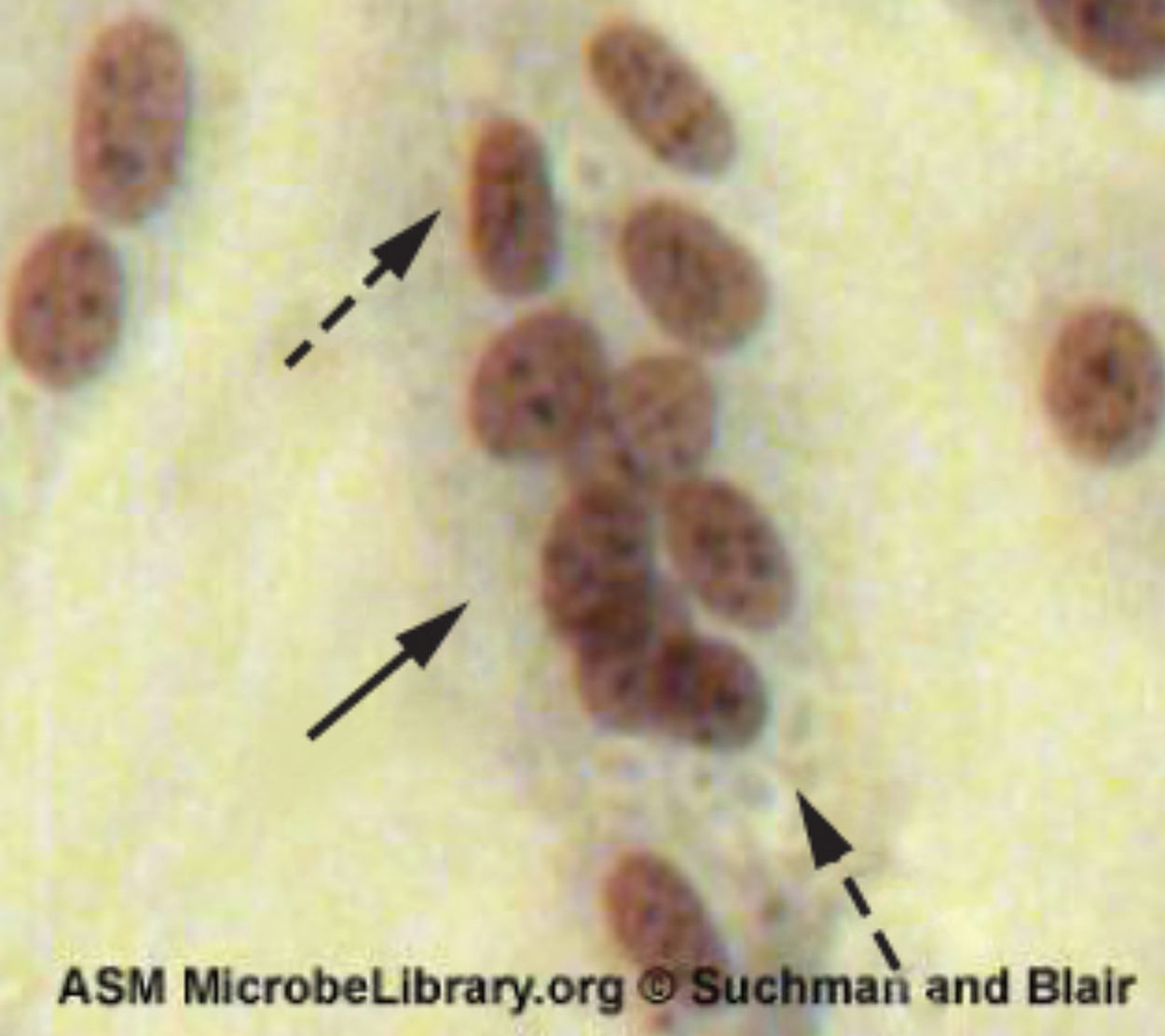
|
| Poxvirus | Pink eosinophilic cytoplasmic inclusion bodies (arrows) and cell swelling |
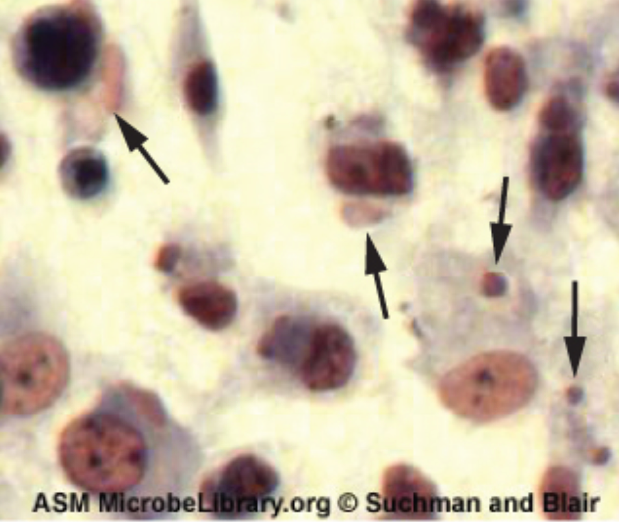
|
| Herpesvirus | Cytoplasmic stranding (arrow) and nuclear inclusion bodies (dashed arrow) |
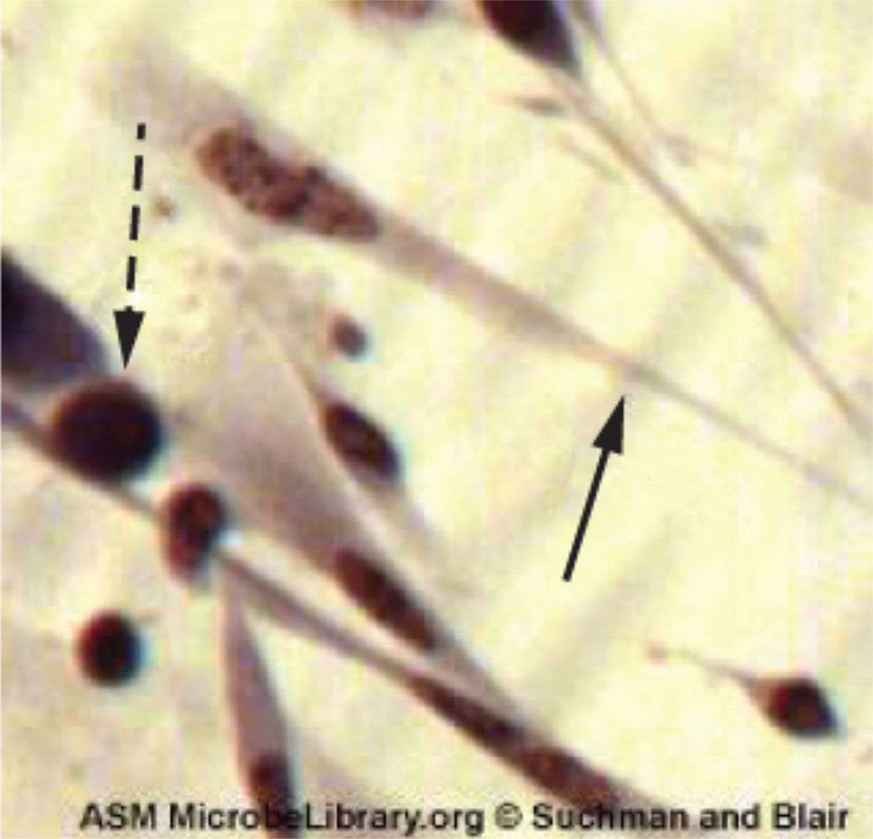
|
| Adenovirus | Cell enlargement, rounding, and distinctive grape-like clusters |
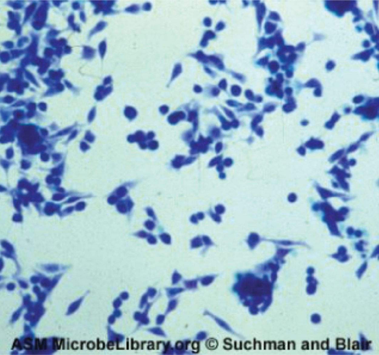
|
A serological assay is a test that detects something, such as the presence of a virus, in body fluids. The term serum specifically refers to blood plasma from which clotting factors have been removed.
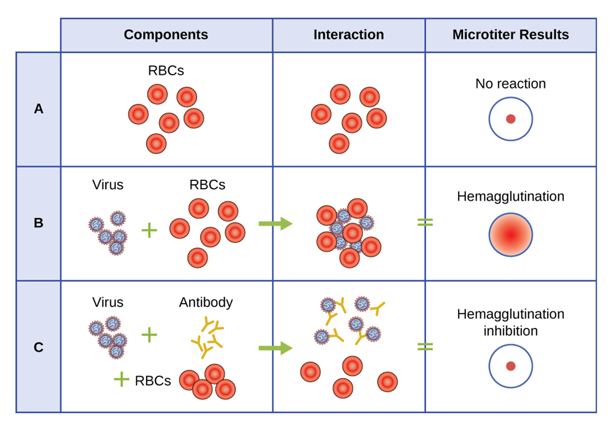
One type of direct serological test is a hemagglutination assay, which measures the clumping together of erythrocytes (red blood cells, abbreviated as RBCs). Agglutination is a general term that describes clumping.
Many viruses produce surface proteins or spikes called hemagglutinins that can bind to receptors on the membranes of erythrocytes. When a virus binds to receptors on more than one erythrocyte, it holds them together and begins to form a clump. These clumps can be observed without using a microscope.
A variety of viruses are capable of hemagglutination, so this test only determines the presence or absence of these types of viruses. It does not determine the identity of a specific pathogen.
Indirect tests can be used to identify specific pathogens. For simplicity, this explanation of indirect tests will focus on mammals and on human testing. When exposed to a pathogen, the human immune system produces antibodies that recognize specific structures (antigens) on the pathogen. You will learn more about antibodies and antigens in other lessons.
It is possible to develop antibodies that bind to components of specific hemagglutinins of a virus of interest. When the virus is exposed to both erythrocytes and these antibodies, the antibodies bind to the hemagglutinins and prevent the virus from binding to erythrocytes. Therefore, agglutination does not occur.
The table below shows how this type of hemagglutination inhibition assay (HAI) can be used to determine whether a specific virus of interest is present. If a virus interacts with erythrocytes (RBCs) to produce hemagglutination but no longer produces hemagglutination once the antibody is added, then this is evidence of the presence of the virus of interest.
Nucleic acid amplification tests (NAATs) are used to make many copies of nucleic acids and then identify specific sequences of interest (e.g., sequences associated with a pathogen). These tests have a wide variety of uses besides pathogen identification.
Polymerase chain reaction (PCR) is a NAAT that can be used to detect viral DNA in a patient’s tissue or body fluid sample. Reverse transcriptase-PCR (RT-PCR) can be used to detect the presence of RNA viruses. RT-PCR is similar to PCR except that DNA is produced using reverse transcriptase to copy RNA to form cDNA.
NAATs can detect very tiny amounts of genetic material, making them sensitive tests for the presence or absence of viruses. You will learn more about NAATs in other lessons.
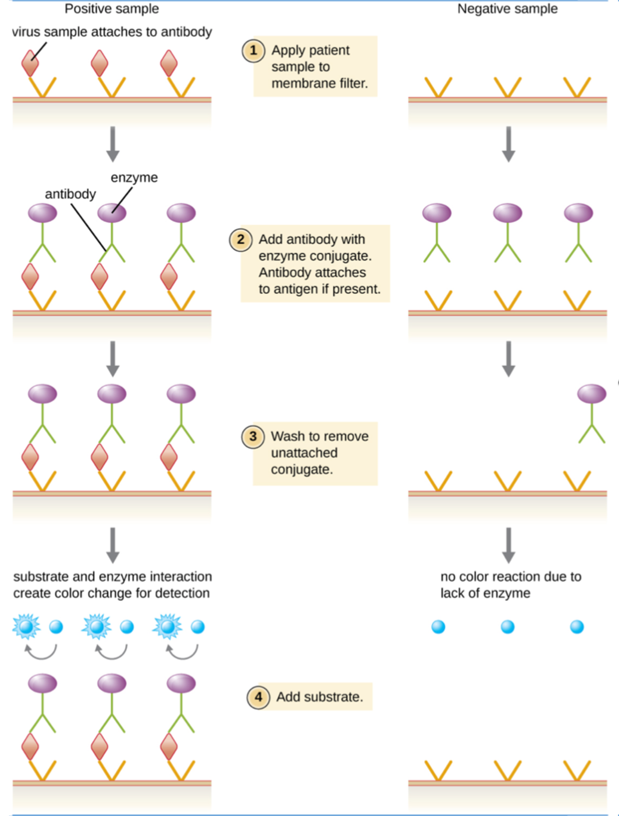
Enzyme immunoassays (EIAs) rely on the ability of antibodies to bind to specific antigens. The detecting antibody attaches to the target antigen with a high degree of specificity.
This type of assay also uses a colorless enzyme attached to the detecting antibody. The enzyme acts as a tag that can interact with a colorless substrate, leading to the production of a colored product. This makes it possible to rapidly detect the presence or absence of the antigen based on whether a color change is observed.
EIAs are often used as preliminary screening tests that can be followed by additional tests for confirmation, such as NAATs. A common type of EIA is called enzyme-linked immunosorbent assay (ELISA).
The steps and image below summarize the basic procedure used in EIAs. However, you will learn more about these tests in other lessons.
Source: THIS TUTORIAL HAS BEEN ADAPTED FROM OPENSTAX "MICROBIOLOGY." ACCESS FOR FREE AT openstax.org/details/books/microbiology. LICENSE: CC ATTRIBUTION 4.0 INTERNATIONAL. Accessed by August 2022.
REFERENCES
Cramer, M. (2021). Henrietta Lacks, whose cells were taken without her consent, is honored by W.H.O. New York Times. Retrieved September 16, 2022, from
www.nytimes.com/2021/10/13/science/henrietta-lacks-cells-who.html
National Public Radio (NPR). (2021). Henrietta Lacks’ estate sued a company saying it used her “stolen” cells for research.
www.npr.org/2021/10/04/1043219867/henrietta-lacks-estate-sued-stolen-cells
Parker, N., Schneegurt, M., Thi Tu, A.-H., Lister, P., & Forster, B. (2016). Microbiology. OpenStax. Access for free at
openstax.org/books/microbiology/pages/1-introduction