Table of Contents |
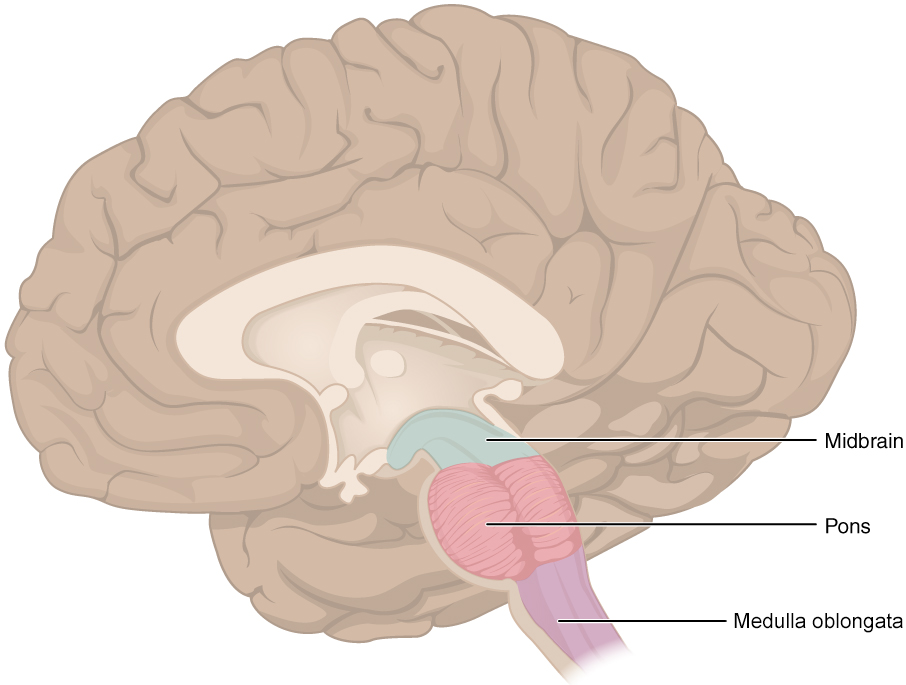
The superior end of the spinal cord connects to the inferior portion of the brain called the brainstem. This region is subdivided into three regions, the medulla oblongata, pons, and midbrain. Collectively, the brainstem provides a pathway for the transport of sensory and motor signals to higher or lower structures and maintains homeostasis, including the cardiovascular and respiratory systems and rates.

The medulla oblongata, commonly referred to as the medulla, is the inferior portion of the brainstem. This region is composed of significant amounts of white matter which is continuous with the white matter of the spinal cord. The medulla serves as a multi-lane information highway with all sensory information traveling to the brain and all motor information leaving the brain traveling through the medulla. This also means that damage to the medulla can severely impair sensory or motor function. A diffuse region of gray matter throughout the brainstem, known as the reticular formation, is related to sleep and wakefulness, such as general brain activity and attention. Additional centers in the medulla regulate homeostatic mechanisms including heart rate and strength as well as breathing rate and depth.
The pons is the middle portion of the brainstem which has a bulbous anterior portion. The word pons comes from the Latin word for bridge. It is visible on the anterior surface of the brainstem as the thick bundle of white matter attached to the cerebellum. The pons is the main connection between the cerebellum and the brainstem. In terms of sensory and motor signals, the pons acts like a point where multiple highways intersect. Like the medulla, the pons allows sensory and motor information to travel through to or from the brain, but it also redirects that information to the cerebellum which sits posteriorly. Additionally, the pons contains centers that regulate those in the medulla.
The midbrain is the superior portion of the brainstem, just below the diencephalon (you will learn about the diencephalon in the next lesson). It is separated into the tectum and tegmentum, from the Latin words for roof and floor, respectively. A canal for cerebrospinal fluid referred to as the cerebral aqueduct passes through the center of the midbrain, such that these regions are the roof and floor of that canal.
The tectum contains four bumps collectively known as the corpora quadrigemina, or four paired bodies (quad, four; geminus, paired; corpora, body). Each bump is referred to as a colliculus (plural, colliculi) which means “little hill” in Latin. The colliculi come in inferior and superior pairs, both process sensory information and both control involuntary reflex movements. The inferior colliculus is the inferior pair of these bumps and is part of the auditory brainstem pathway. The primary function of these structures is to process auditory sensation (sound).
EXAMPLE
If you hear a noise such as a loud bang or someone calling your name, the inferior colliculus receives that signal and controls the movement of the head and neck in order to turn towards the noise reflexively. This response helps you to identify noises quickly.The superior colliculus is the superior pair of bumps. Similar to its inferior counterpart, the superior colliculus controls involuntary reflexive movements of the head, neck, and eyes in order to respond to visual stimuli.
EXAMPLE
If you see something move in your peripheral vision, you may have the reflex to turn towards it while turning on a blinding light that closes the eyes and turns the head away.Additionally, it also combines sensory information about visual space, auditory space, and somatosensory space.
EXAMPLE
If you are walking along the sidewalk on campus and you hear chirping, the superior colliculus coordinates that information with your awareness of the visual location of the tree right above you. That is the correlation between auditory and visual maps. If you suddenly feel something wet fall on your head, your superior colliculus integrates that with the auditory and visual maps and you know that the chirping bird just relieved itself on you. You want to look up to see the culprit but do not.The tegmentum is continuous with the gray matter of the rest of the brainstem. Throughout the midbrain, pons, and medulla, the tegmentum contains the nuclei that receive and send information through the cranial nerves, as well as regions that regulate important functions such as those of the cardiovascular and respiratory systems.
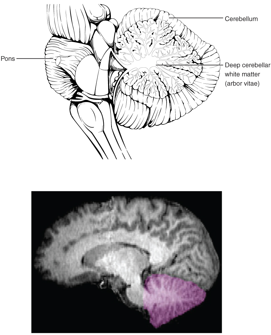
The cerebellum (little brain) is located posterior to the pons of the brainstem and accounts for ~10% of the mass of the brain. The surface of the cerebellum, like the larger cerebrum, is covered in nervous tissue folds called gyri (singular, gyrus) and shallow grooves called sulci (singular, sulcus). As previously mentioned, the cerebellum receives input from the pons which redirects ingoing sensory or outgoing motor signals to or from the cerebrum. The cerebellum is largely responsible for comparing information from the cerebrum with sensory feedback from the periphery through the spinal cord. The various regions of the cerebellum are connected to one another and the brainstem through the white matter tracts collectively known as the arbor vitae (arbor, tree; vitae, life).

The cerebellum receives descending motor signals from the cerebrum and ascending sensory signals from the spinal cord. The cerebellum’s job is to compare motor commands to current sensory information and, when the motor command is inadequate, send out a corrective command to compensate.
EXAMPLE
If walking is not coordinated, perhaps because the ground is uneven or a strong wind is blowing, then the cerebellum sends out a corrective command to compensate for the difference between the original command and the sensory feedback.Source: THIS CONTENT HAS BEEN ADAPTED FROM OPENSTAX "ANATOMY AND PHYSIOLOGY 2E" AT openstax.org/details/books/anatomy-and-physiology-2e