Table of Contents |
Cells in the body replace themselves over the lifetime of a person, but what triggers a cell to divide, and how does it prepare for and complete cell division?
EXAMPLE
The cells lining the inside of the mouth must be frequently replaced when constantly “worn off” by the movement, chewing, and swallowing of food.The cell cycle is the sequence of events from the moment a cell is created until it finishes dividing itself, generating two new cells called daughter cells.
Although there are a few cells in the body that do not undergo cell division (such as gametes—eggs and sperm, red blood cells, most neurons, and some muscle cells), many cells divide regularly. However, before we can discuss the events and phases of cell division, it is important to know what cells undergo this process and more about the DNA involved.
The cells of the body can be divided into two categories, somatic and germ. A somatic cell is a general term for a body cell such as a skin cell, liver cell, neuron, muscle cell, and many more. A germ cell, also referred to as a sex cell, is one that produces or is an egg or sperm cell. Most somatic cells undergo one version of cellular division, mitosis, whereas germ cells undergo a different version of cellular division, meiosis, which will be discussed later in this course.
Human somatic cells contain 23 pairs of chromosomes. Homologous chromosomes are a pair of a single chromosome (one copy from each parent). Because there are two of each individual chromosome, humans are diploid (di, two) organisms, and somatic cells are diploid cells.
One “turn” or cycle of the cell cycle consists of two general phases: interphase and mitosis. Interphase is the period of the cell cycle during which the cell is not dividing. The majority of cells are in interphase most of the time. Mitosis is the division of genetic material, during which the cell nucleus breaks down and two new, fully functional nuclei are formed. Cytokinesis is when the cytoplasm divides into two distinctive cells called daughter cells and occurs during the later portion of the mitotic phase.
Interphase is the period when a cell is not dividing. During this phase, a cell is doing one of two things—performing its specific cellular function(s) or preparing to divide. The time a cell spends in interphase is split into three or four phases, G₀, G₁, S, and G₂, based on what is occurring.
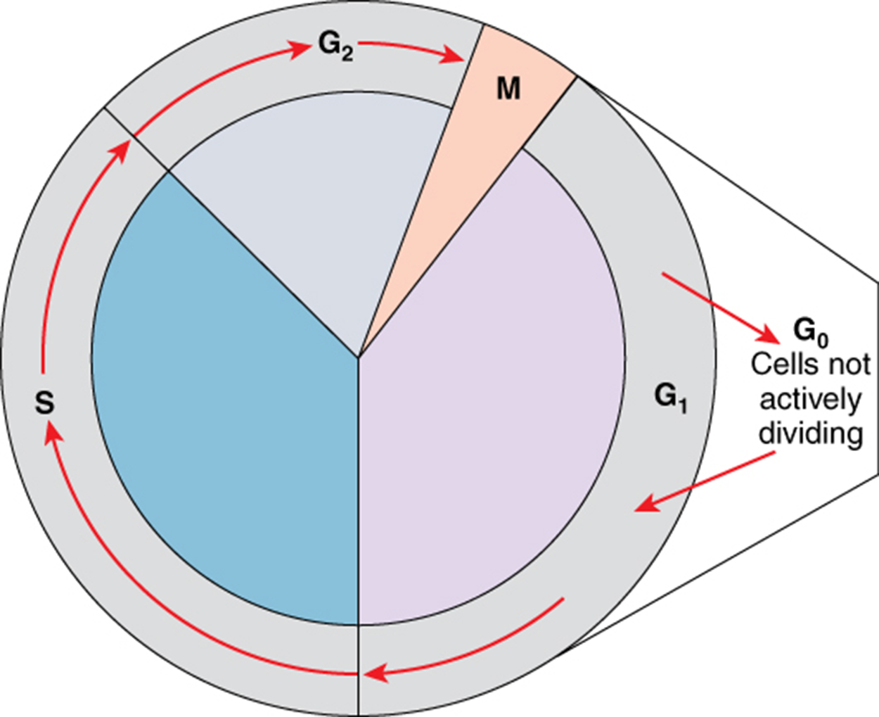
A new cell, once produced, begins to carry out its normal metabolic functions. This is called the G₁ phase (gap 1 phase) and is a growth phase during which the cell builds proteins, makes ATP, and grows in size in early preparation for future divisions. Cells that have temporarily or permanently stopped dividing will continue to perform their metabolic function for extended periods of time without continuing on. This is called the G₀ phase (gap 0 phase). Cells who restart dividing later on will reenter the G₁ phase.
Cells that will be dividing again right away will then enter the S phase (synthesis), in which the cell synthesizes a second set of DNA through DNA replication, which you will learn more about in a future lesson. After the synthesis phase, the cell proceeds through the G₂ phase. In the G₂ phase (gap 2 phase), the cell continues to grow, stores up energy, and makes the necessary preparations for mitosis. Between G₁, S, and G₂ phases, cells will vary the most in their duration of the G₁ phase. It is here that a cell might spend a couple of hours or many days. The S phase typically lasts between 8–10 hours, and the G₂ phase is approximately 5 hours.
Billions of cells in the human body divide every day. During the S phase of interphase, the cell will replicate all DNA, doubling it. Recall that all somatic cells contain 46 chromosomes, 23 from each parent. Each chromosome, whether before or after replication, contains compact DNA connected in the middle by a structure called a centromere. The number of centromeres determines the number of chromosomes present, regardless of how much DNA is attached to them.
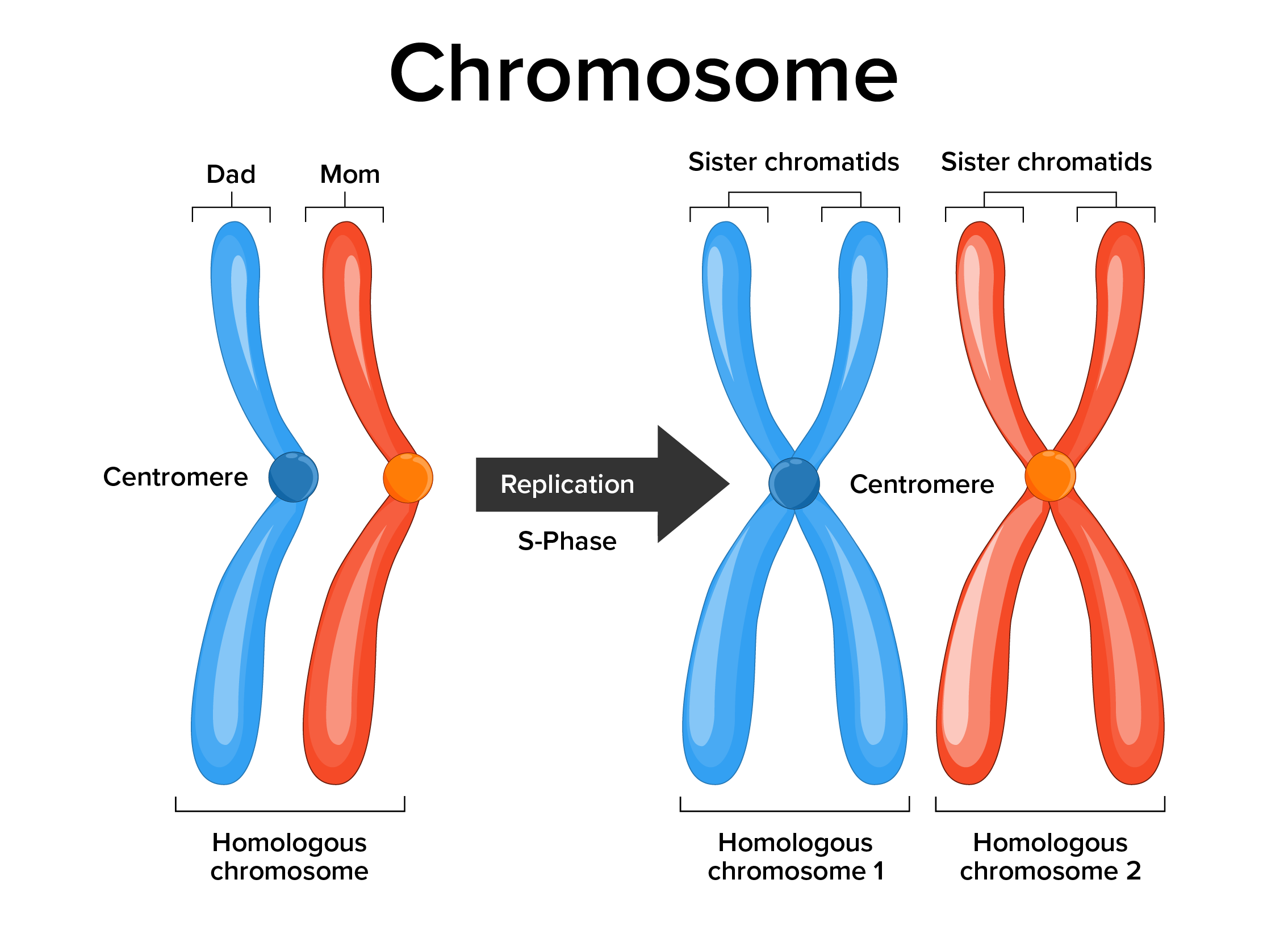
Before the S phase, each chromosome (called an unduplicated chromosome) contains DNA in two arms that extend out from the centromere. After the S phase, each chromosome (called a duplicated chromosome) contains two sets of identical arms, now called sister chromatids, connected to the centromere. Duplicated chromosomes look like the letter “X.” At this point, the cell contains 46 chromosomes with 92 chromatids (46 × 2).
Once the cell has replicated its DNA and prepared the cell, the mitotic phase can begin. The mitotic phase, which typically takes between 1 and 2 hours, undergoes two major processes.
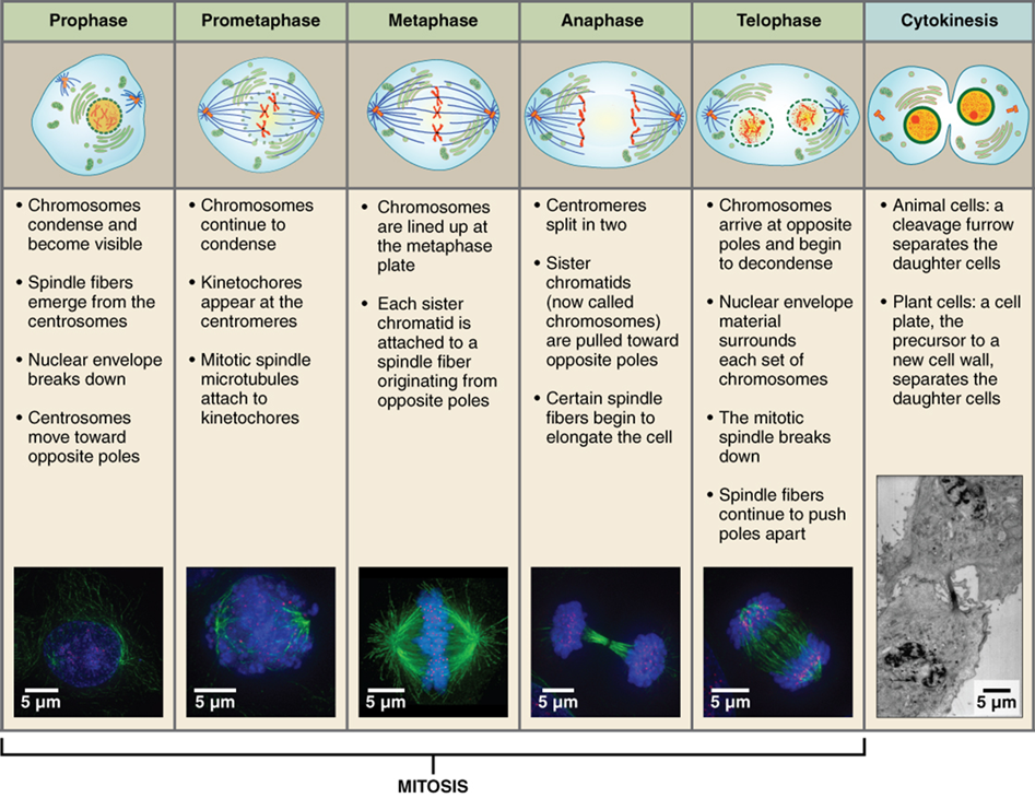
Prophase is the first phase of mitosis, during which the loosely packed chromatin coils and condenses into visible chromosomes. The nucleolus disappears early during this phase, and the nuclear envelope also disintegrates.
A major occurrence during prophase concerns a very important structure that contains the origin site for microtubule growth. A cellular structure called the centrosome (a pair of centrioles) serves as the origin point for microtubules. The cell contains two centrosomes side by side, which begin to move apart during prophase. As the centrosomes migrate to two different sides of the cell, microtubules begin to extend from each like long fingers from two hands extending toward each other. The mitotic spindle is the structure composed of the centrosomes and their emerging microtubules.
Near the end of prophase, the nuclear envelope has disintegrated, and the microtubules from the mitotic spindle invade to attach themselves to the centromeres of each chromosome. The kinetochore is a protein structure on the centromere that is the point of attachment between the mitotic spindle and the sister chromatids. This stage is referred to as late prophase or “prometaphase” to indicate the transition between prophase and metaphase.
Metaphase is the second stage of mitosis. During this stage, the sister chromatids, with their attached microtubules, line up along a linear plane in the middle of the cell. A metaphase plate forms between the centrosomes that are now located at either end of the cell. The metaphase plate is the name for the plane through the center of the spindle on which the sister chromatids are positioned. The microtubules are now poised to pull apart the sister chromatids and bring one from each pair to each side of the cell.
Anaphase is the third stage of mitosis. Anaphase takes place over a few minutes, when the pairs of sister chromatids are separated from one another, forming individual unduplicated chromosomes once again. These chromosomes are pulled to opposite ends of the cell by their kinetochores, which are protein structures on the centromere that are the point of attachment between the mitotic spindle and the sister chromatids, as the microtubules shorten.
Telophase is the final stage of mitosis. Telophase is characterized by the formation of two new nuclei at either end of the dividing cell. These newly formed nuclei surround the genetic material, which uncoils such that the chromosomes return to loosely packed chromatin. Nucleoli also reappear within the new nuclei, and the mitotic spindle breaks apart, each new cell receiving its own complement of DNA, organelles, membranes, and centrioles. At this point, the cell is already beginning to split in half as cytokinesis begins.
The cleavage furrow is a contractile band made up of microfilaments that forms around the midline of the cell during cytokinesis. This contractile band squeezes the two cells apart until they finally separate. Two new cells, called daughter cells, are now formed.
To understand the concept of how the cell cycle is regulated, let’s consider traffic in a city or town.
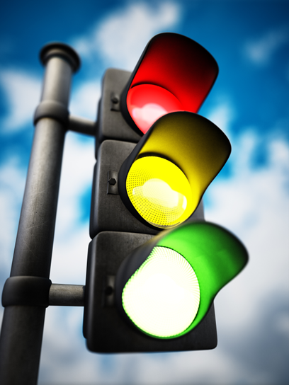
While traveling in, around, or through a town, traffic is not allowed to continue forward at all times. Sometimes it needs to stop. Stopped traffic also eventually needs to move. One of the most common forms of traffic regulation is the traffic light, a series of three lights—green, yellow, and red—which indicate when traffic should go, slow down, and stop. These lights are highly coordinated based on known traffic patterns in order to optimize traffic flow and decrease the risk of dangerous situations.
If you have ever been to a busy intersection when the power goes out (and the traffic lights are out), the risk of a dangerous situation increases—cars not stopping or moving in a coordinated fashion could lead to an accident.
The cell cycle is regulated in a similar way. There are certain locations in the cycle where the cycle is stopped by specific signals. To move on, the cell must receive a specific signal to move again. The coordination of these signals allows the many events in the cell cycle to be coordinated and avoid potentially dangerous situations such as cancer.
As the cell proceeds through its cycle, each phase involves certain processes that must be completed before the cell should advance to the next phase. A checkpoint, like an intersection with a traffic light, is a point in the cell cycle at which the cycle can be signaled to move forward or stopped, and various cell cycle control molecules work together to determine if the cell will progress past each checkpoint.
Many checkpoints exist in the cell cycle. Four of the major checkpoints are in the G₁ phase, S phase, G₂ phase, and metaphase. At the G₁ checkpoint, the cell must have grown in size and have the proper signal to continue onto DNA synthesis before moving forward. Movement beyond this checkpoint means a cell is committed to a full cycle of cell division. At the S phase checkpoint, DNA must be undamaged, and DNA replication must not make any mistakes. At the G₂ checkpoint, the cell must have doubled its size since entering G₁ before moving forward into mitosis. At the metaphase checkpoint, the cell needs to have all sister chromatids properly aligned and attached to microtubules before moving on.
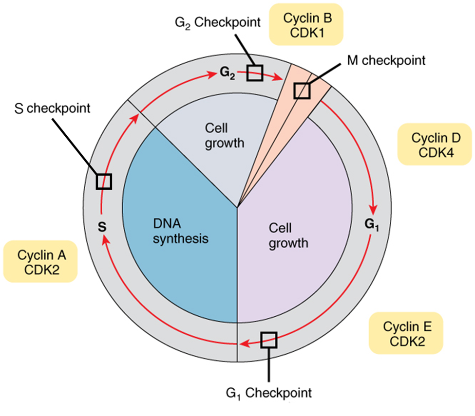
If the cell cycle is unregulated—a lack of stop signal and oversupply of go signal—then the cell is unable to stop at required checkpoints to make sure it is prepared for the next phase. Cells without cell cycle regulation move forward through the cell cycle much like a car with the gas pedal stuck to the floor and no brake pedal. In this case, abnormal cells continue forward despite DNA damage, errors in DNA replication, or incorrect cell size. The primary risk to unregulated cell cycle progression is a tumor or cancer.
If the abnormal cells continue to divide unstopped, they can damage the tissues around them, spread to other parts of the body, and eventually result in death. Failures of control may be caused by inherited genetic abnormalities and/or environmental conditions which damage DNA, leading to altered or absent “stop” and “go” signals.
Uncontrolled cell division creates an excess of cells with no bodily function that is called a tumor. If the tumor remains within the boundary of the original tissue and does not pose a threat to outside tissues, it is considered benign. Benign tumors can typically be removed if not located near other critical body structures (i.e., parts of the brain). If a tumor invades surrounding tissue, it is considered malignant (mal, bad). A malignant tumor poses a threat to other tissues and is therefore diagnosed as a cancer. Any cancerous growth that has entered and traveled by the blood to a secondary location is considered metastatic.
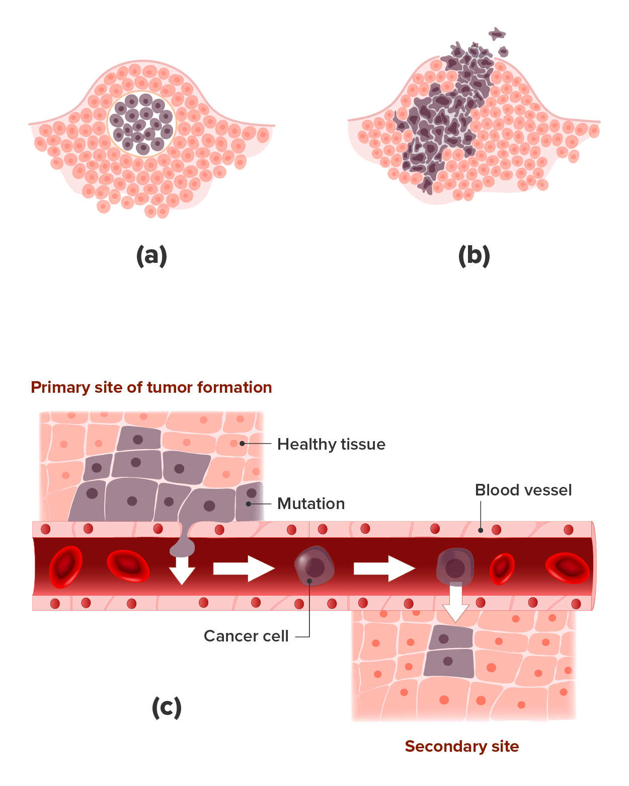
The process of a cell escaping its normal control system and becoming cancerous may actually happen throughout the body quite frequently. Fortunately, certain cells of the immune system are capable of recognizing cells that have become cancerous and destroying them. However, in certain cases, the cancerous cells remain undetected and continue to proliferate.
SOURCE: THIS TUTORIAL HAS BEEN ADAPTED FROM (1) OPENSTAX “BIOLOGY 2E”. ACCESS FOR FREE AT OPENSTAX.ORG/BOOKS/BIOLOGY-2E/PAGES/1-INTRODUCTION (2) OPENSTAX “ANATOMY AND PHYSIOLOGY 2E”. ACCESS FOR FREE AT OPENSTAX.ORG/BOOKS/ANATOMY-AND-PHYSIOLOGY-2E/PAGES/1-INTRODUCTION. LICENSING (1 & 2): CREATIVE COMMONS ATTRIBUTION 4.0 INTERNATIONAL.