Table of Contents |
As explained by the cell theory, cells arise from other cells. Cell division is the process by which this happens.
Prokaryotic cells have cell cycles during which they prepare for and then undergo cell division. Prior to cell division, they grow and increase the number of cell components. Most prokaryotic cells actually divide using a process you were introduced to in the lesson on spontaneous generation, called binary fission. In this process, their single circular chromosome is replicated and then the cell grows apart before forming new outer layers (including cell membranes and cell walls) between the two halves.
“Binary” means two and “fission” means splitting in half, so binary fission is a very general term for division into two parts that can describe more than one type of division. In this course, the term refers to prokaryotic cell division unless otherwise specified.
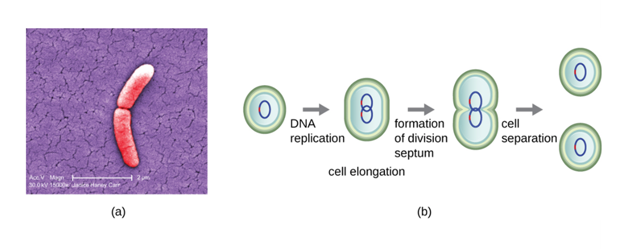
The image below shows a micrograph of a rod-shaped bacterial cell undergoing binary fission (a) and the steps of binary fission (b). It shows how DNA replication is followed by cell elongation, formation of the division septum, and separation of one cell into two complete daughter cells.
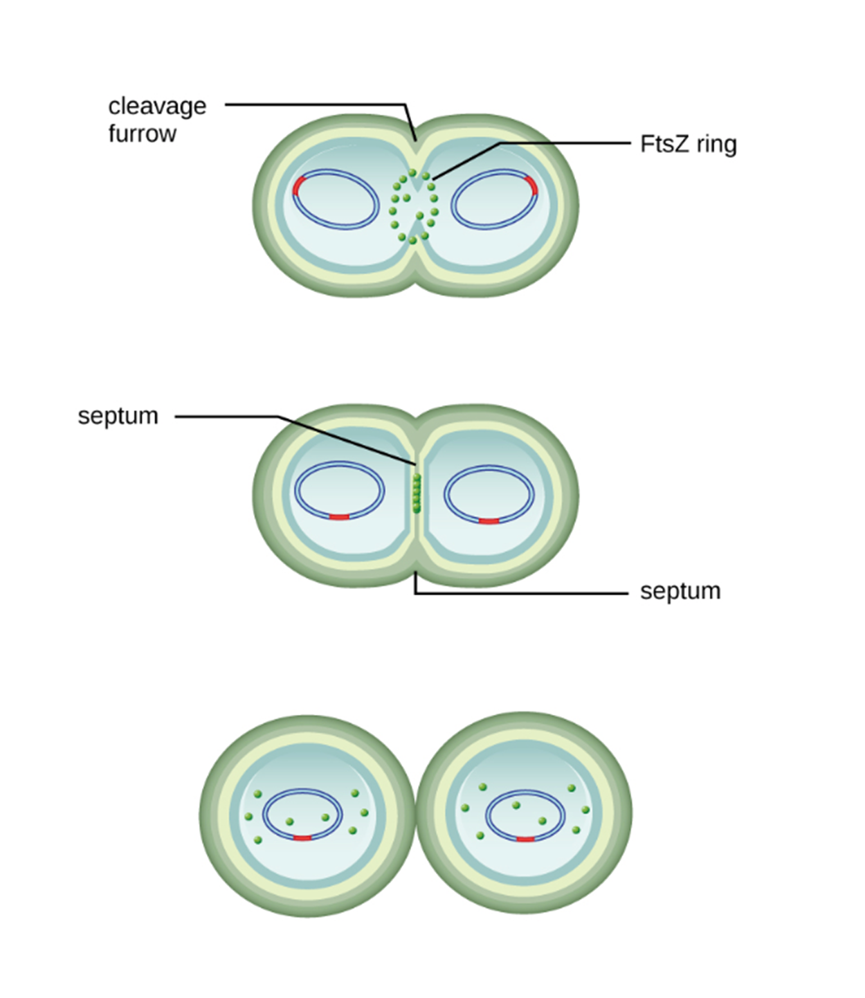
The image below shows how the cleavage furrow is formed by a ring of FtsZ that forms a septum that divides the single cell into two cells.
Division of eukaryotic cells involves some complexities not experienced by prokaryotic cells. In eukaryotic cells, DNA is enclosed in a nucleus and cannot move freely until the nucleus has broken down. Additionally, eukaryotic cells have multiple chromosomes whereas prokaryotic cells usually have a single circular chromosome. This means that the chromosomes must be carefully separated so that each daughter cell has a copy of each necessary chromosome. Eukaryotic cells have specialized processes of cell division that address these challenges.
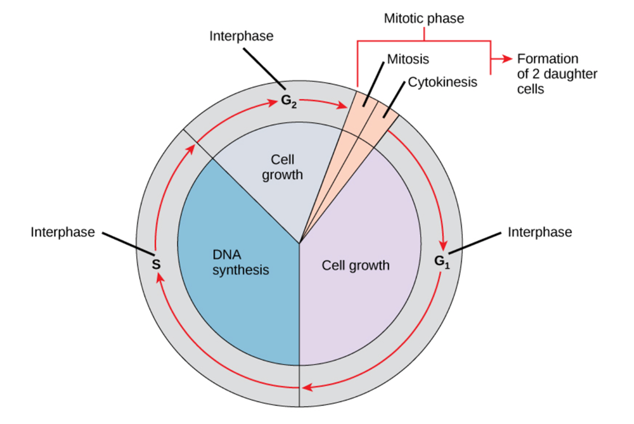
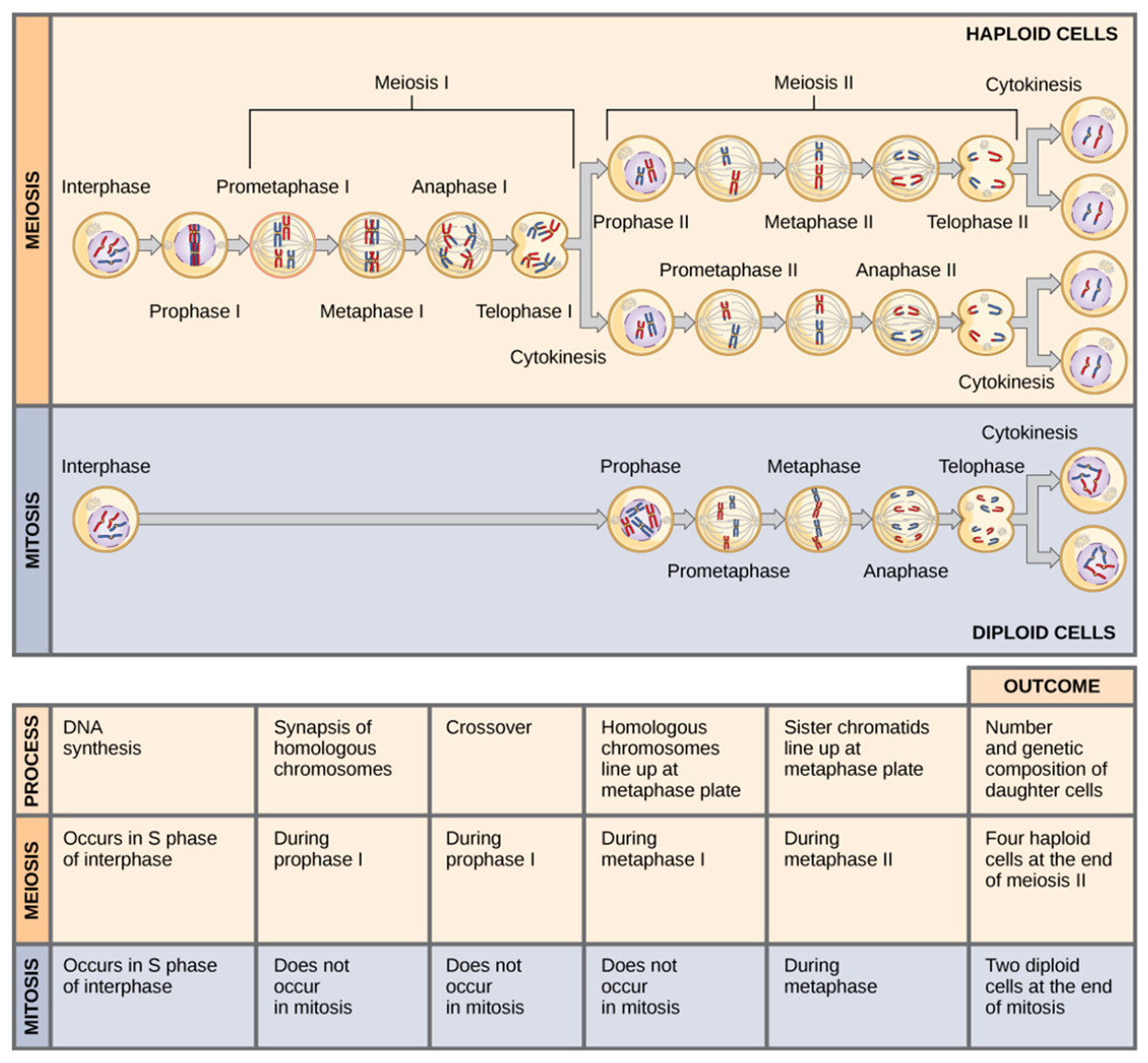
The eukaryotic cell cycle describes the stages of the process by which a cell prepares to divide. As shown in the image below, most of the cell cycle is interphase. This is the period of time in which the cell is not dividing. During interphase, the cell begins in the G1 phase. This is the time during which the cell is growing, performing its normal functions, and preparing itself for subsequent stages. During the next phase, the S phase, the cell copies its DNA in preparation for cell division. Next, the cell enters the G2 phase and continues to grow and replicate organelles in preparation for division. After G2, the cell enters the mitotic phase. During this phase, the cell divides to produce daughter cells. The first part of this phase is mitosis, during which karyokinesis (division of the nucleus into two nuclei) occurs. Next, cytokinesis (the splitting of one cell into two) occurs. After cytokinesis, the newly produced daughter cells enter the G1 phase.
Although the term mitosis is generally used to refer to eukaryotic cell division that produces two identical daughter cells through a specific series of steps, it most specifically refers to karyokinesis. It is possible for mitosis to occur without cytokinesis to produce a multinucleate cell.
This cycle is carefully regulated so that the cell does not divide when it is not ready or when division is inappropriate. When regulation fails, the cell divides at inappropriate times. This leads to cancer.
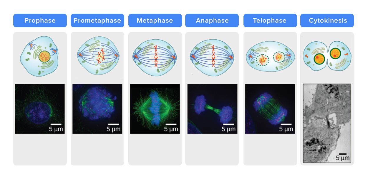
Mitosis involves a series of steps that divide one cell into two identical daughter cells. Most often, a cell with two sets of chromosomes (diploid) divides to produce two identical diploid daughter cells. However, mitosis can also produce identical daughter cells from parent cells with different numbers of sets of chromosomes.
| The Stages of Mitosis | |
|---|---|
| Prophase | Chromosomes condense and become clearly visible. The nuclear envelope breaks down, freeing the chromosomes to move throughout the cell. The nucleolus becomes invisible. Additionally, cytoskeletal spindle fibers emerge to assemble a spindle apparatus that moves chromosomes as needed. |
| Prometaphase | The chromosomes continue to condense. The mitotic spindle microtubules (a type of cytoskeletal element discussed later in this lesson) attach to structures called kinetochores on the chromosomes. In animal cells, structures called centrosomes anchor the spindle fibers and begin to move toward opposite poles of the cell. In other cells, the spindle fibers are anchored differently but the process is similar. |
| Metaphase | The mitotic spindle is fully developed and the chromosomes are lined up on an imaginary line across the center of the cell (the metaphase plate). Once each chromosome has been duplicated prior to mitosis, each duplicated chromosome consists of two sister chromatids joined together at a centromere. In metaphase, the two sister chromatids in a pair are each attached to a spindle fiber originating from an opposite pole. |
| Anaphase | Cohesin proteins binding the sister chromatids together break down and the former sister chromatids (now daughter chromosomes) are pulled toward opposite poles by the spindle fibers. Other spindle fibers lengthen to elongate the cell. |
| Telophase | Chromosomes arrive at opposite poles and begin to decondense. Nuclear envelope material surrounds each set of chromosomes, forming new nuclei. Additionally, the mitotic spindle breaks down. |
| Cytokinesis | One cell splits into two. In animal cells, a cleavage furrow forms that pinches the cells apart. This furrow is formed by actin microfilaments (a type of cytoskeletal element), not FtsZ. In plant cells, a cell plate forms that separates the daughter cells and gives rise to a new cell wall. |
Unlike mitosis, which produces identical daughter cells, meiosis produces varied daughter cells with half the number of chromosomes of the parent cell (in other words, they are haploid). This contributes to genetic variation in organisms that reproduce sexually.
As shown in the image below, mitosis and meiosis differ in several important ways.
EXAMPLE
Mitosis has one division and meiosis has two divisions. Mitosis produces two identical daughter cells that are also identical to the parent cell. If the parent cell is diploid, then both daughter cells are diploid. Meiosis produces four different daughter cells with half the number of chromosomes of the parent cell. If the parent cell is diploid, then the daughter cells are haploid.The two divisions of meiosis are called meiosis I and meiosis II. The stages of each division have the same names as the stages of mitosis with I or II added.
Genetic variation is also increased by a process called crossing over that takes place in meiosis but very rarely in mitosis. In sexually reproducing organisms, a diploid individual has received one set of chromosomes from one parent and a second set of chromosomes from the other parent. If there are 46 chromosomes in an organism, then that means that there are 23 sets of two chromosomes each. During prophase I of meiosis, pairs of homologous chromosomes (meaning different chromosomes of the same type, one inherited from each parent) come into physical contact and exchange genetic material. This is called crossing over and produces new genetic combinations.
EXAMPLE
In humans, there are 23 chromosomes in a set. Each haploid egg and sperm cell contains 23 chromosomes. When an egg and sperm fuse, the resulting zygote (fertilized egg) has 46 chromosomes and is diploid.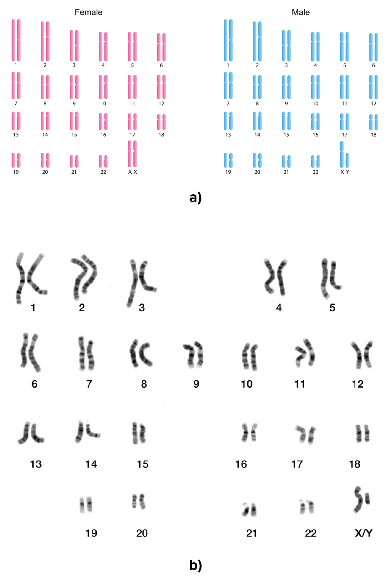
Another important difference between mitosis and meiosis occurs in metaphase I. In mitosis and in metaphase II of meiosis, chromosomes line up on the metaphase plate and then the sister chromatids separate in to move toward opposite sides of the cell. In metaphase I of meiosis, pairs of homologous chromosomes line up on the metaphase plate and then move toward opposite poles.
Source: THIS TUTORIAL HAS BEEN ADAPTED FROM OPENSTAX "MICROBIOLOGY." ACCESS FOR FREE AT openstax.org/details/books/microbiology. LICENSE: CC ATTRIBUTION 4.0 INTERNATIONAL. Accessed by August 2022.
REFERENCES
Karyotype. (2022, October 21). In Wikipedia. https://en.wikipedia.org/wiki/Karyotype
Merriam-Webster. (n.d.). Binary. In Merriam-Webster.com dictionary. Retrieved August 13, 2022, from www.merriam-webster.com/dictionary/binary
Merriam-Webster. (n.d.). Fission. In Merriam-Webster.com dictionary. Retrieved August 13, 2022, from www.merriam-webster.com/dictionary/fission
Parker, N., Schneegurt, M., Thi Tu, A.-H., Lister, P., & Forster, B. (2016). Microbiology. OpenStax. Access for free at openstax.org/books/microbiology/pages/1-introduction
Wikipedia: Karyotype. (2022, October 21). In Wikipedia. https://en.wikipedia.org/wiki/Karyotype