Table of Contents |
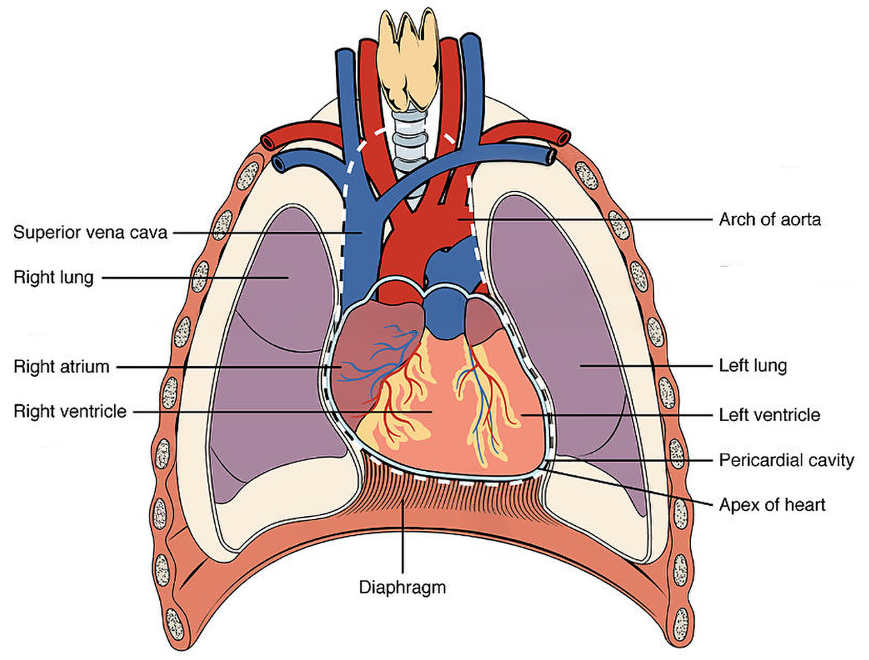
The heart is located within the thoracic cavity, medially between the lungs, and in the space known as the mediastinum. The great veins (the superior and inferior venae cavae) and the great arteries (the aorta and pulmonary artery) are attached to the superior surface of the heart, called the base. The inferior tip of the heart, the apex, lies just to the left of the sternum between the junction of the fourth and fifth ribs. Health care professionals must know the position of the heart when placing a stethoscope to listen to internal body sounds (especially heart and lung sounds), referred to as auscultation.
The heart consists of four chambers: two atria and two ventricles. The right atrium receives deoxygenated blood from the body via the inferior and superior vena cava. Flow of blood from the right atrium to the right ventricle is controlled by an atrioventricular valve (the tricuspid valve). The right ventricle pumps the deoxygenated blood through the pulmonary arteries to the lungs. From the lungs, the newly oxygenated blood travels through the pulmonary veins to the left atrium, which propels the blood through an atrioventricular valve (the bicuspid valve or mitral valve) into the left ventricle. The left ventricle pumps blood to the aorta to travel to the rest of the body. Note that semilunar valves, which are shaped differently from atrioventricular valves, control the blood flow from the heart into the pulmonary artery (the pulmonary valve) and aorta (the aortic valve).
MTA24
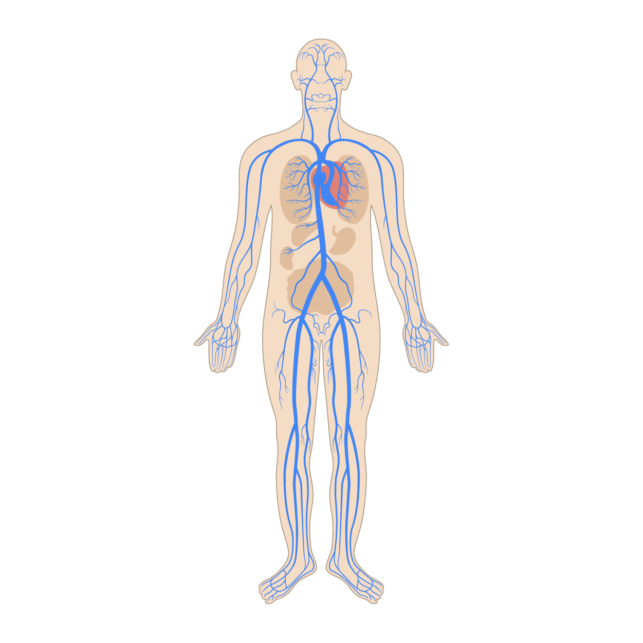
After blood is pumped out of the left ventricle through the aorta, it is carried throughout the body via systemic arteries. An artery is a blood vessel that carries blood away from the heart. Arteries branch into ever-smaller vessels called arterioles and eventually into tiny vessels called capillaries. The figure below shows the systemic arteries that carry oxygenated blood throughout the body to organs and tissues, as indicated by the red color.
![An anterior view of a woman shows complex branching of arteries throughout the body.]](https://cdn.sophia.org/markup_pictures/56158/file/6bb34eaf592f7c5537099a325b8b3258.png)
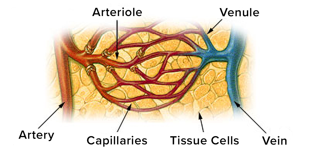
Oxygen and nutrients are exchanged with cells at the capillary level. A capillary is a microscopic channel that supplies blood to the tissue cells. Capillaries connect arterioles and venules, which are small vessels that carry blood toward the heart into larger veins. The figure below shows capillaries supplying blood to tissue cells.
Veins return blood to the heart. Two large veins, the inferior vena cava and superior vena cava, connect to the heart. The figure below shows the systemic veins that carry deoxygenated blood back to the heart, indicated in blue. Medications may be administered by health care professionals into veins, referred to as intravenous (IV) medications.
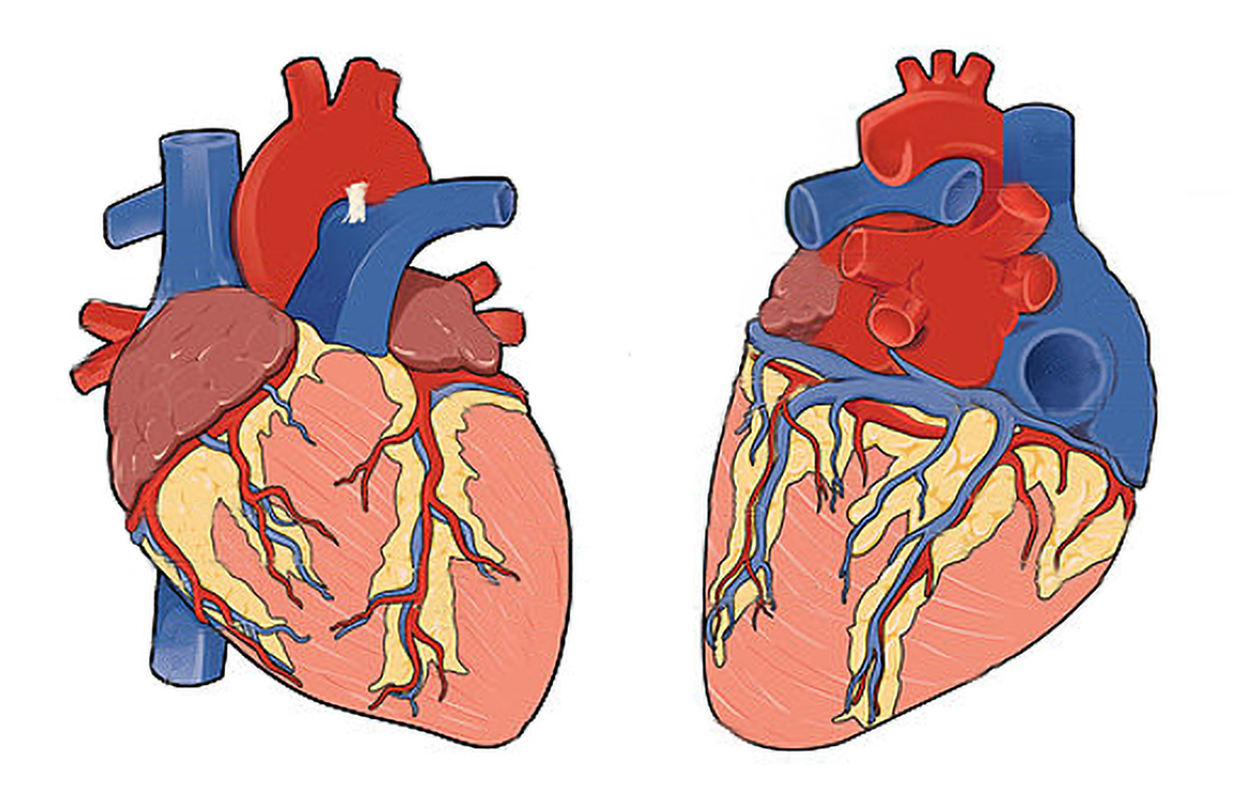
Coronary arteries are arteries that branch off the aorta and carry oxygenated blood to the heart muscle itself. Coronary veins bring deoxygenated blood back to the right atrium. Blood circulates through the coronary arteries and veins with each heartbeat. The figure below shows the coronary arteries and veins. Note how the red arteries and blue veins extend across the myocardium.
IN CONTEXT
Myocardial Infarction
For many people, it’s easy to quickly think of heart attacks when thinking of the cardiovascular system. However, have you ever really thought about what happens when someone has a heart attack?
Technically, a heart attack is called a myocardial infarction (MI). An infarction occurs when tissue dies because it isn’t receiving enough oxygen. A myocardial infarction is an infarction in heart muscle tissue. Damage to the heart muscle can leave the heart unable to pump sufficient blood.
Myocardial infarctions occur when there is a blockage in one or more of the vessels that supply the heart with blood. These blockages can occur when atherosclerotic plaque breaks off and forms a clot (even though these plaques cause narrowing of blood vessels, it’s damage to the plaque that typically causes an MI) (Ojha and Dhamoon, 2023).
If the heart stops beating, that is cardiac arrest. Cardiac arrest can happen with or without a heart attack; it is a distinct medical event. For example, certain medications or even a very hard blow to the chest can cause cardiac arrest (NHLBI, 2022).
There are two distinct, but linked circuits in the human circulation called pulmonary and systemic circuits.
The pulmonary circuit transports oxygenated blood from the right ventricle to the lungs, where carbon dioxide is delivered for exhalation, oxygen is picked up, and this newly oxygenated blood returns to the left atrium of the heart.
The systemic circuit transports oxygenated blood from the left ventricle of the heart, through the aorta, and into the systemic arteries throughout the body where it diffuses into the tissues at the capillaries. Deoxygenated blood travels back to the heart by entering small veins that merge and eventually drain into the superior and inferior vena cava and into the right atrium of the heart. The figure shows blood flow through the pulmonary and systemic circuits.
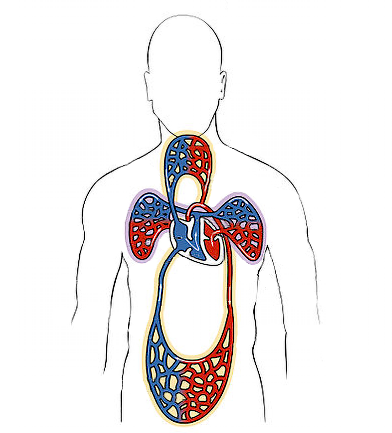
The systemic and pulmonary circuits transport blood and its components for physiological processes that occur throughout the body. The steps below show the path of blood flow through the pulmonary and systemic circuits. Remember that the pulmonary circuit carries blood from the heart to the lungs and back to the heart, and the systemic circuit carries blood from the heart through the entire body and back to the heart.
As blood passes through the body (the systemic circuit), it participates in a variety of activities. Some of the events in the systemic circuit include:
EXAMPLE
Note that the artery has a thicker wall designed to withstand the higher pressure blood that travels through it. Arteries need to be able to expand and bounce back, which becomes more difficult if they stiffen due to plaque buildup. Also note that the lumen is lined by endothelium.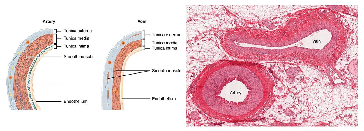
There are important differences in the structures of the walls of arteries, veins, arterioles, venules, and capillaries that allow them to perform their unique functions. Capillaries have very thin walls that typically consist of endothelium and basement membrane, sometimes with a small amount of smooth muscle. This unique structure facilitates the rapid exchange of oxygen, nutrients, and wastes with tissues.
Heart rate (HR) is the number of heart beats per minute (bpm). HR can vary considerably among people based on their age, as well as their fitness level. For an adult, normal resting HR is 60–100 bpm. Although similar to the pulse, the heart rate is auscultated directly using a stethoscope and is a more accurate measurement than the pulse.
Abnormal heart rates are referred to as bradycardia and tachycardia. Bradycardia refers to a slow heart rate, less than 60 bpm in an adult. Tachycardia refers to a fast heart rate, greater than 100 bpm in an adult.
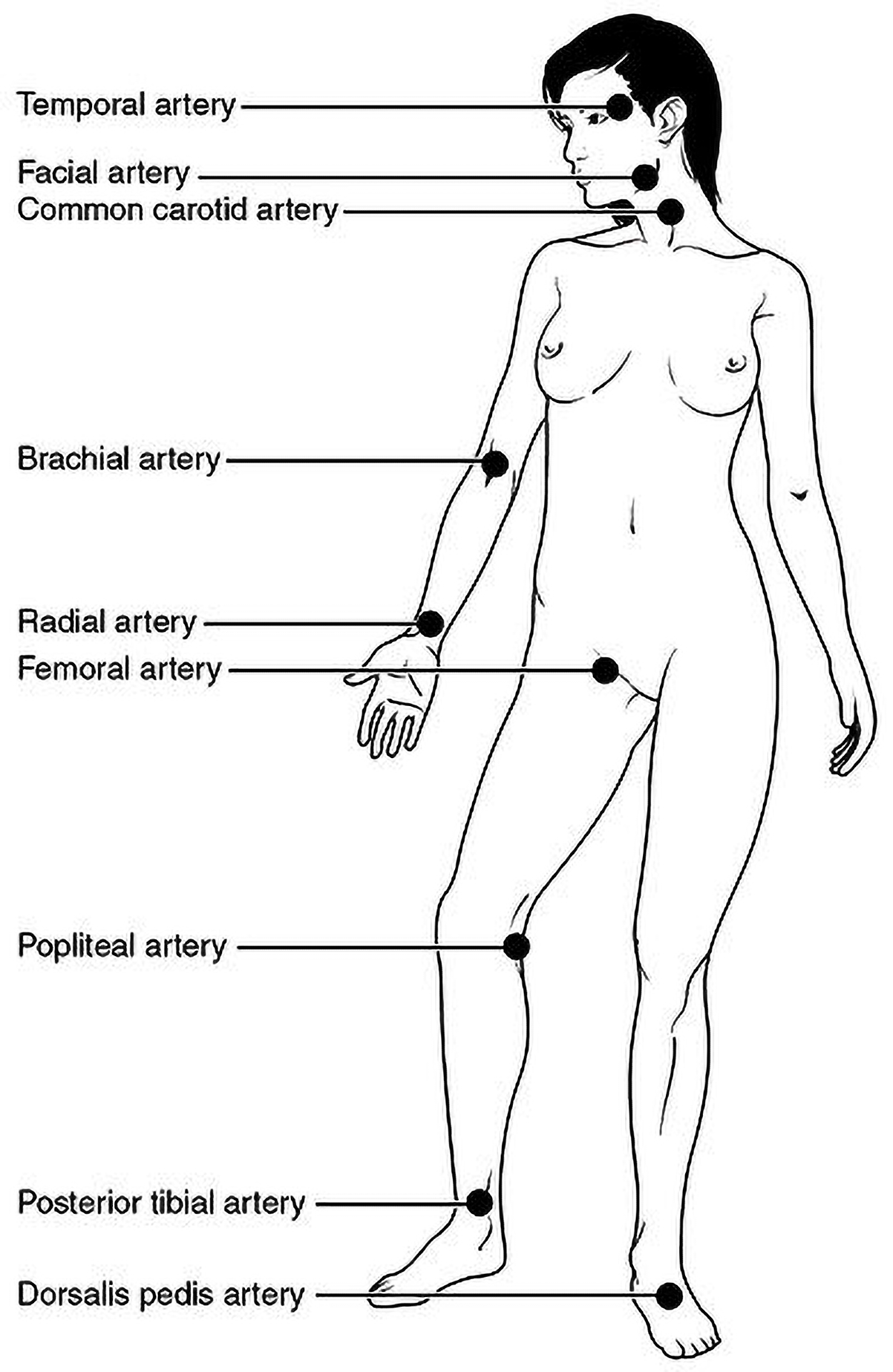
Each time the heart pumps, ejecting blood forcefully into the circulation, the arteries expand and recoil to accommodate the surge of blood moving through them. This expansion and recoiling of the arterial wall are called the pulse and allow for the measurement of heart rate. The pulse can be palpated manually by placing the tips of the fingers across an artery that runs close to the body surface, such as the radial artery (on the thumb side of the wrist) or the common carotid artery (in the neck). These sites and other pulse sites are shown in the figure below.
Both the rate and the strength of the pulse are important clinically. A high pulse rate can be temporarily caused by physical activity, but an extended fast or irregular pulse may indicate a heart condition. The pulse strength indicates the strength of ventricular contraction, cardiac output, and perfusion. Cardiac output is the amount of blood pumped by the heart per minute. Perfusion is the passage of blood through the blood vessels.
If the pulse is strong, then cardiac output is high and perfusion to that site is good. If the pulse is weak, cardiac output is low or perfusion is impaired, and medical intervention may be warranted.
The period of time that begins with contraction of the atria, continues through ventricular contraction, and ends with ventricular relaxation is known as the cardiac cycle. The phase of the cardiac cycle when ventricles contract and eject blood is called systole. The phase of the cardiac cycle where the heart muscles relax, allowing the chambers to fill with blood, is called diastole. When the electrical activity of the heart stops, that is called asystole (Mayo Clinic, 2022).
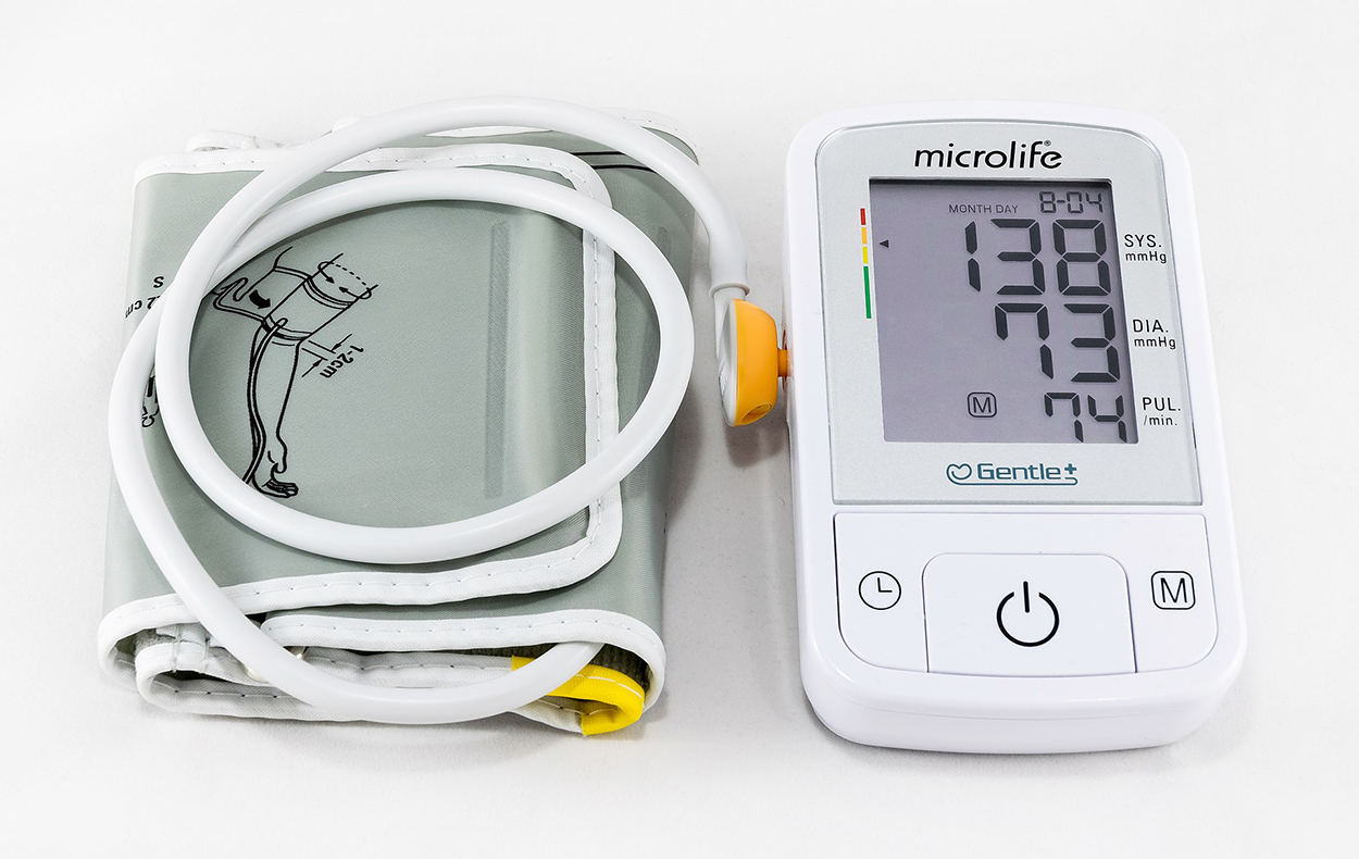
Blood pressure (BP) is the force of the blood against the vessel walls. In clinical practice, blood pressure is typically measured using a sphygmomanometer, commonly called a blood pressure cuff. The cuff is placed over the brachial artery of the patient’s upper arm and inflated with air to temporarily occlude (block) blood flow and measure blood pressure. Blood pressure is recorded as a ratio of two numbers expressed as systolic pressure over diastolic pressure, measured in millimeters of mercury (mm Hg).
EXAMPLE
120/80 mm Hg is a normal adult blood pressure.The systolic pressure is the higher value and reflects the arterial pressure resulting from the ejection of blood during ventricular contraction called systole. The diastolic pressure is the lower value and represents the arterial pressure of blood during ventricular relaxation called diastole.
The picture below shows a modern electronic sphygmomanometer.
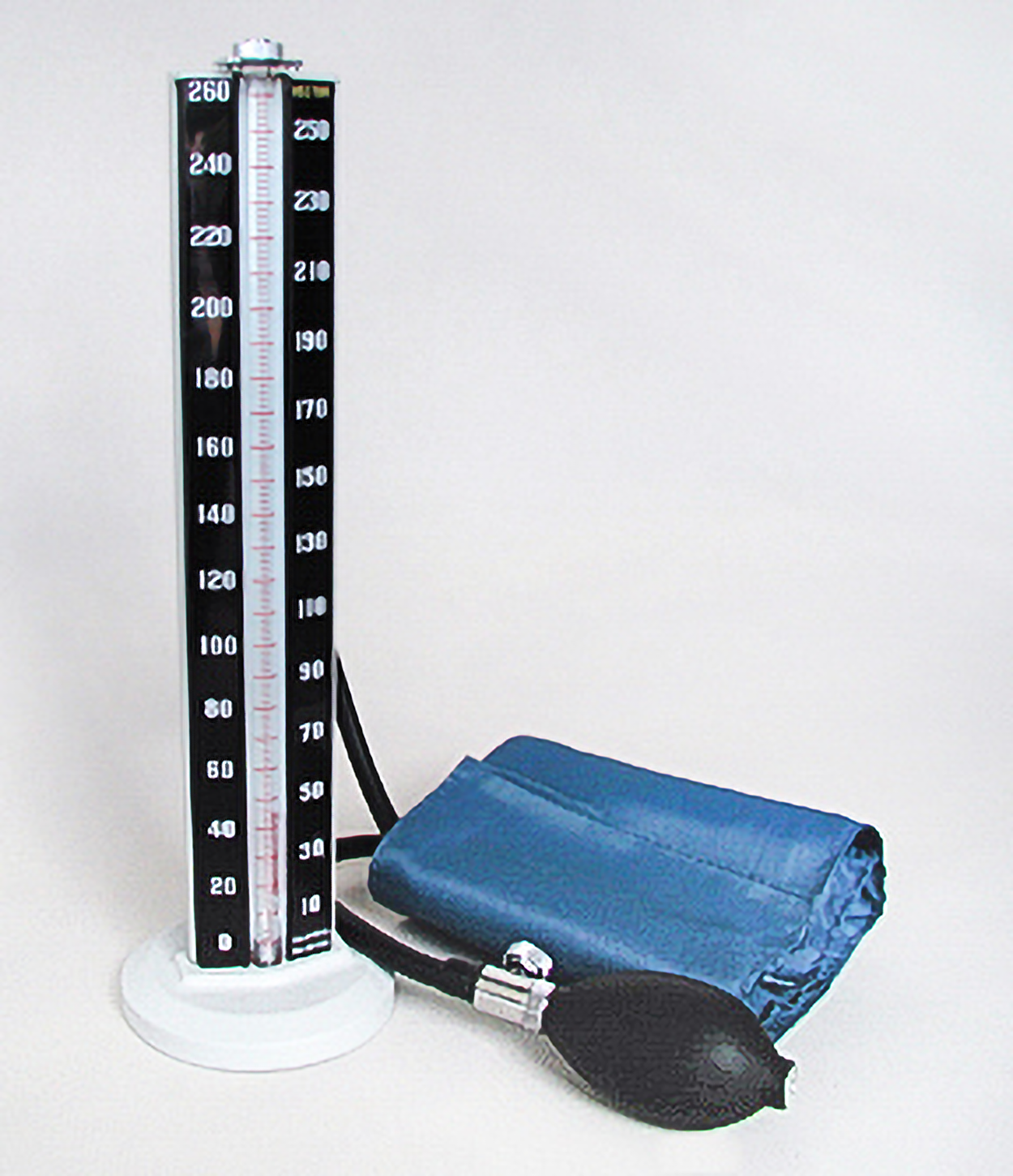
The image below shows a traditional sphygmomanometer. This type of device has a cuff to use around a person’s arm and a bulb to squeeze to increase the pressure. The vertical bar has a column of mercury that rises to measure pressure. That is why the measurement is given in millimeters of mercury (mm Hg). To take a reading, the cuff is inflated beyond the point that the pulse can be heard, then the cuff is deflated more slowly. The point at which the pulse is heard again is the systolic pressure, and the point at which the pulse ceases to be heard is the diastolic pressure.
Low blood pressure, called hypotension, has several causes, such as dehydration, vomiting, diarrhea, or medications used to treat hypertension. Treatment for hypotension typically includes fluid replacement. High blood pressure is called hypertension (HTN). Hypertension has a variety of causes, with risk factors such as obesity, lack of exercise, smoking, high salt diet, diabetes, and kidney disease.
| Term | Definition | Audio |
|---|---|---|
| Systole | The phase of the cardiac cycle when ventricles contract and eject blood | AUDIO |
| Diastole | The phase of the cardiac cycle where the heart muscles relax, allowing the chambers to fill with blood | AUDIO |
| Asystole | All electrical activity in the heart stops | AUDIO |
SOURCE: THIS TUTORIAL HAS BEEN ADAPTED FROM (1) “OPEN RN | MEDICAL TERMINOLOGY – 2E” BY ERNSTMEYER & CHRISTMAN AT OPEN RESOURCES FOR NURSING (OPEN RN). (2) "MEDICAL TERMINOLOGY" BY HOBBS & CASTEEL AT PHOENIX COLLEGE NURSING. ACCESS FOR FREE AT WTCS.PRESSBOOKS.PUB/MEDTERM/ AND OPEN.MARICOPA.EDU/MEDICALTERMINOLOGY/FRONT-MATTER/INTRODUCTION/. LICENSING: CREATIVE COMMONS ATTRIBUTION 4.0 INTERNATIONAL.
REFERENCES
Ojha N, Dhamoon AS. (2023 Aug 8). Myocardial Infarction. Treasure Island (FL): StatPearls Publishing. www.ncbi.nlm.nih.gov/books/NBK537076/.
Cardiac Arrest Causes and Risk Factors. (2022, May 19). National Heart, Lung, and Blood Institute (NHLBI), National Institutes of Health (NIH). Cardiac Arrest - Causes and Risk Factors | NHLBI, NIH. https://www.nhlbi.nih.gov/health/cardiac-arrest/causes .
Angina. (2024, March 24). Mayo Clinic. Angina - Symptoms and causes - Mayo Clinic. https://www.mayoclinic.org/diseases-conditions/angina/symptoms-causes/syc-20369373.
Asystole. (2022, May 3). Cleveland Clinic. Asystole: Causes, Symptoms and Treatment. https://my.clevelandclinic.org/health/symptoms/22920-asystole .