Table of Contents |
Recall that bone (osseous) tissue is a rigid connective tissue that provides protection to internal organs and supports the body. Osseous tissue forms most of the adult skeleton, the support structure of the body. In the areas of the skeleton where bones move (i.e., the ribcage and joints), cartilage, a semi-rigid form of connective tissue, provides flexibility and smooth surfaces for movement. Recall that the skeletal system is the organ system composed of bones and cartilage that performs the following critical functions for the human body:
The table below shows some especially important combining forms related to the skeletal system:
| Term | Definition | Example | Definition of Example |
|---|---|---|---|
| Arthr/o | Pertaining to a joint or joints | Arthropathy | Joint disease |
| Chondr/o | Pertaining to cartilage | Chondrocyte | Cartilage cell |
| Myel/o | Pertaining to bone marrow | Myelofibrosis | A type of bone cancer that results in restructuring of bone |
| Oste/o | Pertaining to bone | Osteogenesis | Bone formation |
Important medical terminology related to bones includes:
Some combining forms relate to particular bones. Examples of these are shown in the table below.
| Term | Bone Referenced |
|---|---|
| Calcane/o | Calcaneus (heel bone) |
| Clavicul/o | Clavicle |
| Ili/o | Ilium (pelvic bone) |
| Ischi/o | Ischium (pelvic bone) |
| Metacarp/o | Metacarpal or metacarpals (hand bones) |
| Metatars/o | Metatarsal or metatarsals (foot bones) |
| Pelv/o | Pelvic bone |
| Phalang/o | One or more bones of the fingers and toes (phalanges) |
| Spondyl/o | Vertebra or vertebrae |
| Vertebr/o | Vertebra or vertebrae |
There are also many general terms that relate to the skeletal system but are often used to describe other systems as well. The terms below are examples of these types of terms.
| Term | Bone referenced |
|---|---|
| Brachi/o | Pertaining to the arm |
| Cephal/o | Pertaining to the head |
| Dactyl/o | Pertaining to the digits (fingers or toes) |
| Pod/o | Pertaining to the foot |
| Pub/o | Pertaining to the pubic area or pubic bones |
For example, pod/o can be used to form the word podiatrist (pod/o = foot + -ist, a suffix used to describe a certain type of practitioner, such as a pharmacist). A podiatrist is a specialist in foot conditions and has a doctorate in podiatric medicine (DPM). A podiatrist treats all sorts of foot conditions, including those involving the skeletal system.
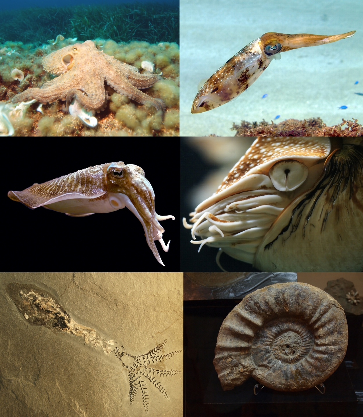
The picture below shows examples of cephalopods. The picture on the upper left shows an octopus, which is a type of cephalopod. Look at the octopus’s anatomy and think about how the name cephalopod describes it. Can you see the same pattern in the other cephalopods?
Although you will be focusing on medical terms related to bones, it is important to have a basic understanding of bone structure and function to more fully understand the medical terms.
The image below is a long bone. Long bones are useful for studying the parts of bones in general because you can see the major parts clearly. A long bone has two parts: the diaphysis and the epiphysis. The diaphysis is the tubular shaft that runs between the proximal and distal ends of the bone. The epiphysis (plural epiphyses) is the rounded end of a bone. The hollow region in the diaphysis is called the medullary cavity, which is filled with yellow marrow. The walls of the diaphysis are composed of dense and hard compact bone.
Compact bone (cortical bone) is the dense outer layer that provides strength. Spongy bone (trabecular or cancellous bone) is the porous inner layer containing marrow. It can be found under the periosteum and in the diaphyses of long bones, where it provides support and protection. It is made up of concentric rings of tissue, called osteons. Spongy bone (cancellous bone) is the porous interior region, and some spongy bone contains bone marrow. Bone marrow produces blood cells (hematopoiesis).
You can see these bone parts in the figure below. Note the articular cartilage at the ends of the bone, where it articulates with other bones.
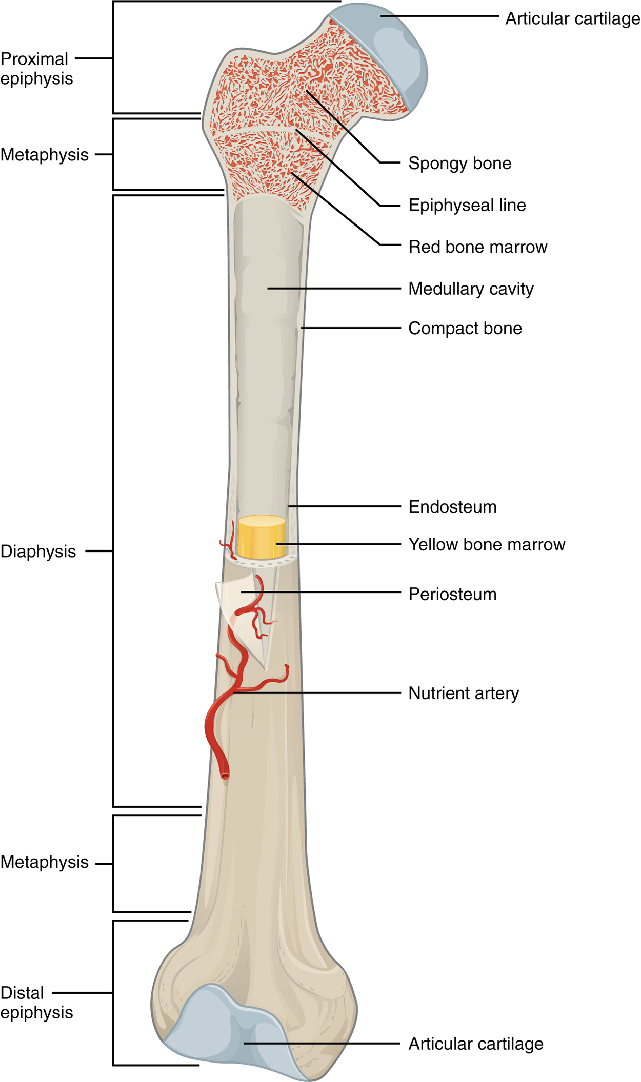
The medullary cavity has a delicate membranous lining called the endosteum (end- = “inside”; oste/o- = “bone”), where bone growth, repair, and remodeling occur. The outer surface of the bone is covered with a fibrous membrane called the periosteum (peri- = “around” or “surrounding”). The periosteum contains blood vessels, nerves, and lymphatic vessels that nourish compact bone. The periosteum covers the entire outer surface, except where the epiphyses meet other bones to form joints. In this region, the epiphyses are covered with articular cartilage, a thin layer of cartilage that reduces friction and acts as a shock absorber.
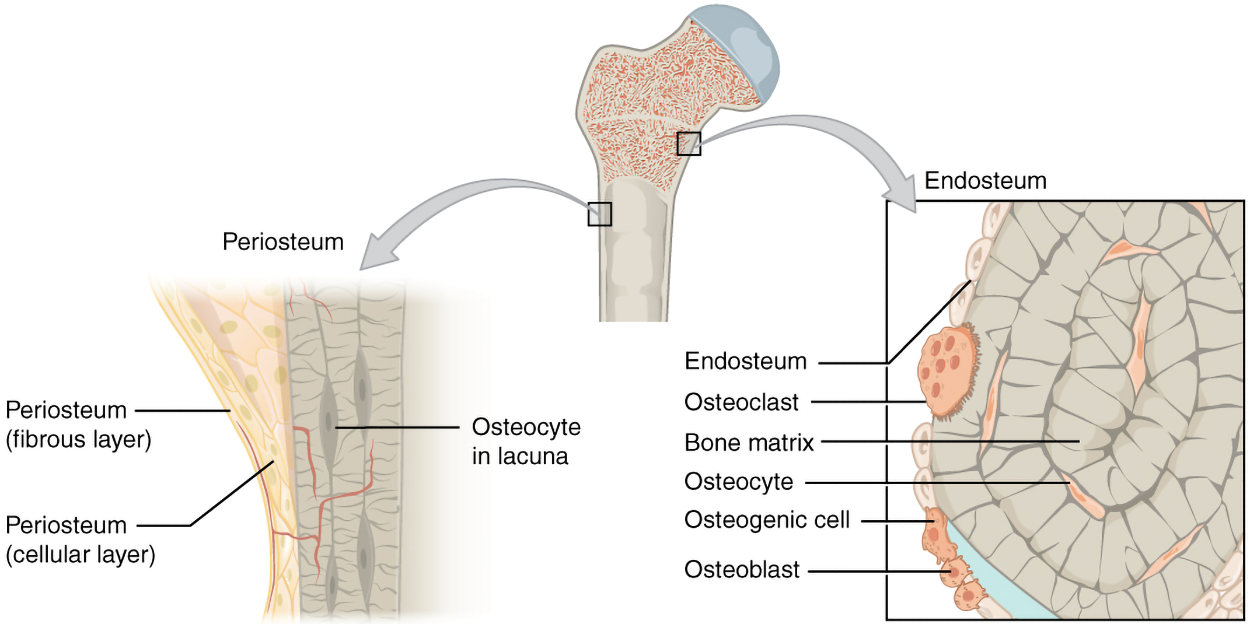
Tendons and ligaments also attach to bones at the periosteum. Both tendons and bones are types of connective tissue. Tendons connect muscles to bones, and ligaments connect bones to other bones at joints. The figure below shows examples of ligaments and tendons.
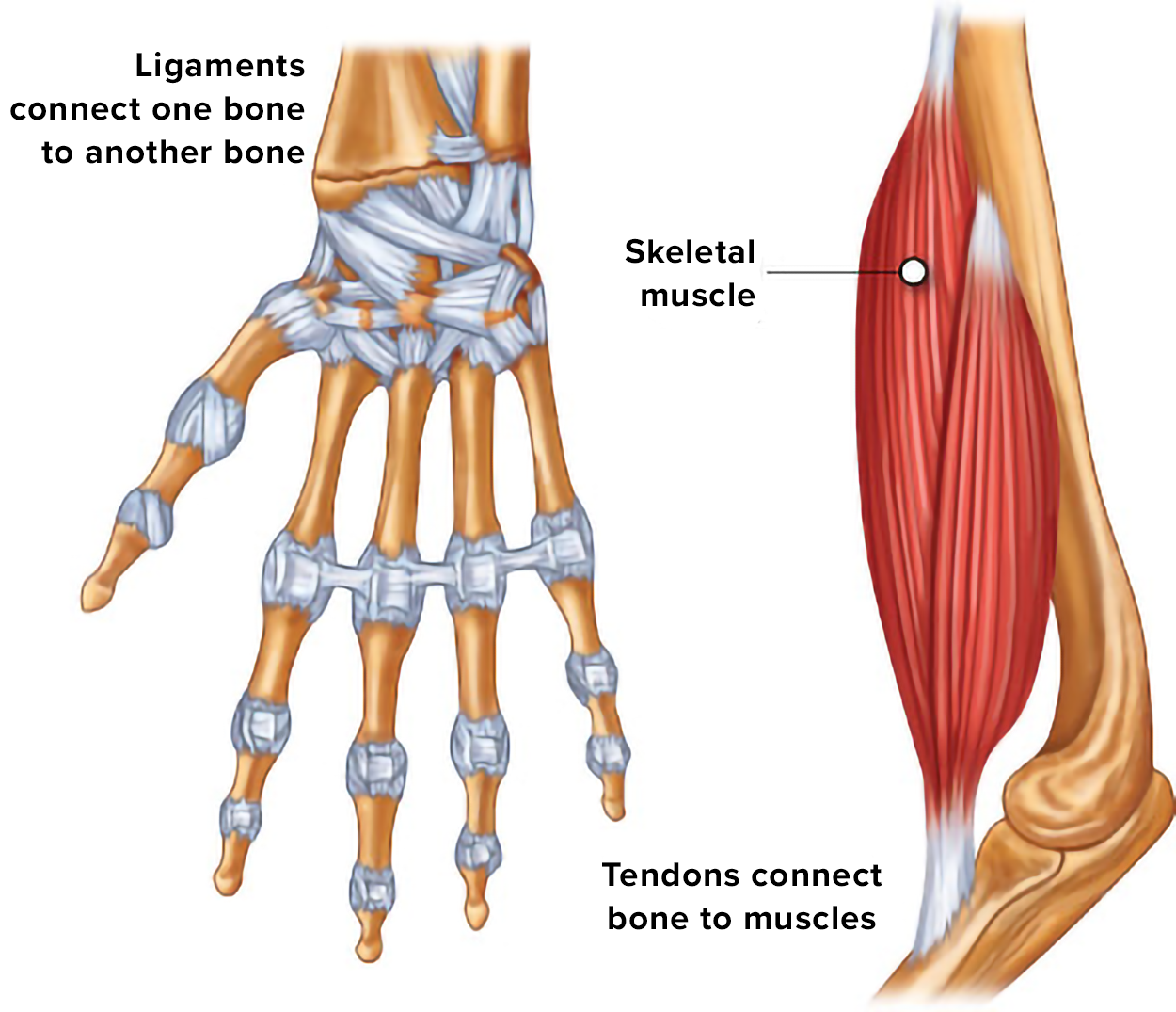
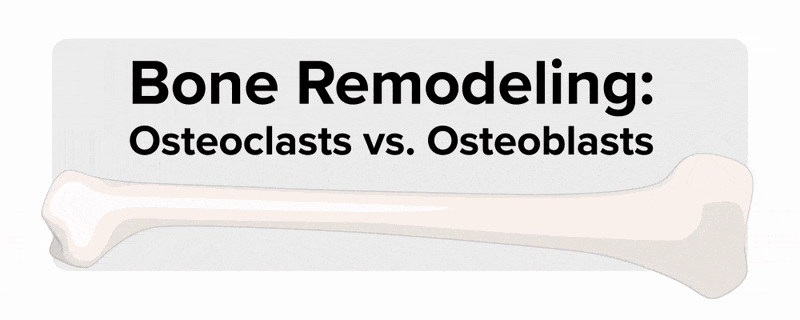
The figure above also shows bone cells. Look for osteoblasts, osteoclasts, and osteocytes. Osteoblasts build bone, osteoclasts break down bone, and osteocytes are mature bone cells. The table below provides more information about these cell types. Together, they work to remodel bone and regulate calcium homeostasis. When bone is broken down, calcium is released into the blood. When there is plenty of calcium in the blood, calcium can be stored in bone.
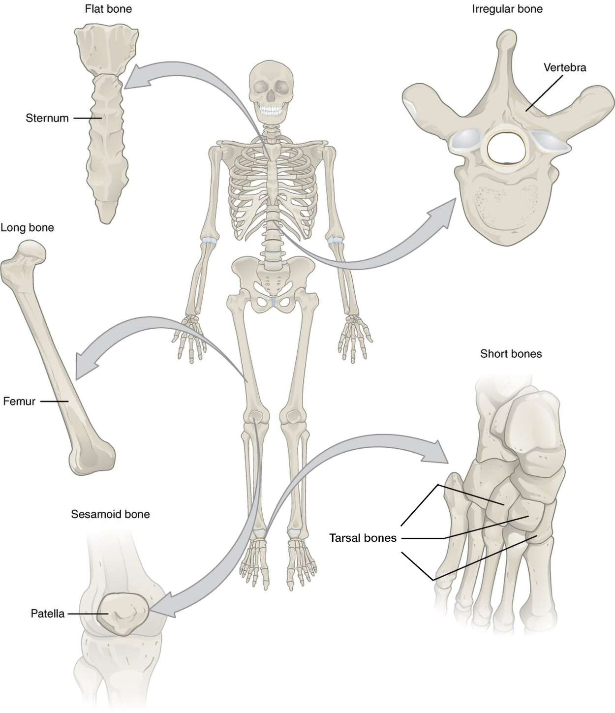
An anterior view of a human skeleton highlights five bones. The sternum (breastbone) lies along the midline of the thorax and connects to the ribs. It is classified as a flat bone and is shaped somewhat like a tie, with an enlarged upper section and a long, flattened, tapering lower section, classified as a flat bone. The femur (thigh bone) is a long bone with a broad lower end connecting to the knee and a ball-shaped upper end fitting into the hip socket. The patella (kneecap) is a small, wedge-shaped sesamoid bone on the front of the knee. The tarsal bones in the foot are small, square-shaped short bones located between the toes and the shin. Lastly, a lumbar vertebra, an irregular bone, is shown with its kidney-shaped body and projecting spines.
| Cell type | Function | Location |
|---|---|---|
| Osteogenic cells | Develop into osteoblasts | Deep layers of the periosteum and the marrow |
| Osteoblasts | Bone formation | Growing portions of bone, including periosteum and endosteum |
| Osteocytes | Maintain mineral concentration of matrix | Entrapped in matrix |
| Osteoclasts | Bone resorption | Bone surfaces and at sites of old, injured, or unneeded bone |
Although it is beyond the scope of this course to discuss bone development in full, there are some important terms for you to know. Ossification (osteogenesis) is the process of bone formation. Epiphyseal plates (growth plates) are areas where bones grow in length. They are located at epiphyses. These plates close at maturity, so examination of the epiphyseal plates can be helpful in assessing a person’s development.
Throughout life, bone is constantly being remodeled. It can be reabsorbed and then rebuilt. This allows bone to respond to stresses and strains.
Bones can be further classified by shape and function. Long bones are involved in support and movement. A long bone is longer than it is wide and functions as a lever. These include important leg and arm bones such as the femur (upper leg), tibia (lower leg), humerus (upper arm), and radius (lower arm).
Short bones are involved in shock absorption and stability. A short bone is approximately equal in length, width, and thickness and provides stability, support, and limited movement. These include the carpals in your hand and the tarsals in the equivalent part of your foot.
Flat bones play important roles in protection and muscle attachment. A flat bone is thin and curved, and provides an attachment point for muscles as well as protection. For example, these include the sternum (breastbone), ribs, scapula (shoulder blade), and skull bones.
Some bones are irregular and have a complex structure. These include the vertebrae in your vertebral column (spine) and the bones of your pelvis.
Sesamoid bones are embedded in tendons. A sesamoid bone is small, round, and sesamoid shaped and protects tendons from compressive forces. An example of a sesamoid bone is the patella (kneecap).
You can see examples of these bone types in the image below.
| Term | Definition | Audio |
|---|---|---|
| Diaphysis | The tubular shaft that runs between the proximal and distal ends of the bone |
|
| Epiphysis | The rounded end of a long bone |
|
| Hematopoiesis | Blood cell production |
|
| Epiphyseal plate | Areas where bones grow in length |
|
Source: THIS TUTORIAL HAS BEEN ADAPTED FROM (1) OPEN RN "MEDICAL TERMINOLOGY 2E". ACCESS FOR FREE AT wtcs.pressbooks.pub/medterm/. (2) Openstax "Anatomy and Physiology 2E". Access for free at OPENSTAX.ORG/DETAILS/BOOKS/ANATOMY-AND-PHYSIOLOGY-2E. LICENSING (1&2): CREATIVE COMMONS ATTRIBUTION 4.0 INTERNATIONAL. Accessed by March 2025.
REFERENCES
Osteogenesis imperfecta. (2024, August 18). Cleveland Clinic. Osteogenesis Imperfecta: What It Is, Symptoms & Types. https://my.clevelandclinic.org/health/diseases/osteogenesis-imperfecta-brittle-bone-disease.
Drake, M. T., & Cremers, S. C. (2010). Bisphosphonate therapeutics in bone disease: the hard and soft data on osteoclast inhibition. Molecular Interventions, 10(3), 141–152. doi.org/10.1124/mi.10.3.5
Do, W. S., Park, J. K., Park, M. I., Kim, H. S., Kim, S. H., & Lee, D. H. (2012). Bisphosphonate-induced Severe Hypocalcemia - A Case Report. Journal of Bone Metabolism, 19(2), 139–145. doi.org/10.11005/jbm.2012.19.2.139
Hypocalcemia. (2022, May 31). Cleveland Clinic. Hypocalcemia: Causes, Symptoms & Treatment. https://my.clevelandclinic.org/health/diseases/23143-hypocalcemia.