Table of Contents |
There are four pairs of abdominal muscles that cover the anterior and lateral abdominal region and meet at the anterior midline. These muscles of the anterolateral abdominal wall can be divided into four groups:
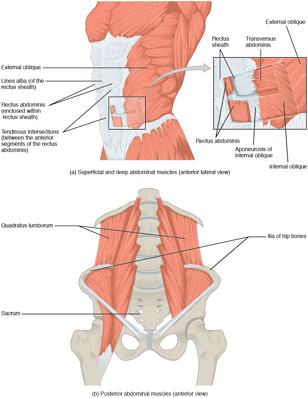
There are three flat skeletal muscles in the antero-lateral wall of the abdomen. The external oblique is closest to the surface and extends inferiorly and medially (V-shape), in the direction of sliding one’s four fingers into pants pockets. Perpendicular and deep to it is the internal oblique, extending superiorly and medially (an upside down V-shape), the direction the thumbs usually go when the other fingers are in the pants pocket. These two abdominal obliques perform flexion, lateral flexion, and rotation of the spine. The transversus abdominis is the deep muscle and is arranged transversely around the abdomen, similar to the front of a belt on a pair of pants. This muscle provides compression to the abdomen. This arrangement of three bands of muscles in different orientations allows various movements and rotations of the trunk. The three layers of muscle also help to protect the internal abdominal organs in an area where there is no bone.
The linea alba (linea, line; alba, white) is a white, fibrous band along the anterior abdominal midline that is made from the joining of the bilateral rectus sheaths (rectus, straight), or abdominal aponeuroses of the transversus abdominis and abdominal obliques. These sheaths enclose the rectus abdominis muscles, a pair of long, linear muscles, commonly called the “sit-up” muscles. Each muscle is segmented by three transverse bands of collagen fibers or tendinous intersections. This results in the look of “six-pack abs,” as each segment hypertrophies on individuals at the gym who do many sit-ups.
The posterior abdominal wall is formed by the lumbar vertebrae, parts of the ilia of the hip bones, and the quadratus lumborum muscle. This part of the core plays a key role in stabilizing the rest of the body and maintaining posture through controlling lateral flexion of the spine.
| Muscle | Action | Origin | Insertion |
|---|---|---|---|
| External Oblique | Flexes, laterally flexes, and rotates the spine | Ribs 5–12; ilium | Linea alba, iliac crest, pubis |
| Internal Oblique | Flexes, laterally flexes, and rotates the spine | Iliac crest | Linea alba, ribs 10–12 |
| Quadratus Lumborum | Extends and laterally flexes the spine | Iliac crest | Rib 12; transverse process of L1–L4 |
| Rectus Abdominis | Flexes the spine | Pubis | Xiphoid process; ribs 5 and 7 |
| Transversus Abdominis | Compresses the abdomen, rotates the spine | Iliac crest; ribs 7–12 | Linea alba, pubis |
The muscles of the chest serve to facilitate breathing by changing the size of the thoracic cavity. When you inhale, your chest rises because the cavity expands. Alternately, when you exhale, your chest falls because the thoracic cavity decreases in size.
| Muscle | Action | Origin | Insertion |
|---|---|---|---|
| Diaphragm | Expands the thoracic cavity | Xiphoid process of sternum; ribs 7–12; vertebral bodies of L1–L4 | Central tendon of diaphragm |
| External Intercostals | Elevate the ribs, expand the thoracic cavity | Rib superior to each intercostal muscle | Rib inferior to each intercostal muscle |
| Innermost Intercostals | Depresses the ribs, compress the thoracic cavity | Rib inferior to each intercostal muscle | Rib superior to each intercostal muscle |
| Internal Intercostals | Depress the ribs, compress the thoracic cavity | Rib inferior to each intercostal muscle | Rib superior to each intercostal muscle |
The change in volume of the thoracic cavity during breathing is due to the alternate contraction and relaxation of the diaphragm. This thoracic muscle separates the thoracic and abdominal cavities and is dome-shaped at rest. The superior surface of the diaphragm is convex, creating the elevated floor of the thoracic cavity. The inferior surface is concave, creating the curved roof of the abdominal cavity.
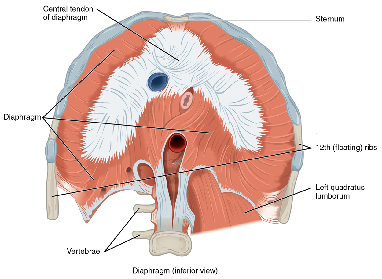
Defecating, urination, and even childbirth involve cooperation between the diaphragm and abdominal muscles (this cooperation is referred to as the “Valsalva maneuver”). You hold your breath with a steady contraction of the diaphragm; this stabilizes the volume and pressure of the peritoneal cavity. When the abdominal muscles contract, the pressure cannot push the diaphragm up, so it increases pressure on the intestinal tract (defecation), urinary tract (urination), or reproductive tract (childbirth).
There are three sets of muscles, called intercostal muscles, which span each of the intercostal spaces. The principal role of the intercostal muscles is to assist in breathing by changing the dimensions of the rib cage.
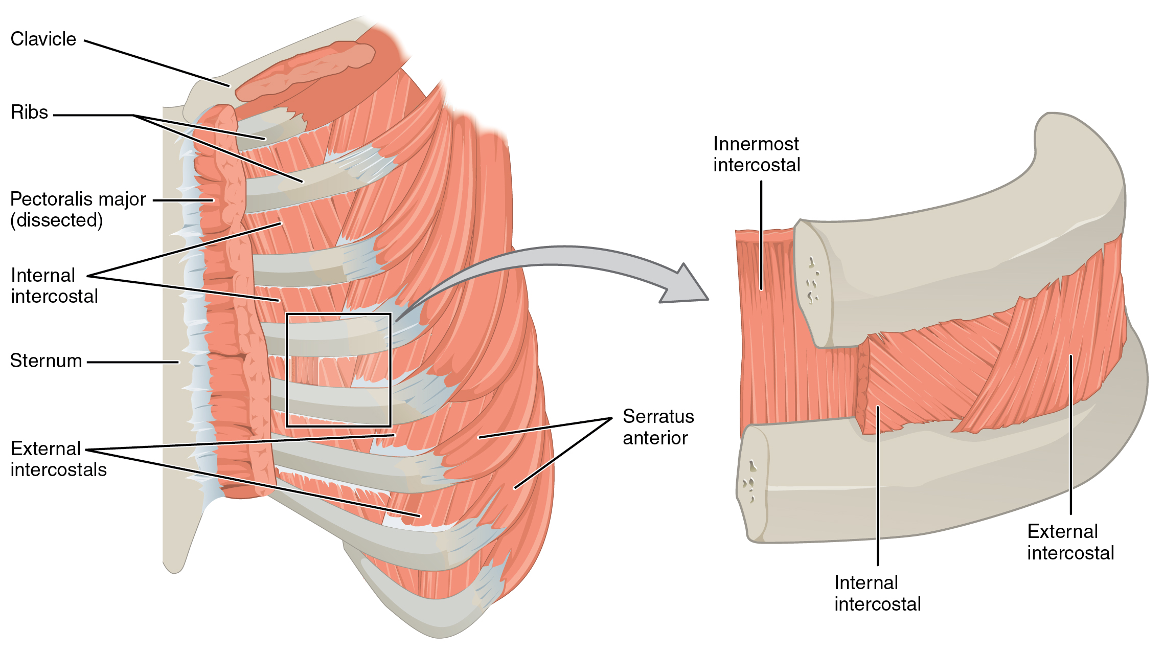
The 11 pairs of superficial external intercostal muscles aid in the inspiration of air during breathing because when they contract, they raise the rib cage, which expands it. The 11 pairs of internal intercostal muscles, just under the externals, are used for expiration because they draw the ribs together to constrict the rib cage. The innermost intercostal muscles are the deepest, and they act as synergists for the action of the internal intercostals.
The pelvic floor is a muscular sheet that defines the inferior portion of the pelvic cavity. The pelvic diaphragm, spanning anteriorly to posteriorly from the pubis to the coccyx, comprises the levator ani and the ischiococcygeus. Its openings include the anal canal and urethra, and vagina in females.
The large levator ani consists of two skeletal muscles and is considered the most important muscle of the pelvic floor because it supports the pelvic viscera. It resists the pressure produced by the contraction of the abdominal muscles so that the pressure is applied to the colon to aid in defecation and to the uterus to aid in childbirth. The levator ani is assisted by the ischiococcygeus, also referred to as the coccygeus, which pulls the coccyx anteriorly. This muscle also creates skeletal muscle sphincters at the urethra and anus.
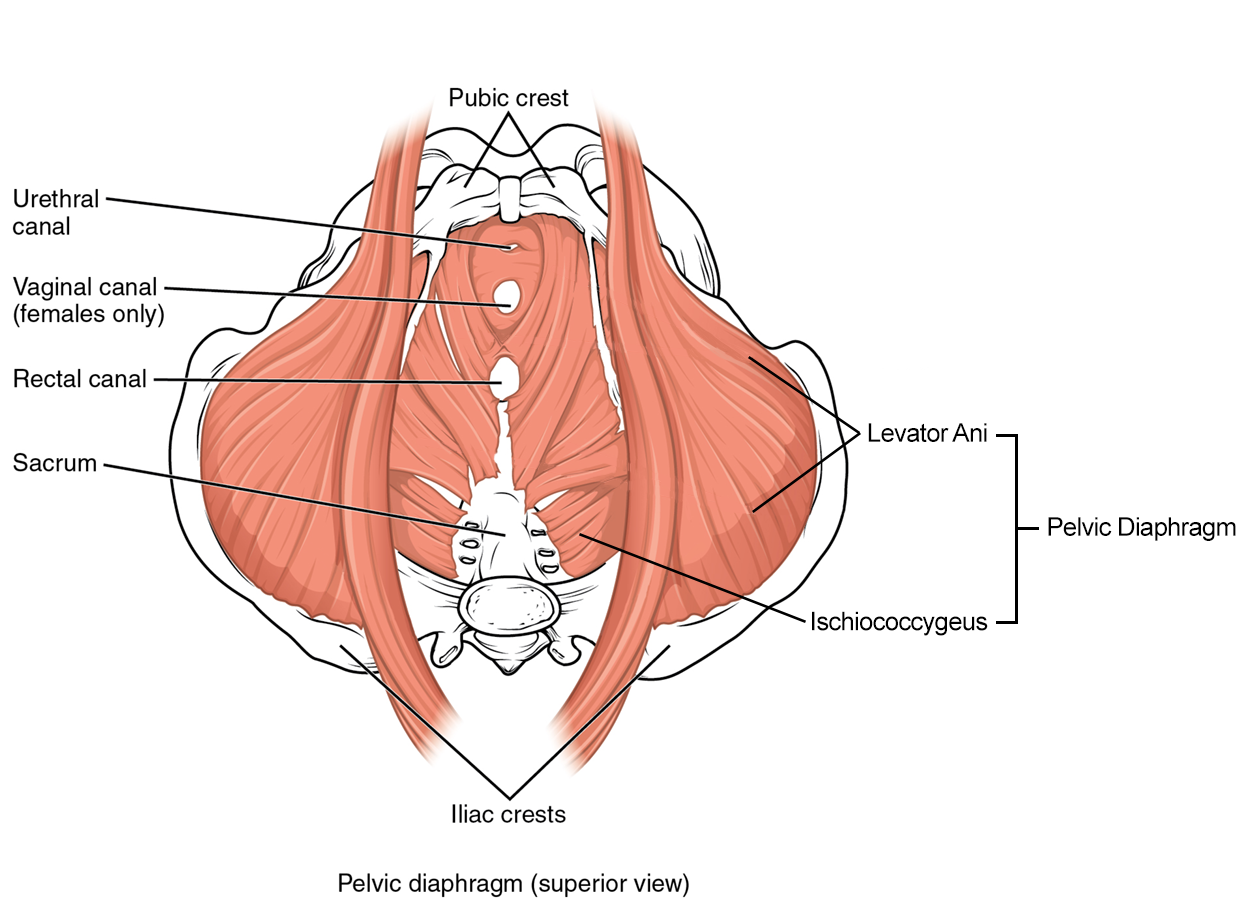
The perineum is the diamond-shaped space between the pubic symphysis (anteriorly), the coccyx (posteriorly), and the ischial tuberosities (laterally), lying just inferior to the pelvic diaphragm (levator ani and coccygeus). Divided transversely into triangles, the anterior is the urogenital triangle, which includes the external genitals. The posterior is the anal triangle, which contains the anus. The perineum is also divided into superficial and deep layers with some of the muscles common to people of any sex. The muscles of this region function to compress the urethra to block urination (peeing), close the vagina (female only), and promote ejaculation (male only). You will learn about these muscles in the digestive, urinary, and reproductive systems if you take the A&P II course.
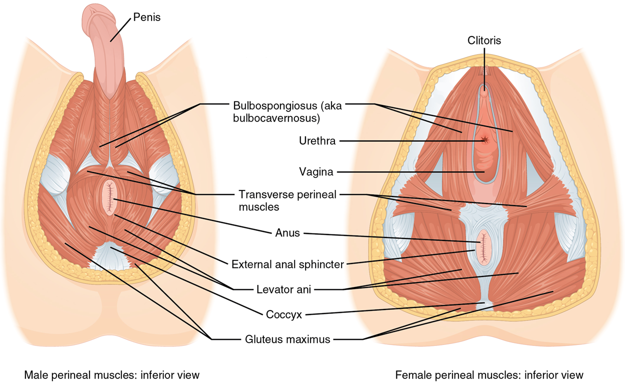
| Muscle | Action | Origin | Insertion |
|---|---|---|---|
| Ischiococcygeus | Flexes the coccyx | Ischium | Sacrum, coccyx |
| Levator Ani | Stabilizes the pelvic floor by resisting intra-abdominal pressure | Pubis, ischium | Coccyx, prostate, vagina |
Source: THIS TUTORIAL HAS BEEN ADAPTED FROM OPENSTAX “ANATOMY AND PHYSIOLOGY 2E.” ACCESS FOR FREE AT HTTPS://OPENSTAX.ORG/DETAILS/BOOKS/ANATOMY-AND-PHYSIOLOGY-2E. LICENSE: CC ATTRIBUTION 4.0 INTERNATIONAL.