Table of Contents |
The skeletal system is often divided into two major parts: the axial skeleton and the appendicular skeleton. The axial skeleton runs along the main superior to inferior body axis, from the head through the vertebral column.
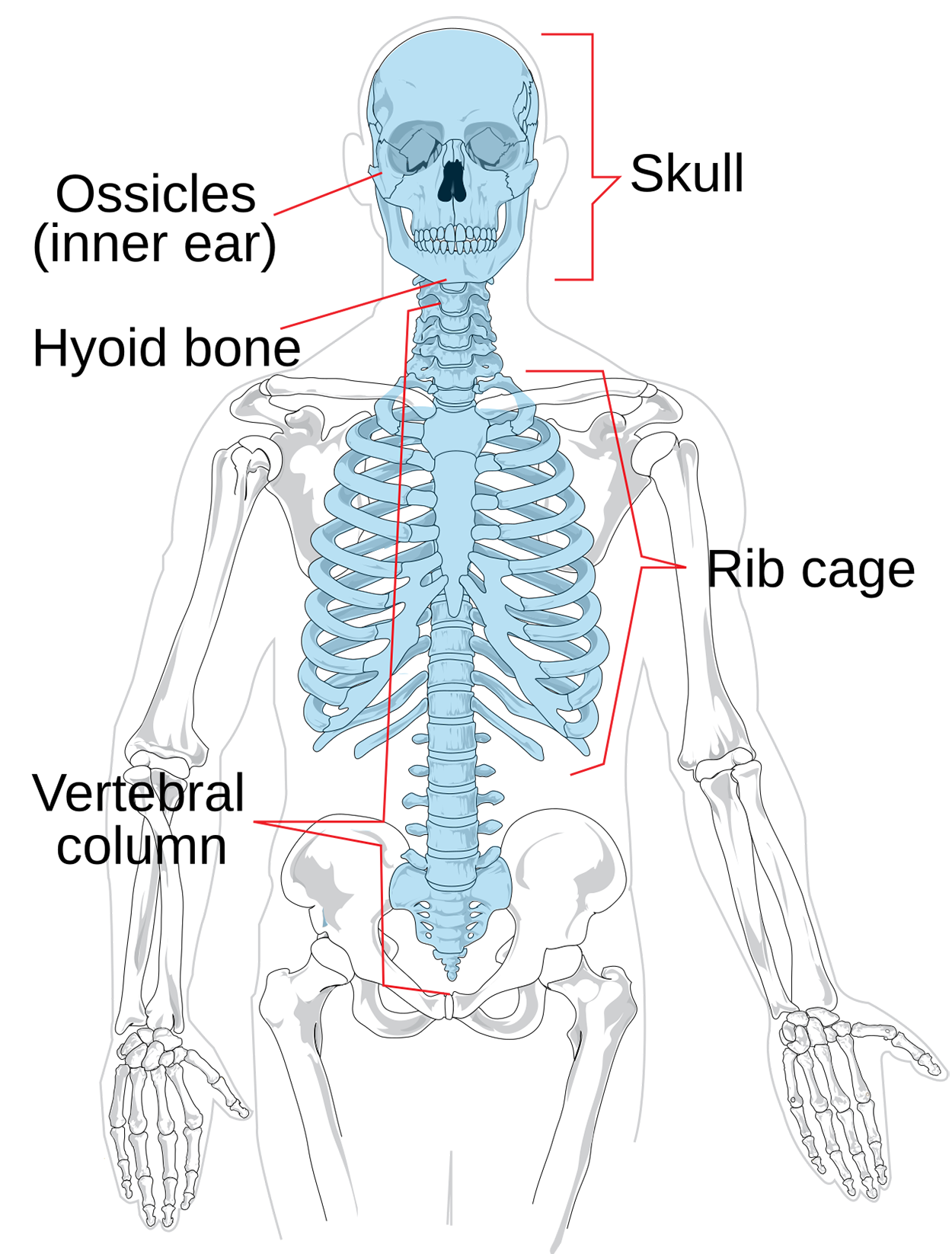
The figure below shows the axial skeleton. Note that it includes the skull, vertebral column, and rib cage.
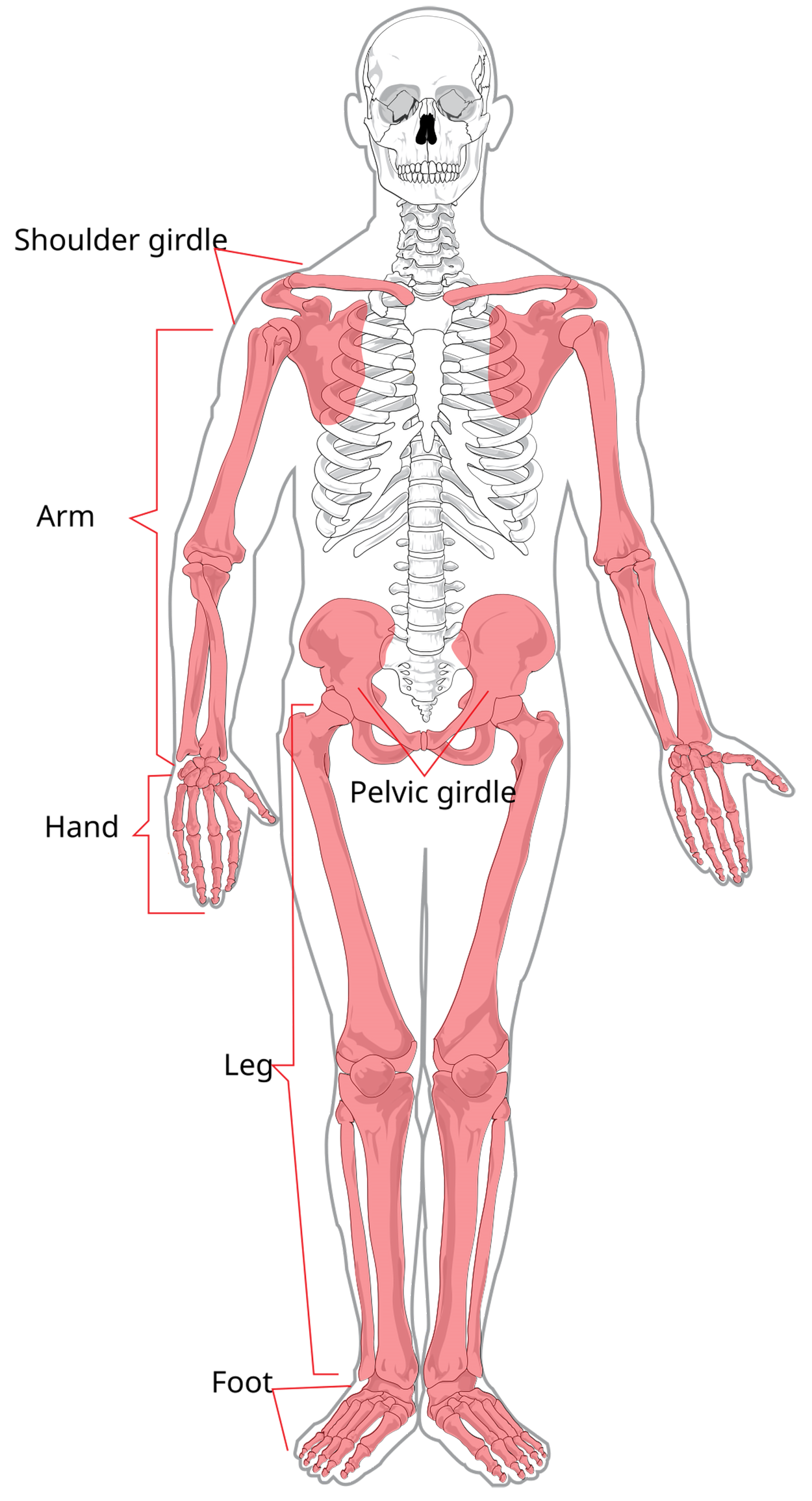
The figure below shows the appendicular skeleton. Note how it includes the appendages (limbs) and the girdles that support them. The appendicular skeleton includes the limbs, shoulder girdle, and pelvis.
Note several combining forms related to the skeletal divisions:
- Crani/o (pertaining to the cranium/skull)
- Vertebr/o (pertaining to the vertebrae of the vertebral column/spine)
- Cost/o (pertaining to the ribs)
- Pelv/i (pertaining to the pelvis)
When learning about the skeleton, you will also see terms that describe bone features and markings. These terms may appear in descriptions used for imaging or surgical procedures, and you will often see them if you take an anatomy class or otherwise do work that requires understanding anatomical structures. These describe structures such as protrusions to which muscles attach and openings (such as an opening through which something can pass).
Examples of these terms include:
- Articulation: a place where two bones meet (e.g., knee joint).
- Head: a prominent rounded surface (e.g., femoral head).
- Condyle: a rounded surface (e.g., medial condyle).
- Process: a prominence feature extending from bone (e.g., mastoid process or spinous process).
- Foramen: a hole through bone (e.g., the foramen magnum in the skull).
- Sinus: an air-filled space in bone (e.g., the paranasal sinuses).
- Spine: a sharp process (e.g., the scapular spine).
- Fossa: an elongated basin (e.g., olecranon fossa and cranial fossae).
- Tuberosity: a rough, elevated surface (e.g., ischial tuberosity and tibial tuberosity).
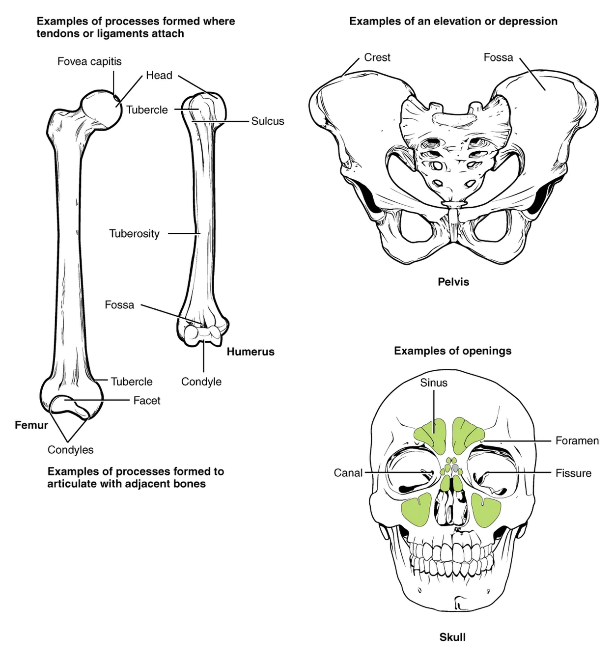
The illustration below shows examples of some of these bone features. Note some of the major examples of processes formed where tendons or ligaments attach and processes formed to articulate with adjacent bones. For example, the rounded head of the femur has a small depression labeled as fovea capitis and rounded protrusions at the bottom labeled as condyles with a depression labeled as facet between them. Note that the humerus also has a rounded head on top. On the head of the humerus, note the tubercle protruding toward the viewer and the small groove labeled as sulcus. It is also important to note the example of elevation (the rim of the pelvis protruding toward the viewer) and depression (the shallow hollow interior below the crest labeled as fossa. The sinuses are examples of openings in the skull.
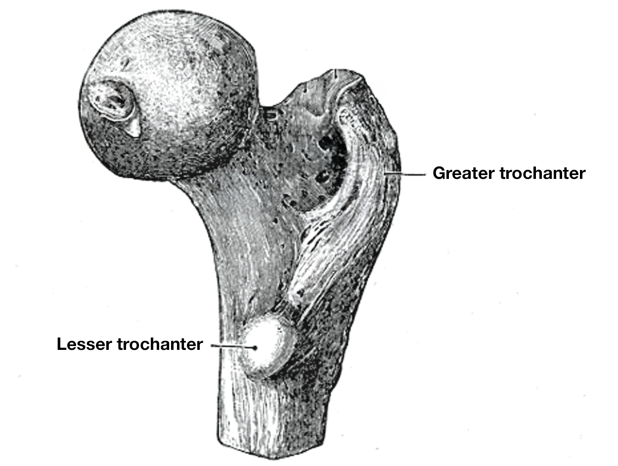
The femur has two notable protrusions just below where it articulates with the hip: the greater and lesser trochanters. Muscles attach to these protrusions. Some people have an additional third trochanter (Bolanowski et al., 2005). You can see the greater trochanter and lesser trochanter in the image below.
As mentioned, the axial skeleton includes the cranium, vertebral column, and associated structures. It forms the central axis of the body and includes the bones of the head, neck, chest, and back. It serves to protect the brain, spinal cord, heart, and lungs. It also serves as the attachment site for muscles that move the head, neck, back, shoulder, and hip joints. The axial skeleton includes the cranium, hyoid, vertebral column, and thoracic cage.
Although learning all of the bones of the skull is beyond this course, it is worth knowing certain bones. For example, the mandible is the lower jaw, the maxilla is the upper jaw, and the zygomatic bone is the cheekbone. You can see these and other bones in the figure below.
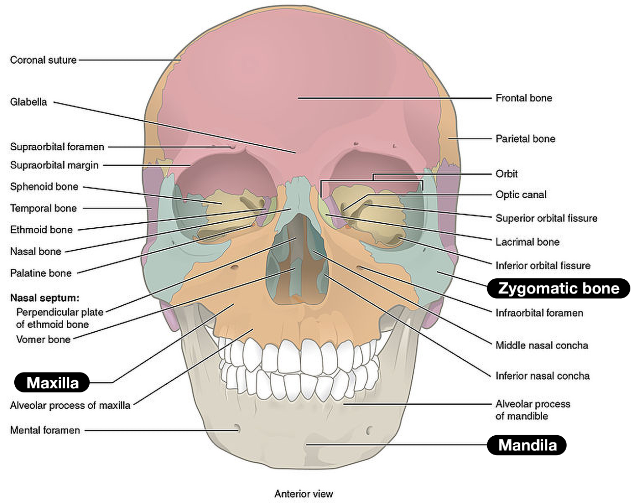
Important skull bones include:
- Zygomatic: Cheekbone
- Maxillary: Upper jaw and hard palate
- Palatine: Pair of L-shaped bones between the maxilla and sphenoid that form the hard palate, walls of the nasal cavity, and the orbital floor of the eye
- Nasal: Pair of bones that form the bridge of the nose
- Lacrimal: Walls of the inner (medial) orbit (i.e., eye socket)
- Inferior conchae: Lower lateral walls of the nasal cavity
- Vomer: Bone that separates the left and right nasal cavity
- Mandible: Lower jawbone and only movable bone of the skull
Note that newborns have membranous gaps between the skull bones called fontanelles. These are sometimes informally called soft spots. Fontanelles allow the skull to move during birth and typically close by 18 months of age (Children’s Hospital Colorado, 2022). Click through the images below to see a view of anterior and posterior fontanelles from above, the fontanelles from the side, and the top of a 1-month-old baby’s head with a healthy, normal anterior fontanelle visible.
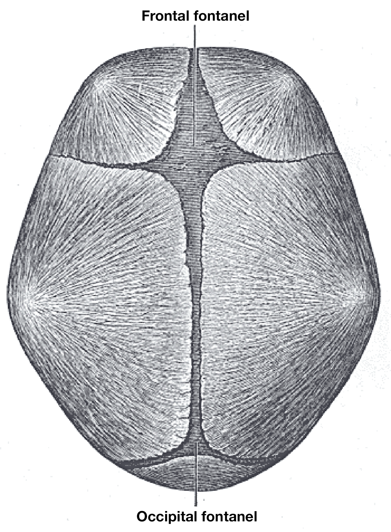
Anterior and posterior fontanelles
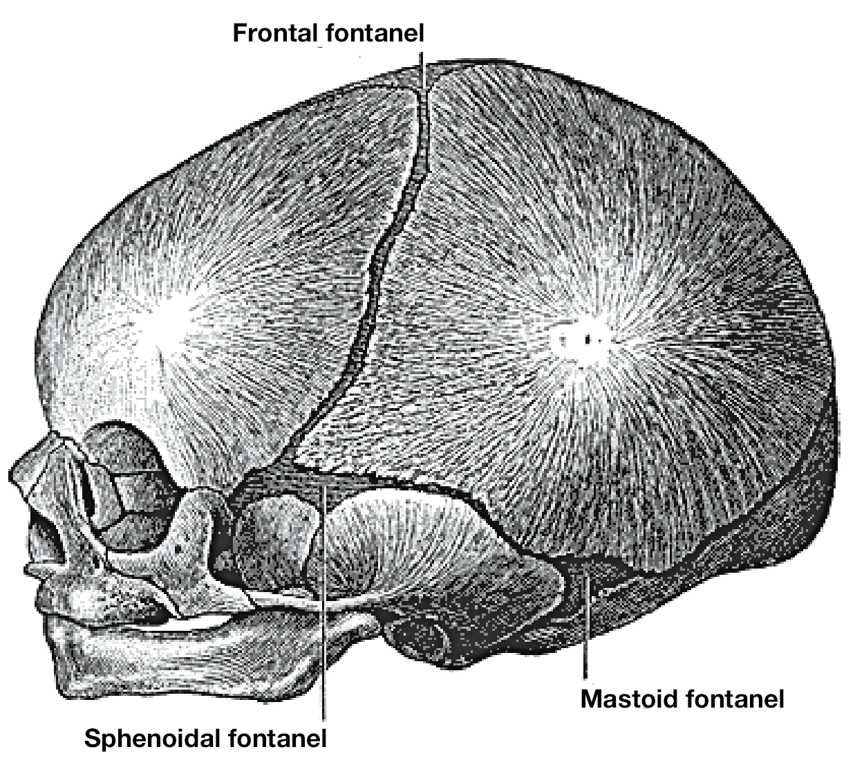
Side view of fontanelles
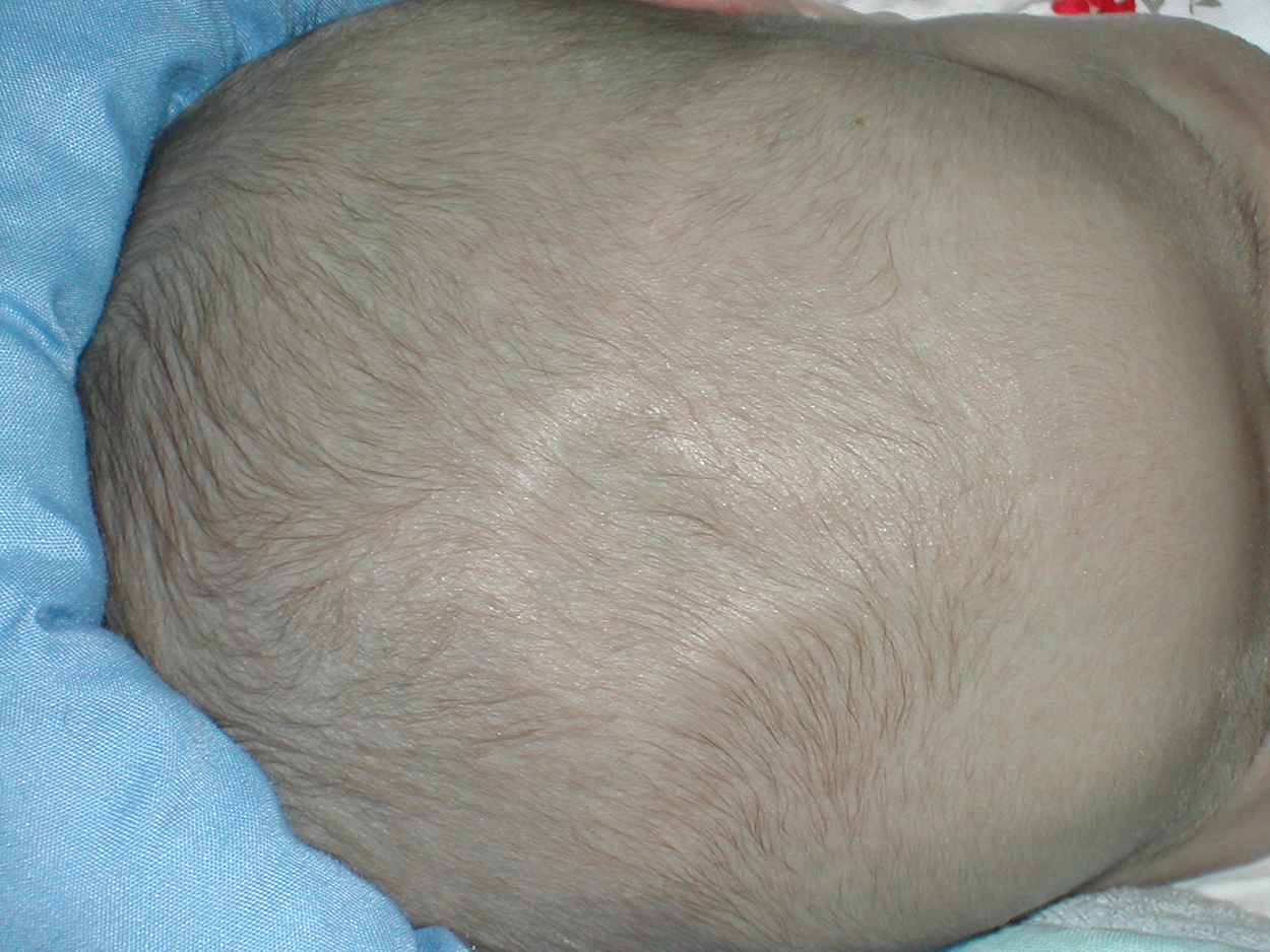
Normal infant anterior fontanelle
Another feature of the skull is the paranasal sinuses. Sinuses are openings. In this case, the paranasal sinuses are openings that contain air. You may be familiar with them from having a sinus infection at some point, as these infections are common.
Click through the images below to see views of the paranasal sinuses and other features of the skull, including additional bones.
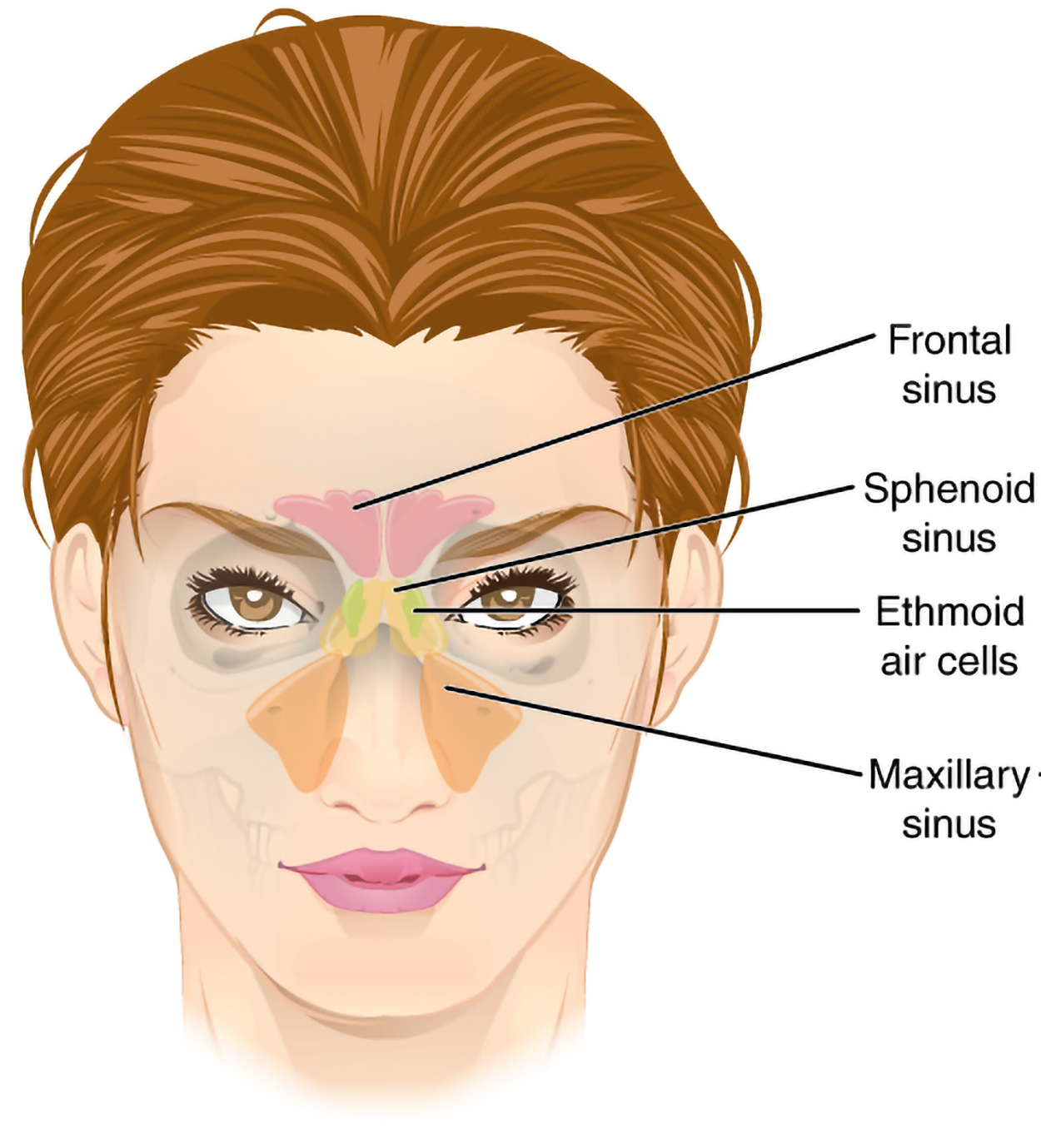
Anterior view of paranasal sinuses
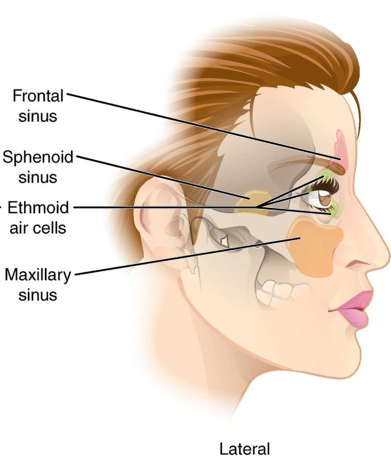
Side view of paranasal sinuses
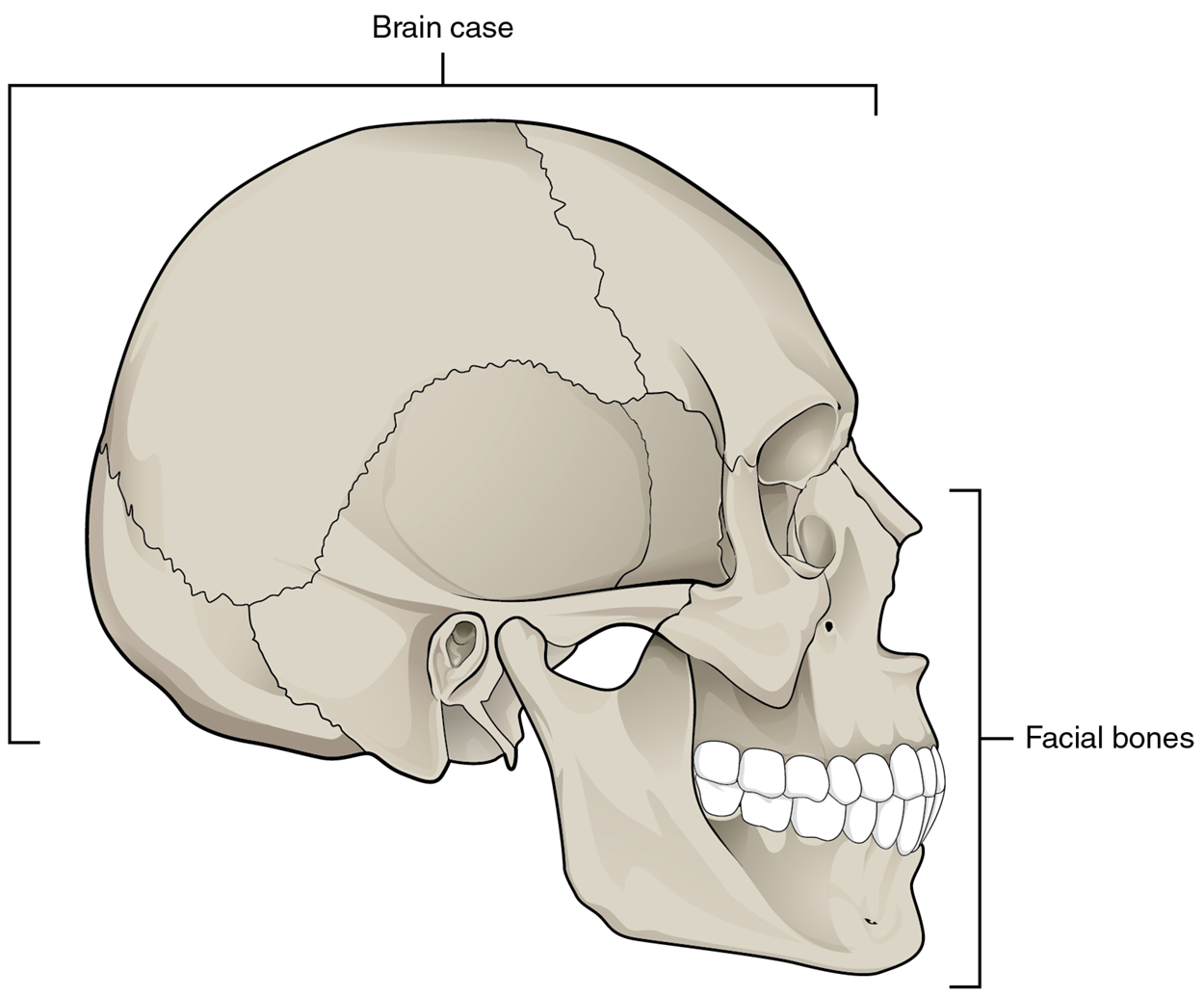
Side view of facial bones
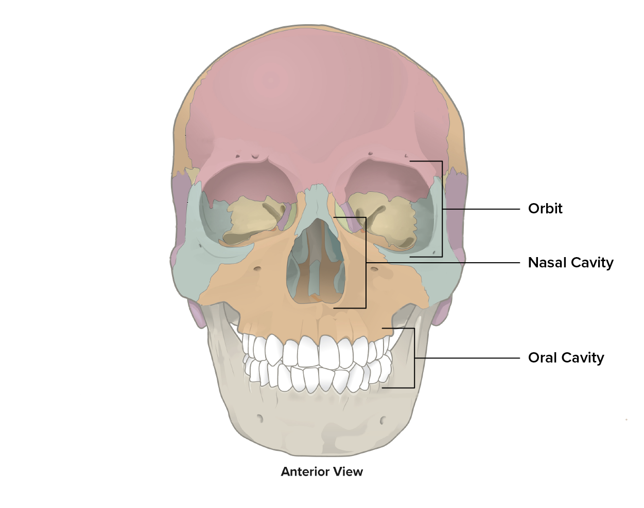
Front view of facial bones
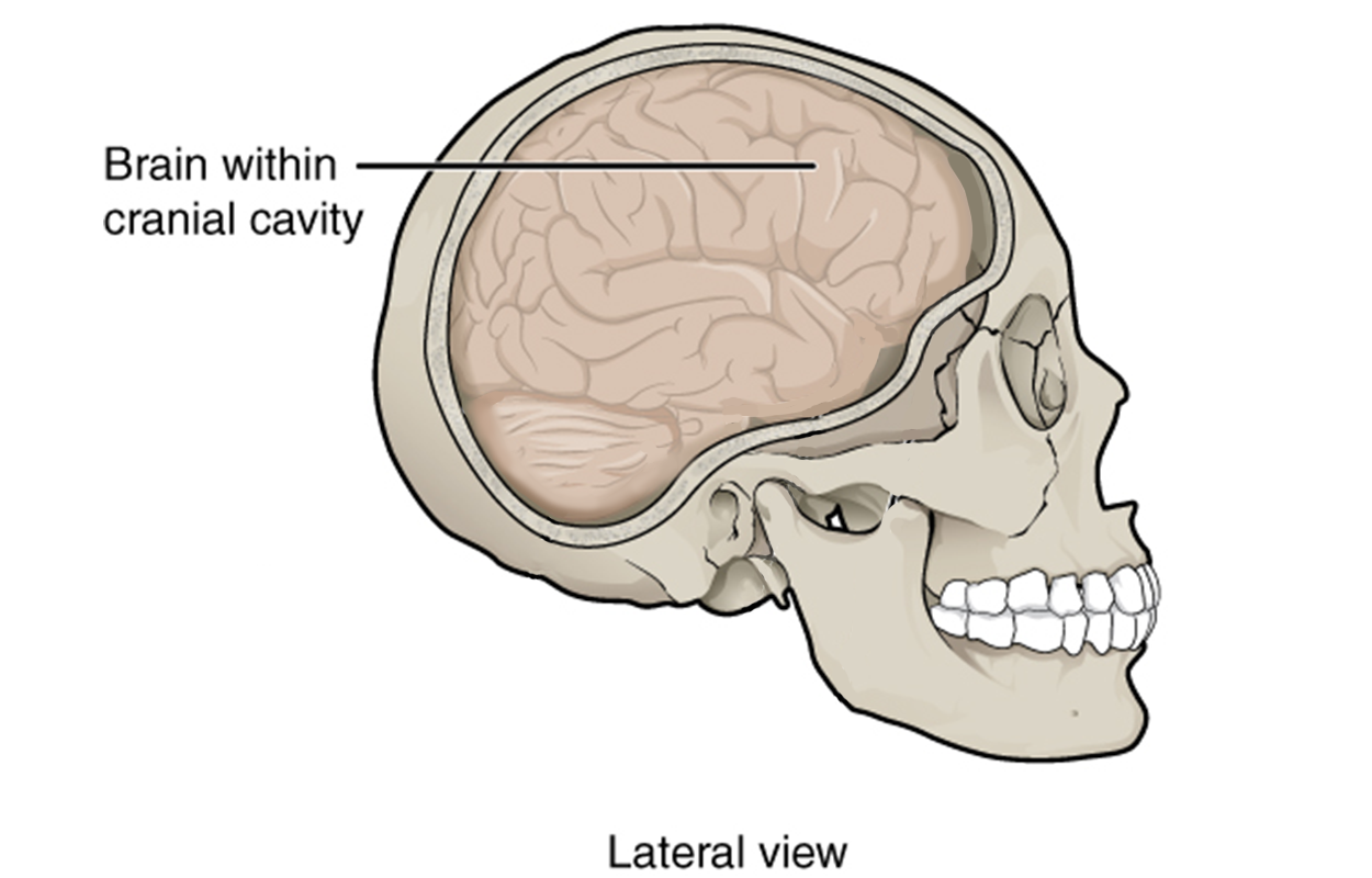
Side view of cranial cavity
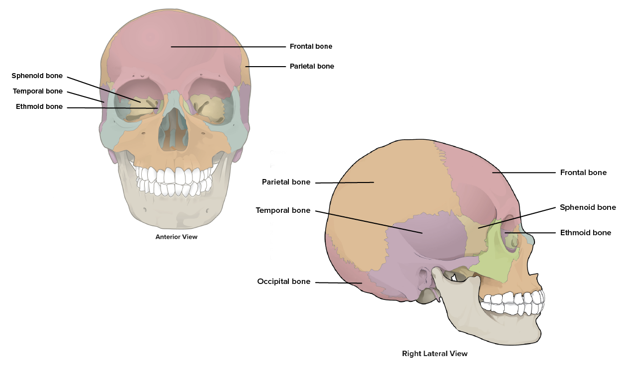
Skull bones
The temporomandibular joint (TMJ) is a hinge joint between the temporal bone and the mandible that allows for the opening, closing, protrusion, retraction, and lateral movement of the lower jaw. Protrusion moves the mandible forward, and retraction pulls the mandible backward towards the neck.
The vertebral column is divided into regions, as you can see in the figure below. There are five major regions of vertebral columns: the cervical region, the thoracic region, the lumbar region, the sacral region, and the caudal region. The caudal region is found in the tail, so humans do not have these vertebrae.
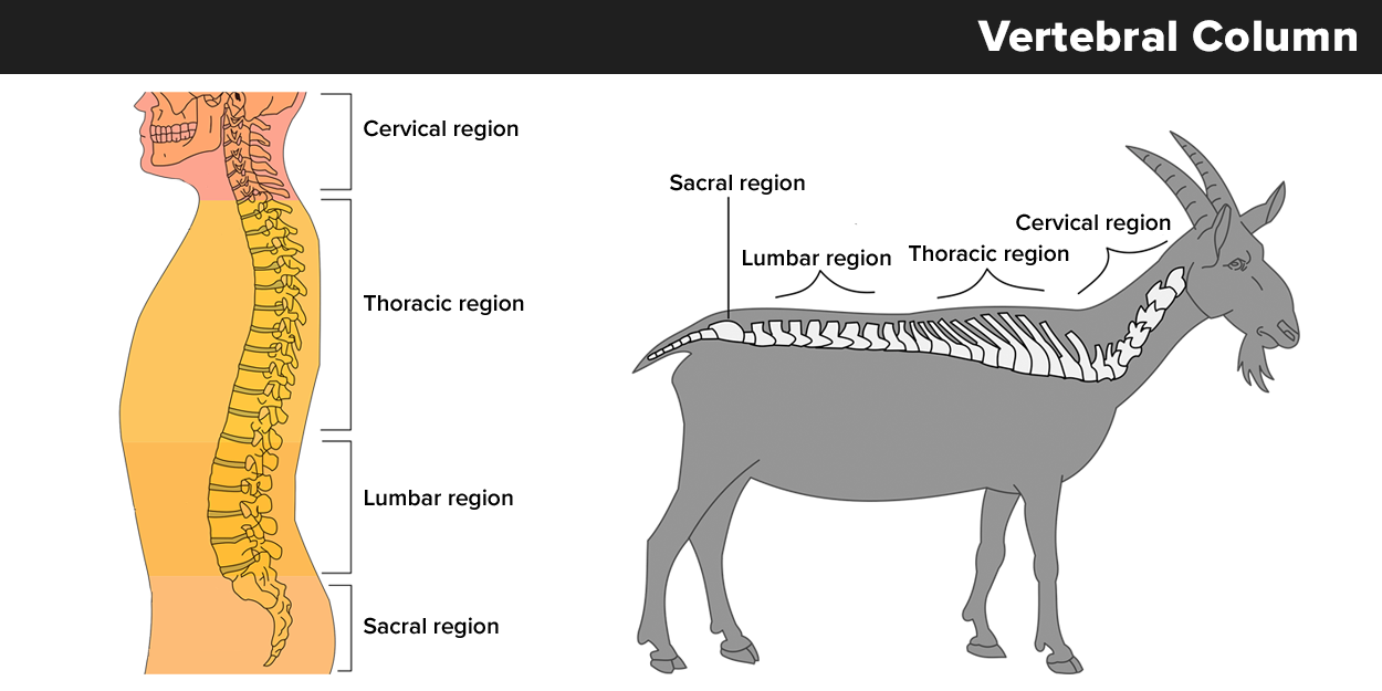
In medical documentation, the vertebrae are labeled based on their region and location. For example, the cervical vertebrae are labeled C1–C7, with C1 closest to the skull. The thoracic vertebrae are labeled T1–T12 and are distinguished from lumbar vertebrae because they have attached ribs. The lumbar vertebrae are labeled L1–L5. These are located in the lower back and do not have attached ribs. The sacrum and coccyx are at the base of the spine and are not numbered. The sacrum has fused vertebrae that join with the pelvic girdle. The coccyx also has fused vertebrae.
Summary of vertebral regions:
- Cervical: The first 7 vertebrae in the neck region, C1 to C7
- Thoracic: The next 12 vertebrae that form the outward curvature of the spine, T1 to T12
- Lumbar: The next 5 vertebrae that form the inner curvature of spine, L1 to L5
- Sacrum: The triangular-shaped bone at the base of the spine, formed by the fusion of five sacral vertebrae, a process that does not begin until after the age of 20.
- Coccyx: The tailbone, formed by the fusion of four very small coccygeal vertebrae.
At the very top of the spine, there are two specialized cervical vertebrae. Click through the images below to see the location of the atlas at the top of the vertebral column (highlighted) with the axis immediately below it, as well as a view of the atlas and a view of the axis.
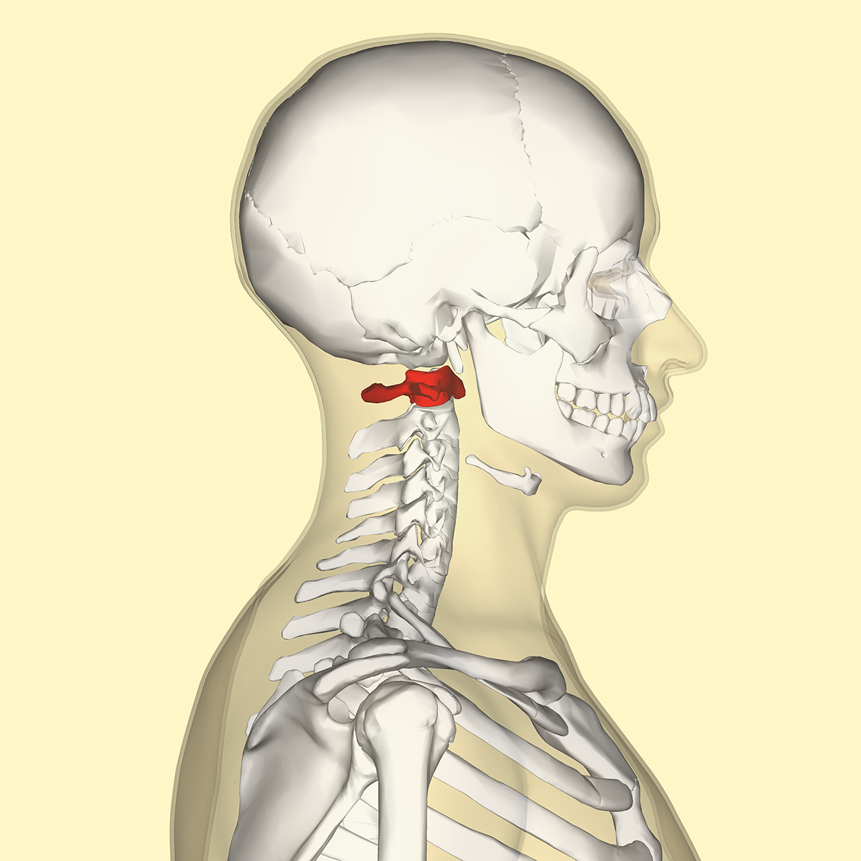
Axis vertebra at top of spine
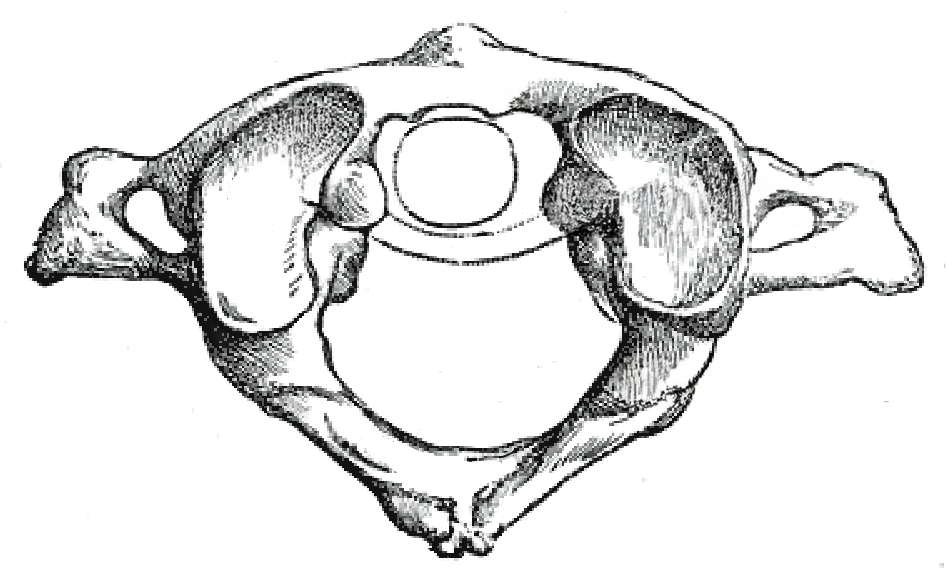
Atlas
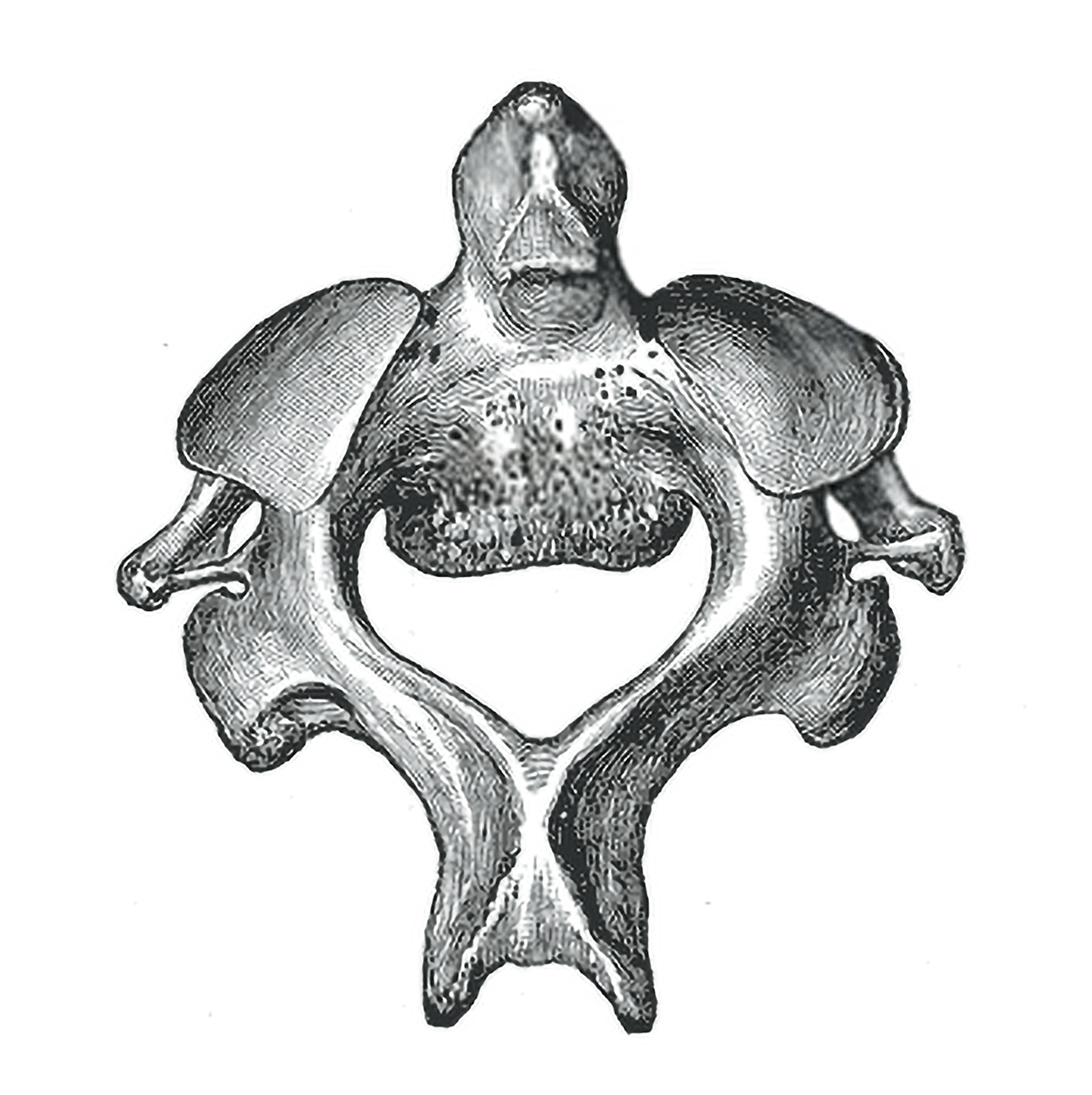
Axis
Here are some word parts related to the regions discussed above:
- Cervic/o refers to the neck region.
- Thorac/o refers to the thoracic region.
- Lumb/o refers to the lumbar region.
You already learned that cost/o refers to the ribs. The sternum, or breastbone, at the front of the rib cage is represented by the word part stern/o.
There are cartilaginous intervertebral discs between the vertebrae, providing cushioning.
The thoracic cage, commonly known as the rib cage, forms the chest (thorax). It consists of the sternum and 12 pairs of ribs, along with their costal cartilages. The ribs are anchored posteriorly to the 12 thoracic vertebrae (T1–T12). The thoracic cage protects the heart and lungs.
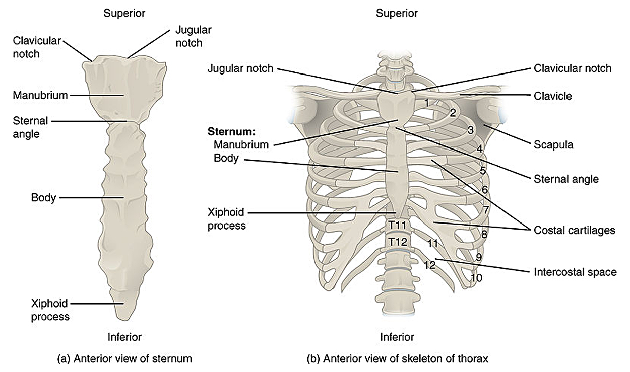
Note that there are three major types of ribs. True ribs articulate with the costal cartilage that articulates with the sternum. False ribs, below the true ribs, articulate with the costal cartilages of ribs above them instead of connecting directly to their own costal cartilages. The bottom two ribs are floating ribs and do not articulate with anything. You can see each of these types of ribs in the figure above. To help distinguish the different types of ribs, note that the costal cartilages are slightly lighter in color than the ribs themselves.
The appendicular skeleton includes the bones of the appendages (the arms and legs) and their supporting girdles (the pectoral and pelvic girdles, respectively). Look at the bones in the figure below. Pay particular attention to the clavicle (across the neck) and scapula (shoulder blade). Notice that there is no bony connection between the upper arms and vertebral column; in mammals, the pectoral girdle that supports the arms is held in place by muscles rather than attached directly to the vertebral column like the pelvic girdle.
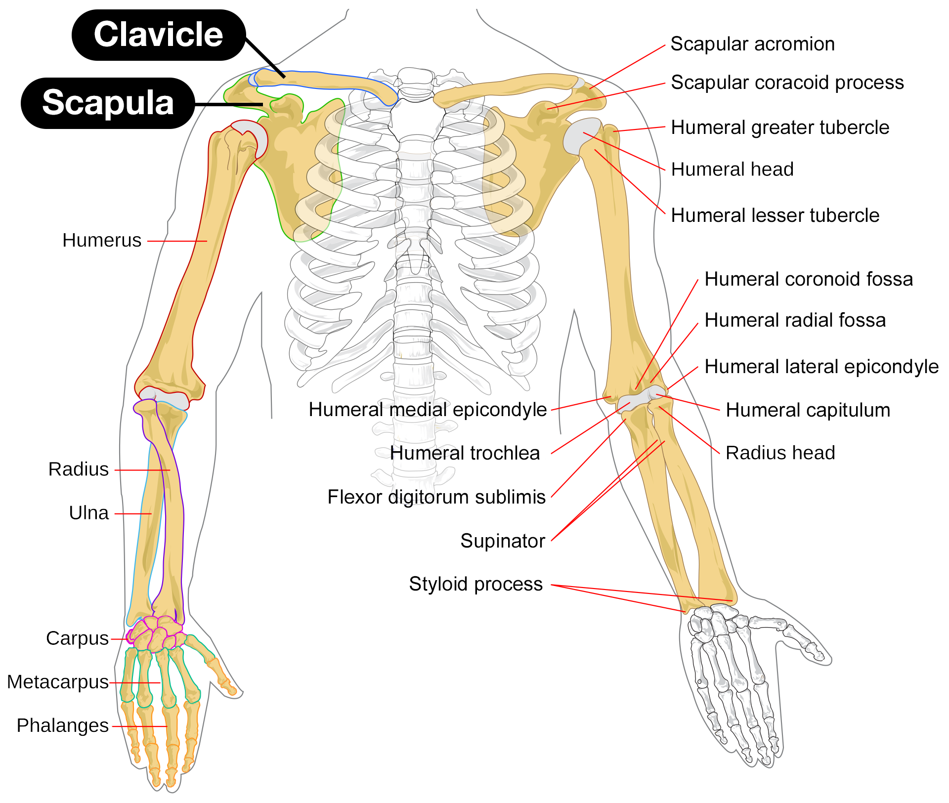
There are useful word parts that use the names of these bones. Word parts associated with upper limb bones include:
- Humer/o means associated with the humerus (upper arm bone).
- Radi/o means associated with the radius (lower arm bone).
- Uln/o means associated with the ulna (lower arm bone).
- Clavicul/o means associated with the clavicle.
Note that the radius and ulna are both in the forearm.
Next, look at the figure below showing a human hand. The bones of the wrist are carpals, the bones of the hand are metacarpals, and the bones of the fingers are phalanges (singular phalanx). The same pattern is used for foot bones: the bones of the ankle are metatarsals, the bones of the foot are tarsals, and the bones of the toes are phalanges.
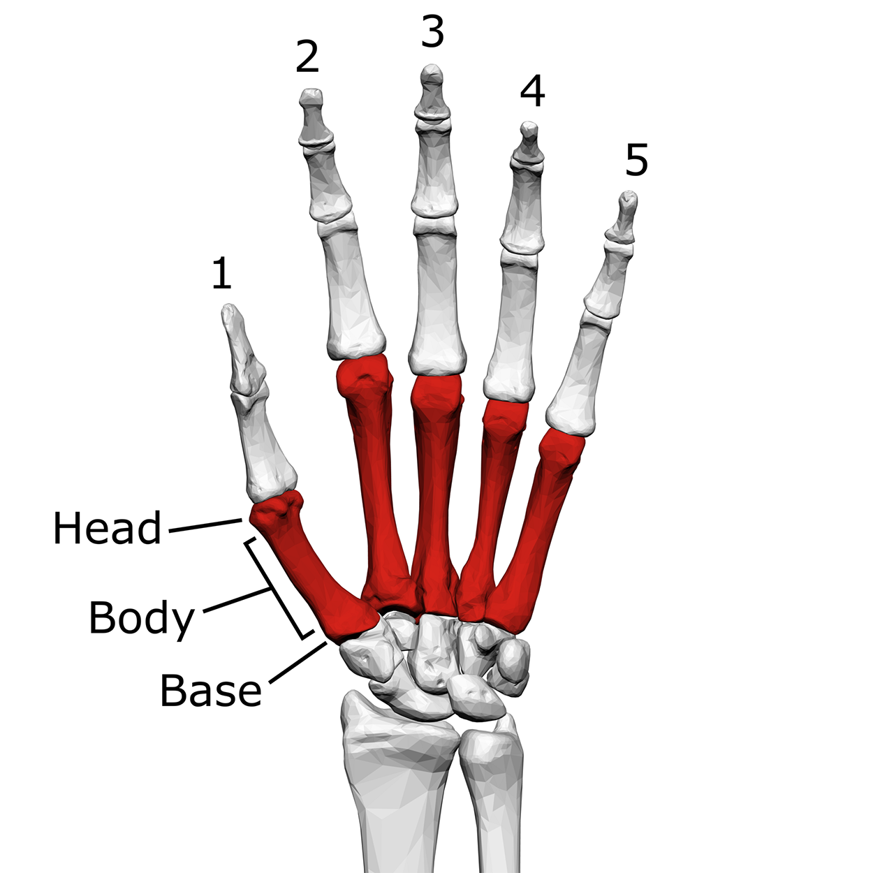
The combining form carp/o refers to the carpals.
Next, look at the figure below showing leg bones. The femur, the large upper leg bone, articulates with the pelvic girdle above (remember that the pelvic girdle includes the ilium, ischium, and pubis). Pay particular attention to the femur (upper leg), patella (kneecap), and tibia and fibula (lower leg).
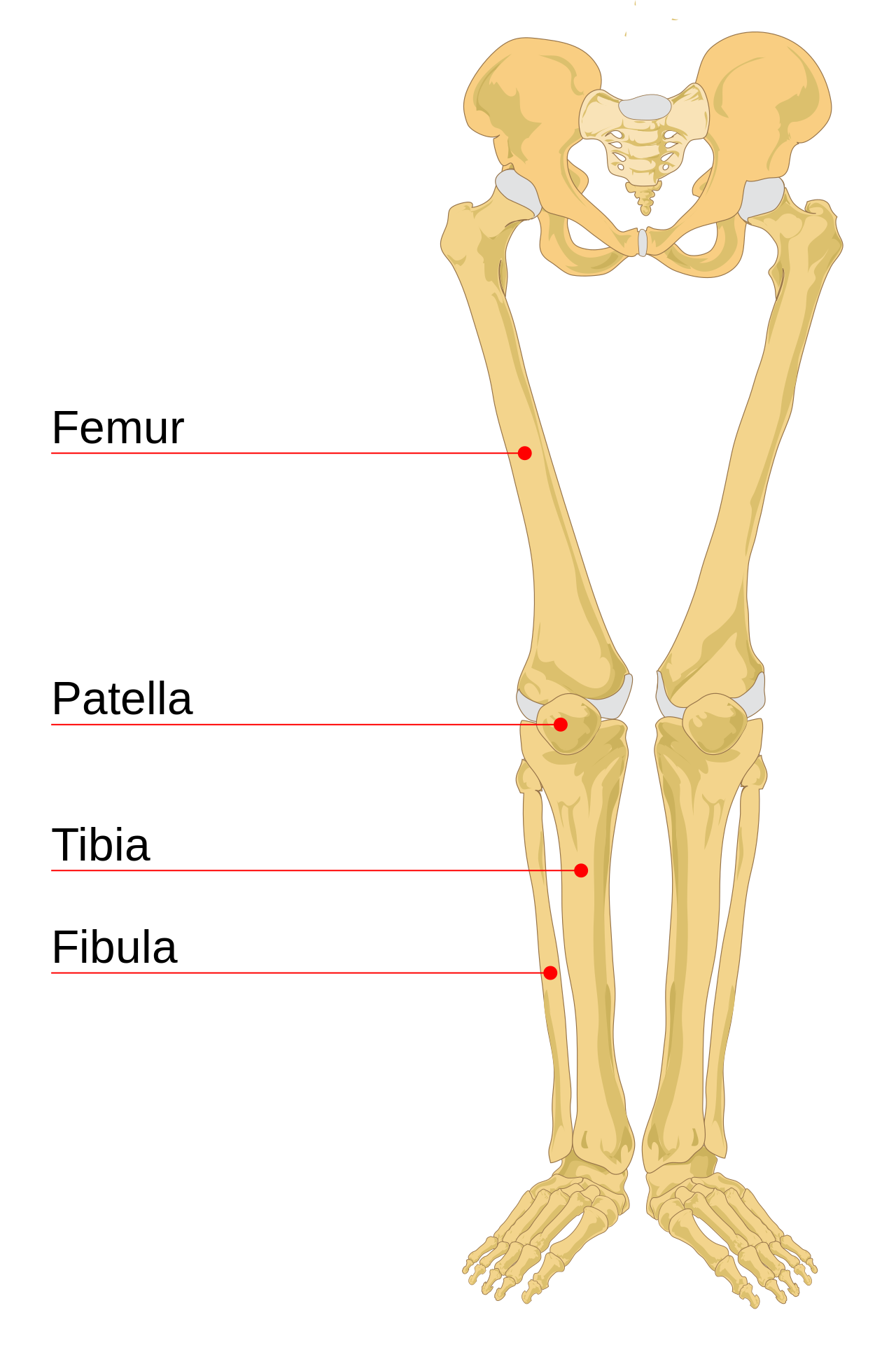
In the image above, you can see where the two pubic bones come together at the bottom of the pelvic girdle. This joining is called the pubic symphysis.
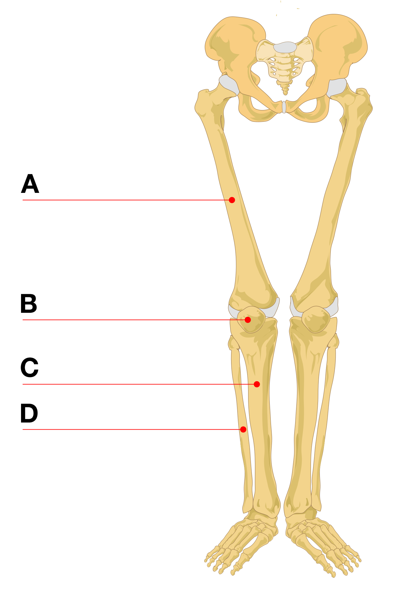
There are useful word parts that use the names of these bones as well. Word parts associated with lower limb bones include:
- Femor/o means associated with the femur (upper leg bone)
- Patell/o means associated with the kneecap
- Tibi/o means associated with the tibia (lower leg bone)
- Fibul/o means associated with the fibular (lower leg bone)
Source: THIS TUTORIAL HAS BEEN ADAPTED FROM (1) OPEN RN "MEDICAL TERMINOLOGY 2E". ACCESS FOR FREE AT wtcs.pressbooks.pub/medterm/. (2) Openstax "Anatomy and Physiology 2E". Access for free at OPENSTAX.ORG/DETAILS/BOOKS/ANATOMY-AND-PHYSIOLOGY-2E. LICENSING (1&2): CREATIVE COMMONS ATTRIBUTION 4.0 INTERNATIONAL. Accessed by March 2025.
REFERENCES
Bolanowski, W., Smiszkiewicz-Skwarska, A., Polguj, M., & Jedrzejewski, K. S. (2005). The occurrence of the third trochanter and its correlation to certain anthropometric parameters of the human femur. Folia morphologica, 64(3), 168–175.
Anterior and Posterior Fontanelle Closures. Children’s Hospital Colorado. Fontanelle Closures | Children's Hospital Colorado
Abbass, M. M. S., Rady, D., El Moshy, S., Ahmed Radwan, I., Wadan, A. S., Dörfer, C. E., & El-Sayed, K. M. F. (2024). The Temporomandibular Joint and the Human Body: A New Perspective on Cross Talk. Dentistry Journal, 12(11), 357. doi.org/10.3390/dj12110357
Samanta, A., Lufkin, T., & Kraus, P. (2023). Intervertebral disc degeneration-Current therapeutic options and challenges. Frontiers in Public Health, 11, 1156749. doi.org/10.3389/fpubh.2023.1156749