Table of Contents |
The upper respiratory system refers to the nose, nasal cavities, sinuses, pharynx, and larynx. You will often see a reference to these structures through the term upper respiratory infection (URI), which refers to a viral infection of one or more of these structures.
The entrance and exit for the respiratory system are through the nose. The nostrils are the opening to the nose, also referred to as nares or external nares (the internal nares are the internal openings into the nasal cavity). The nares and nasal cavities are lined with mucous membranes, containing sebaceous glands and hair follicles that serve to prevent the passage of large debris, such as dirt, through the nasal cavity.
The word root for “nose” is rhin. For example, rhinorrhagia refers to bleeding from the nose, also called epistaxis. Rhinitis refers to inflammation of the nasal mucosa. The nares open into the nasal cavity, which is separated into left and right sections by the nasal septum.
The floor of the nasal cavity is composed of the hard palate and the soft palate. As the names suggest, the hard palate is hard and the soft palate is more flexible. The nasal cavities are lined with mucous membranes that produce mucus, a substance created for lubrication and protection. Rhinorrhea, commonly referred to as a “runny nose,” is a medical term for excess mucus production by the nasal cavity.
Adjacent to the nasal cavity are the sinuses that serve to warm and humidify incoming air. There are four sinuses named for their adjacent bones: frontal sinus, maxillary sinus, sphenoidal sinus, and ethmoidal sinus. Sinusitis refers to inflammation of the sinus cavities. Air moves from the nasal cavities and sinuses into the pharynx.
The pharynx connects the nasal cavity and oral cavity to the trachea and esophagus. It is divided into three major regions: the nasopharynx, the oropharynx, and the laryngopharynx.
The figure below shows the regions of the pharynx.
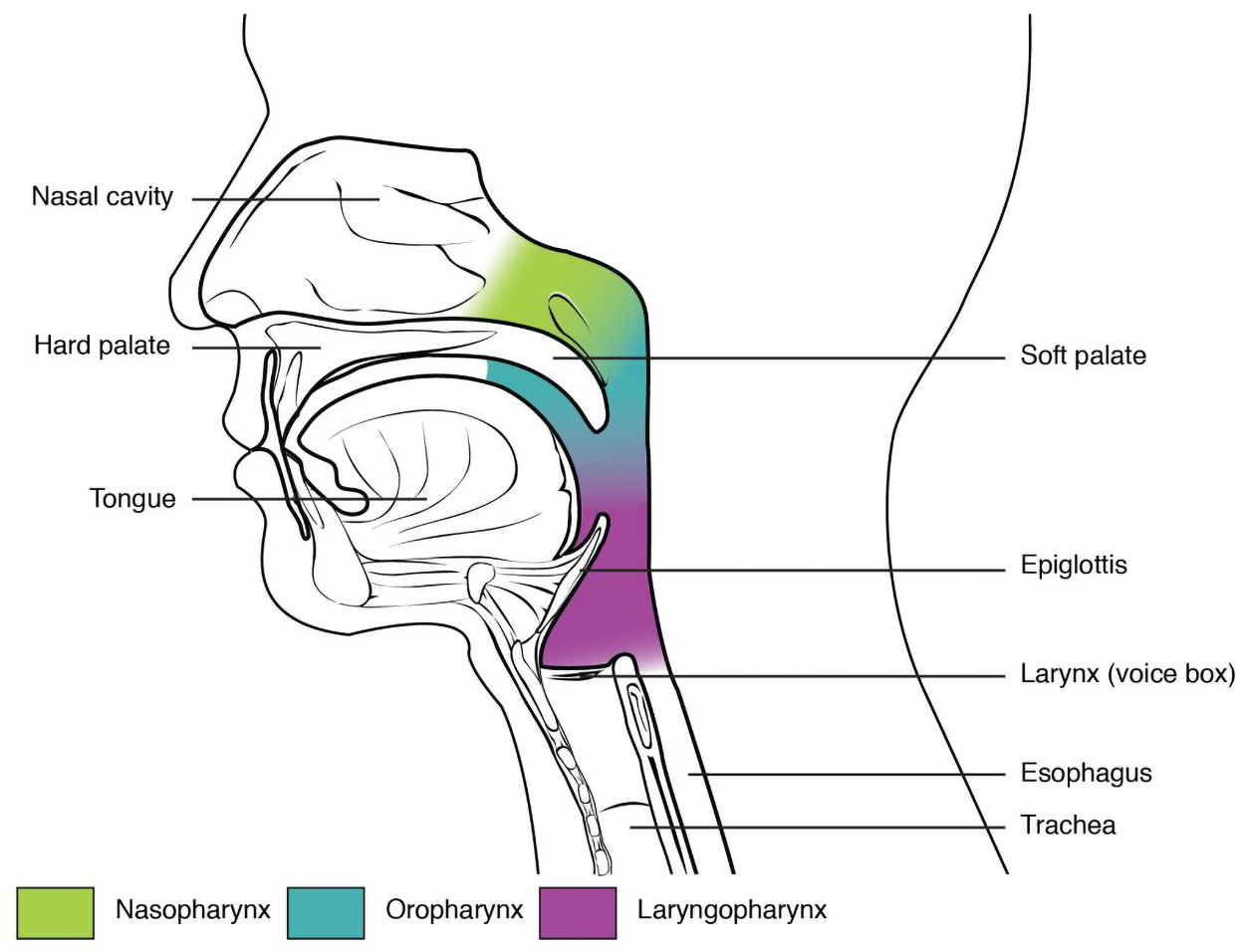
At the top of the nasopharynx is the pharyngeal tonsil, also called the adenoid, which is a collection of lymphatic tissue found at the back of the nasal cavity in the nasopharynx. The function of the pharyngeal tonsil or adenoid is to trap and destroy invading pathogens that enter the airway during inhalation.
The oropharynx is bordered superiorly by the nasopharynx and anteriorly by the oral cavity. The oropharynx contains two distinct sets of tonsils called the palatine tonsils (located on the sides of the oropharynx) and lingual tonsils that also trap and destroy pathogens entering the body through the oral or nasal cavities (Meegala, 2023).
The laryngopharynx is located just below the oropharynx. It is the part of the pharynx (throat) located behind (posterior) the larynx. The laryngopharynx separates into the trachea (the tube going to the larynx) and the esophagus (the tube going into the stomach). The epiglottis prevents food and fluid from entering the trachea while swallowing.
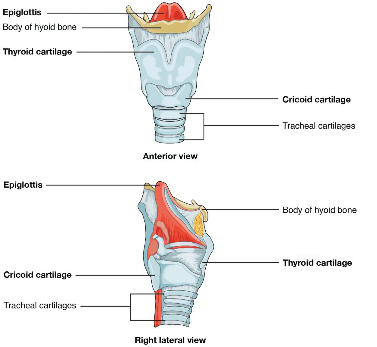
The structure of the larynx is formed by several pieces of cartilage. Three large cartilage pieces form the major structure of the larynx, called thyroid cartilage (the larger piece of cartilage on the anterior side), epiglottis (at the top of the larynx), and cricoid cartilage (just inferior to the thyroid cartilage).
The figure below shows an anterior view and a right lateral view of the larynx. Note the epiglottis at the top with the body of the hyoid bone below. The thyroid cartilage is the large cartilage piece across the upper part of the larynx, with the smaller cricoid cartilage below. The trachea, surrounded by C-shaped tracheal cartilages, is below. The tracheal cartilages provide structural support.
The epiglottis is a flap of tissue that covers the trachea during swallowing to prevent aspiration, the inhalation of food or fluids into the trachea and lower respiratory tract. The act of swallowing causes the pharynx and larynx to lift upward, allowing the pharynx to expand and the epiglottis of the larynx to swing downward, closing the opening to the trachea.
Vocal cords are white, membranous folds attached by muscle to the cartilages of the larynx on their outer edges. The inner edges of the vocal cords are free, allowing oscillation as air passes through to produce sound for speaking. The word root phon refers to sound or voice, so the medical term dysphonia refers to the medical condition of difficulty speaking (i.e., voice).
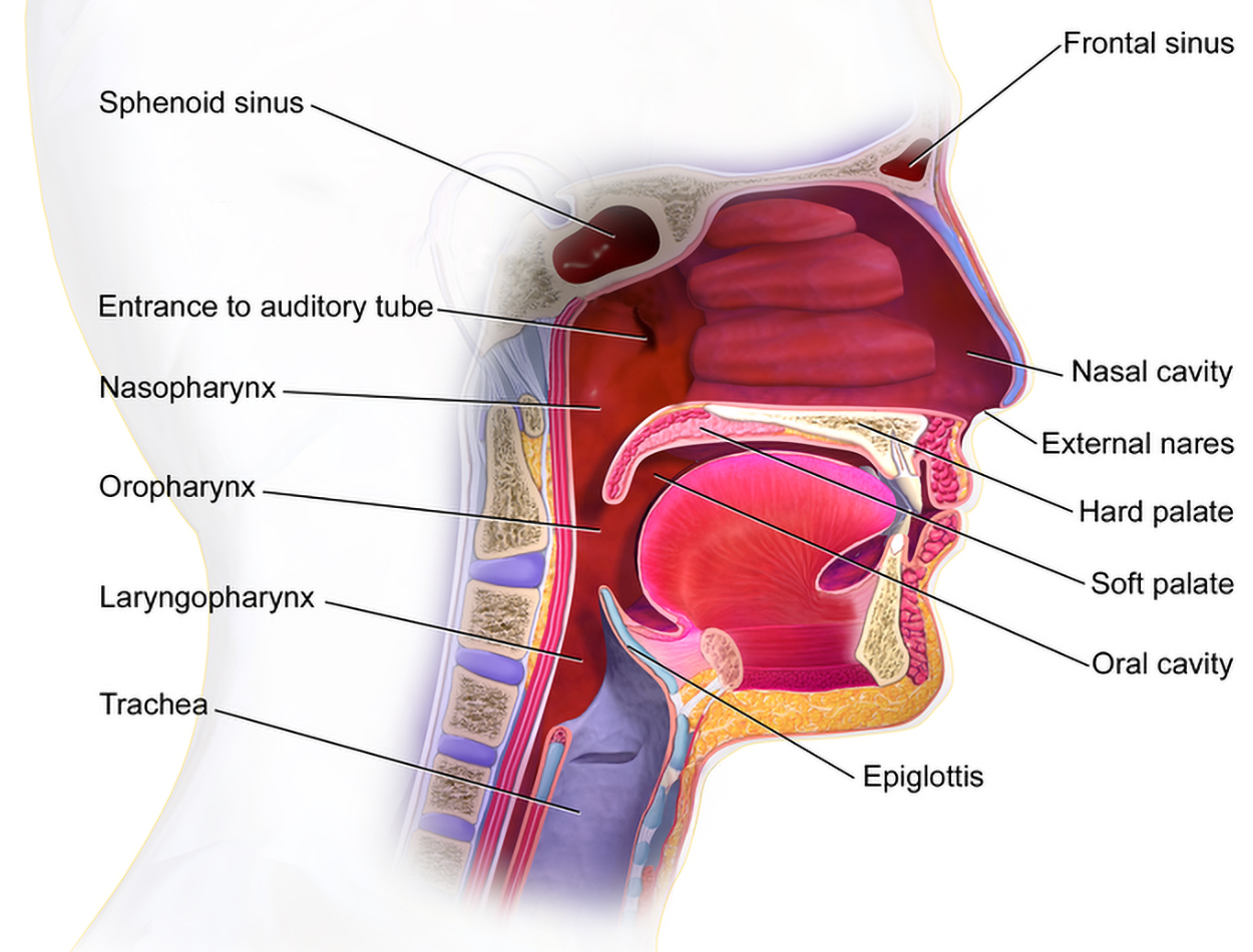
The figure below summarizes components of the upper respiratory system.
The lower respiratory tract consists of the trachea, bronchi, bronchioles, alveoli, and lungs.
The trachea is formed by stacked, C-shaped pieces of cartilage that are connected by dense connective tissue. The trachea stretches and expands slightly during inhalation and exhalation, whereas the rings of cartilage provide structural support and prevent the trachea from collapsing. The trachea is lined with cilia and mucus-secreting cells to trap debris and move it towards the pharynx to be swallowed or spit out.
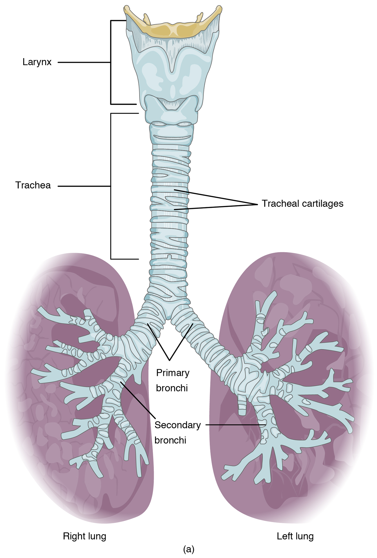
Bronchi are the main air passageways of the lungs. The trachea branches into the right and left primary bronchi at the carina. The carina is a raised structure that contains specialized nervous system tissue that induces violent coughing if a foreign body, such as food, is present. Rings of cartilage, similar to those of the trachea, support the structure of the bronchi and prevent their collapse. The bronchi of each lung continue to branch up to 26 times, creating the bronchial tree, which looks similar to the branching of an actual tree.
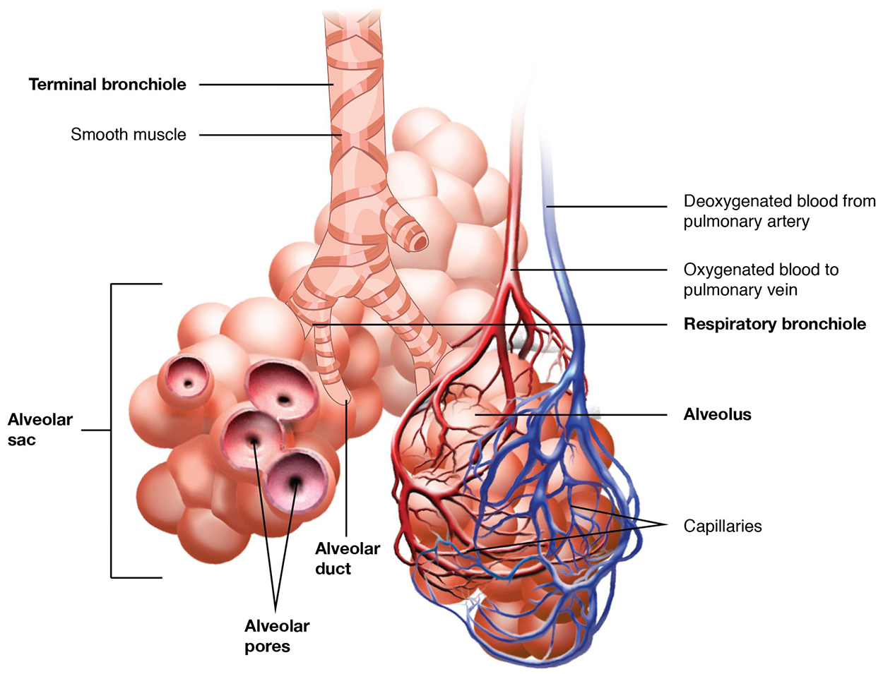
Bronchioles are the smallest branches of the bronchi that lead to the alveolar sacs, each of which contains a group of alveoli. The muscular walls of these tiny bronchioles do not contain cartilage like those of the bronchi, so the muscular wall can change the size of the bronchia to increase or decrease airflow to the alveoli.
The figure below shows a terminal bronchiole encircled by smooth muscle. The terminal bronchiole branches into respiratory bronchioles that extend to alveolar ducts that enter three grape-like clusters of alveoli, each representing an alveolar sac. Some individual alveoli are cut open to show alveolar pores. Deoxygenated blood from the pulmonary artery travels to an alveolar sac, then branches into capillaries. The capillaries become oxygenated as they travel along the alveoli, then join to provide oxygenated blood to the pulmonary vein.
The trachea, bronchi, and bronchioles are lined with mucous membranes that create mucous secretions that can be expelled through the mouth, also referred to as sputum.
Alveoli are small, grapelike sacs where gas exchange occurs. Alveoli have elastic walls that allow the alveolus to stretch during air intake, which greatly increases the surface area available for gas exchange. Alveoli secrete surfactant, a slippery substance that prevents the lungs from collapsing (Atelectasis is a medical term that refers to the collapse of alveoli and/or small passageways of the lungs that can result in a partially or completely collapsed lung).
Alveol is the root word for alveolus, the singular form of alveoli.
EXAMPLE
The medical term alveolar means pertaining to the alveolus.The lungs are connected to the trachea by the main (primary) bronchi that branches to the right and left bronchi. On the inferior surface, the lungs are bordered by the diaphragm. The cardiac notch, a medial indentation found only on the left lung, allows space for the heart. The apex of the lung is the superior region, whereas the base is the distal region near the diaphragm.
The figure below shows how the trachea extends down to the lungs and branches to the left and right main (primary) bronchi. Each bronchus branches further to form lobar (secondary) bronchi, which branch even more into segmental (tertiary) bronchi. The top lobe of each lung is labeled as the superior lobe. The right lung has three lobes, a superior, middle, and inferior lobe, while the left lung has two lobes, a superior and inferior lobe. On the medial side of the left lung, there is an indentation (the cardiac notch).
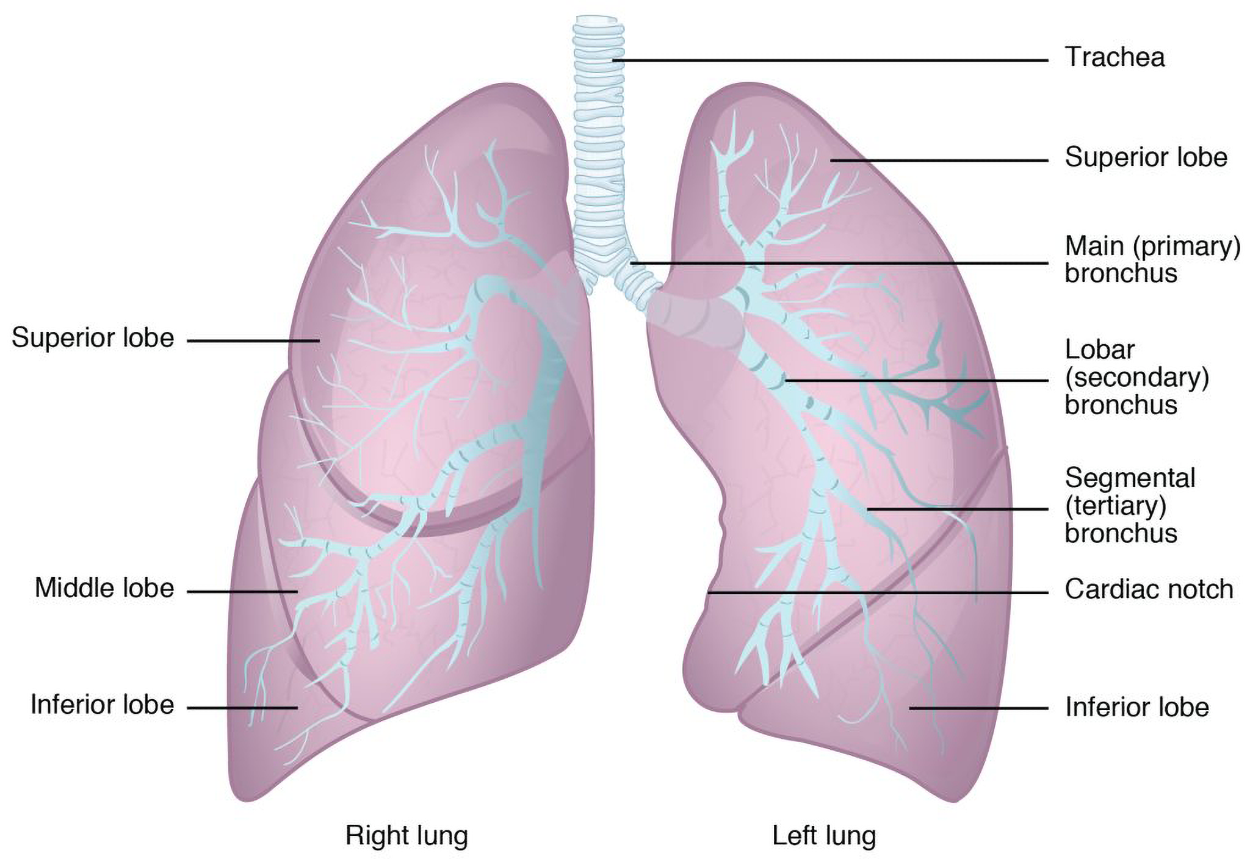
Each lung is composed of smaller units called lobes. The right lung consists of three lobes: the superior, middle, and inferior lobes. The left lung is smaller and only contains two lobes, superior and inferior, as it shares space with the heart. Each lobe receives its own large bronchus that has multiple branches. A lobectomy refers to the surgical removal of a lobe of the lung.
The word roots for lungs and/or air are pneum or pneumon.
EXAMPLE
The medical term pneumonia refers to a diseased state of the lung.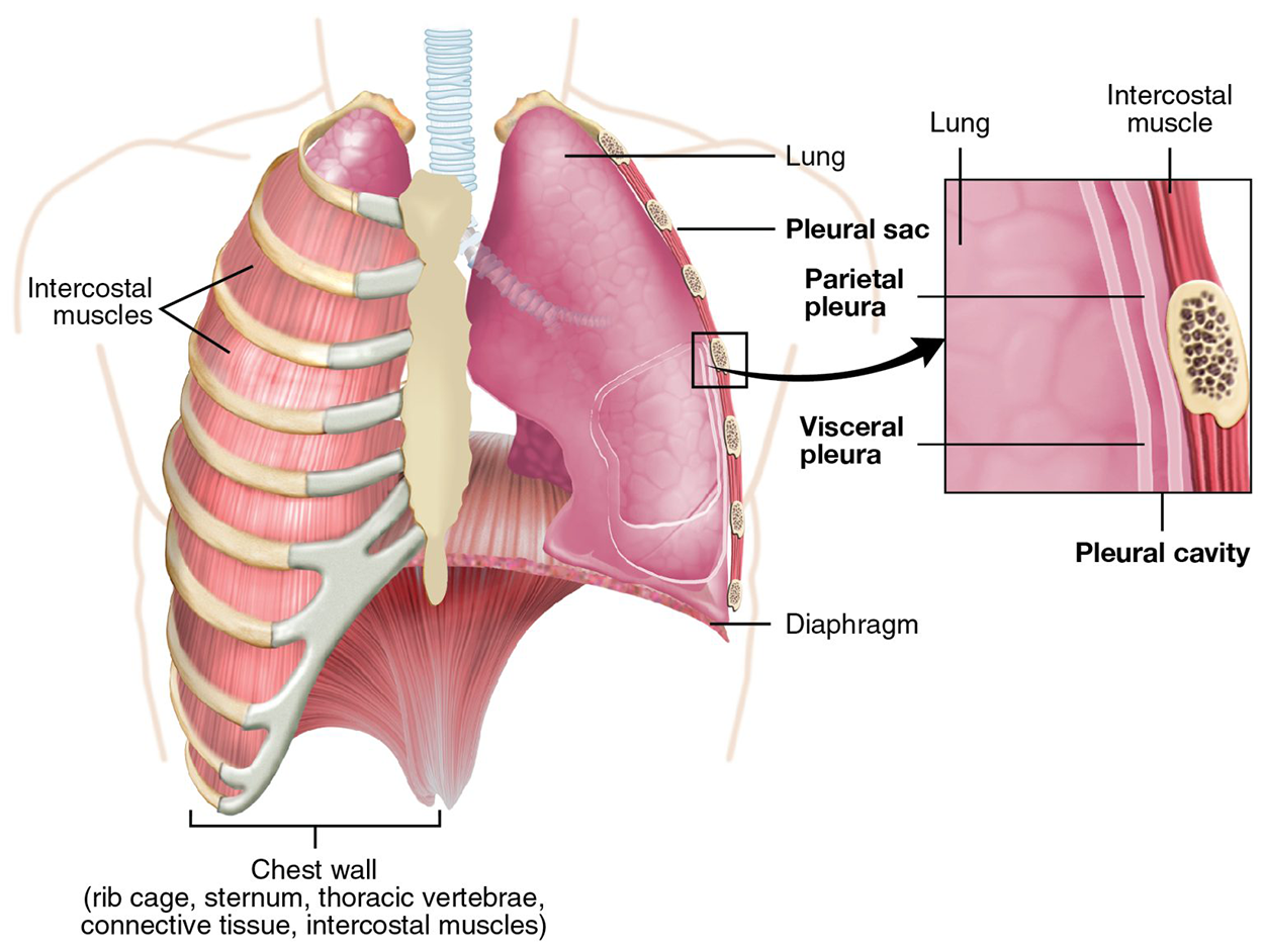
There are two pleural membranes (pleura) enclosing the lungs. The visceral pleura is a thin membrane on the surface of the lungs. The parietal pleural lines the inside of the thoracic cavity. Between these two membranes is the pleural cavity that contains pleural fluid to reduce friction and also sticks to the lungs to keep them inflated. Pleur/o is the combining form of the word root for pleural membranes.
EXAMPLE
Pleural effusion is a medical term that refers to excessive fluid between the pleural membranes caused by disease or trauma.Source: THIS TUTORIAL HAS BEEN ADAPTED FROM OPEN RN "MEDICAL TERMINOLOGY 2E". ACCESS FOR FREE AT wtcs.pressbooks.pub/medterm/ LICENSING: CREATIVE COMMONS ATTRIBUTION 4.0 INTERNATIONAL. Accessed by March 2025.
REFERENCES
Meegalla N, Downs BW. (2023, June 5). Anatomy, Head and Neck, Palatine Tonsil (Faucial Tonsils). StatPearls Publishing.ww.ncbi.nlm.nih.gov/books/NBK538296/