Table of Contents |
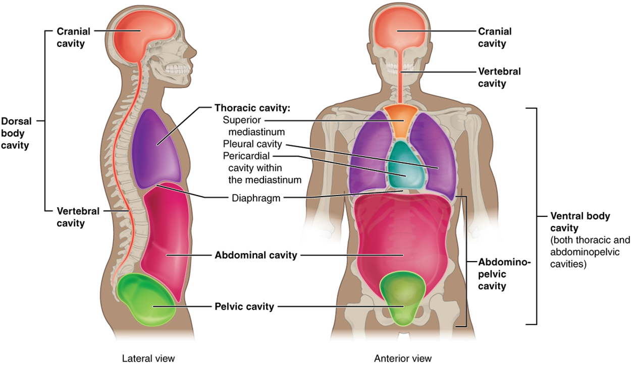
The body maintains its internal organization by means of membranes, sheaths, and other structures that separate compartments. The dorsal (posterior) cavity and the ventral (anterior) cavity are the largest body compartments (see image below). These cavities contain and protect delicate internal organs. The dorsal (posterior) cavity for example, contains and protects the brain and spinal cord. The ventral (anterior) cavity contains and protects organs too but also allows for significant changes in the size and shape of the organs as they perform their functions. The lungs, heart, stomach, and intestines, for example, can expand and contract without distorting other tissues or disrupting the activity of nearby organs.

The dorsal (posterior) and ventral (anterior) cavities are each subdivided into smaller cavities. In the posterior (dorsal) cavity, the superior cranial cavity houses the brain, and the inferior spinal cavity (or vertebral cavity) encloses the spinal cord. Just as the brain and spinal cord make up a continuous, uninterrupted structure, the cranial and spinal cavities that house them are also continuous. The brain and spinal cord are protected by the bones and other tissues of the skull and vertebral column.
The ventral (anterior) cavity has two main subdivisions: the thoracic cavity and the abdominopelvic cavity (see the image above). The thoracic cavity is the more superior subdivision of the anterior cavity, and it is enclosed by the rib cage. The thoracic cavity contains the lungs and the heart. A breathing muscle called the diaphragm forms the floor of the thoracic cavity and separates it from the inferior abdominopelvic cavity. The abdominopelvic cavity is the inferior subdivision of the anterior cavity and the largest cavity in the body. Although no membrane physically divides the abdominopelvic cavity, it can be useful to distinguish between the abdominal cavity, the division that houses the digestive organs, and the pelvic cavity, the division that houses the organs of reproduction.
During a visit to the doctor, if you tell them of pain in your abdomen, they are guaranteed to ask you a standard question—“Can you point to the pain?”—Do you know why they ask that?
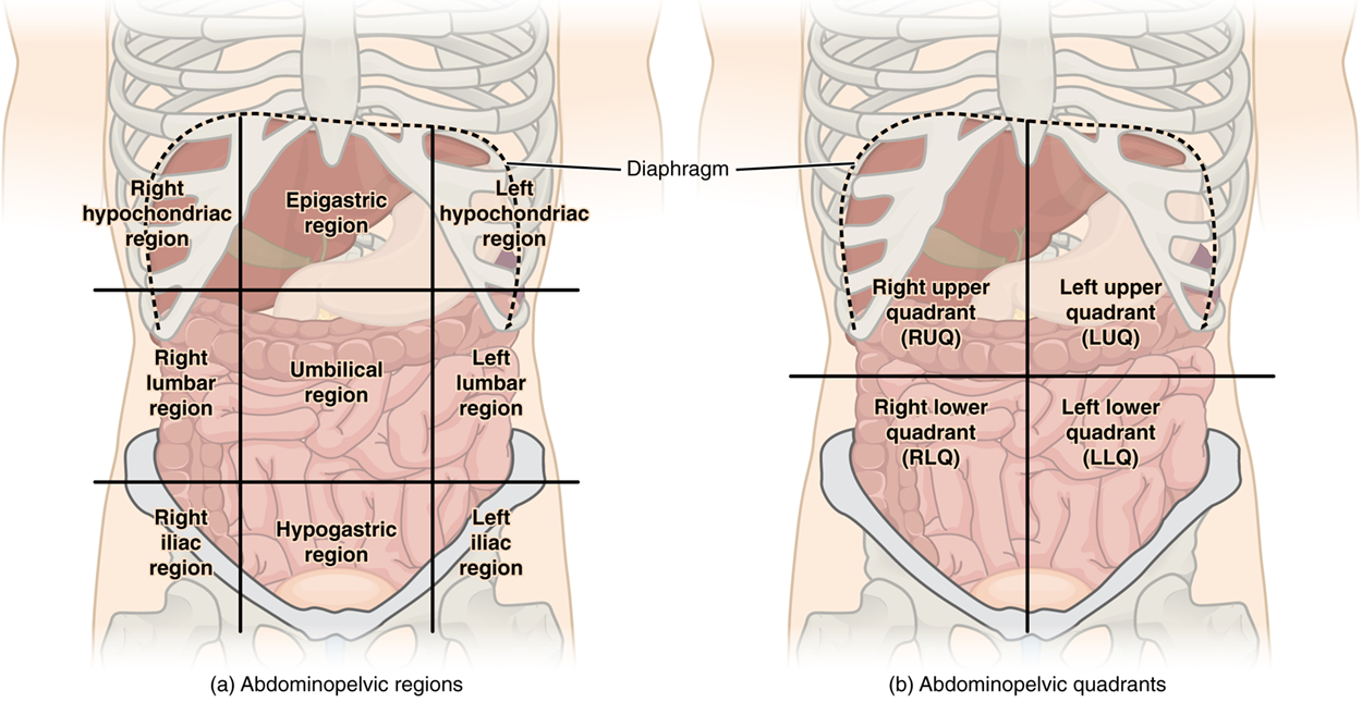
To promote clear communication, healthcare providers typically divide up the abdominopelvic cavity using one of the two systems:

The simpler system for dividing the abdomen uses two lines to create four quadrants and is more commonly used in medicine. The quadrants subdivide the cavity with one horizontal and one vertical line that intersects at the patient’s umbilicus (navel or belly button). The resulting quadrants are named based on if they are on the right or left half and upper or lower half - right upper quadrants (RUQ), left upper quadrant (LUQ), right lower quadrant (RLQ), and left lower quadrant (LLQ).
The more detailed approach subdivides the cavity into nine regions using four lines arranged as a tic-tac-toe board on the abdomen; one horizontal line immediately inferior to the ribs, one horizontal line immediately superior to the pelvis, and two vertical lines drawn as if dropped from the midpoint of each clavicle (collarbone). The resulting nine regions are laid out in three columns of three. The right and left columns share similar names while the central column has unique names. The top center region is epigastric (epi, above; gastric, stomach). The middle center region is the umbilical (where the umbilical cord is attached at the navel or belly button). The bottom center region is the hypogastric (hypo, below; gastric, stomach) or pubic. The top left and right regions are the left and right hypochondriac (hypo, below; chondriac, cartilage). The middle left and right regions are the left and right lumbar (loin, lower back), named after that anatomical region. The bottom left and right regions are the left and right iliac (upper bone of the pelvis), named after that anatomical region.
A serous membrane (also referred to as serosa) is one of the three thin, double-layer membranes that cover the walls and organs in the thoracic and abdominopelvic cavities. These membranes function to reduce friction inside the body as the internal organs they surround expand, contract, and move.
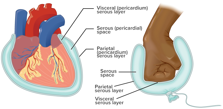
Each serous membrane is made of two continuous layers separated by a fluid-filled space. As the illustration below depicts, imagine a fist pressed into a partially filled balloon. In this analogy, the fist is the internal organ (i.e., heart, lung, intestine, etc) and the balloon is the serous membrane. The balloon material that makes contact with the fist is the deep layer of the serous membrane, called the visceral layer (viscera, internal organ). The balloon material that does not make contact with the fist (and instead is in contact with the inner cavity wall) is the superficial layer of the serous membrane, called the parietal layer (parietal, wall). Observe that these two layers are continuous with one another meaning there is no disconnect between them. The balloon space between the visceral and parietal layers is filled with fluid and is called the serous space.

There are three serous membranes; the pleura, pericardium, and peritoneum. The pleura is the serous membrane that surrounds the lungs. The pericardium is the serous membrane that surrounds the heart. The peritoneum is the serous membrane that surrounds several organs in the abdominopelvic cavity. For specificity when talking about a layer or space of a particular serous membrane, each is named for the specific membrane. The superficial layers are the parietal pleura, parietal, pericardium, and parietal peritoneum. The deep layers are the visceral pleura, visceral pericardium, and visceral peritoneum. The spaces are called the pleural space, pericardial space, and peritoneal space.
The serous membranes form fluid-filled sacs that are meant to cushion and reduce friction on internal organs when they move, such as when the lungs inflate or the heart beats. Both the parietal and visceral serosa secrete serous fluid, a slippery liquid, within the serous spaces. The pleural cavity produces pleural fluid to reduce friction between the lungs and the body wall. Likewise, the pericardial cavity produces pericardial fluid to reduce friction between the heart and the wall of the pericardium. The peritoneal cavity produces peritoneal fluid to reduce friction between the abdominal and pelvic organs and the body wall. Therefore, serous membranes provide additional protection to the viscera they enclose by reducing friction that could lead to inflammation of the organs.
Source: THIS TUTORIAL HAS BEEN ADAPTED FROM OPENSTAX “ANATOMY AND PHYSIOLOGY 2E.” ACCESS FOR FREE AT HTTPS://OPENSTAX.ORG/DETAILS/BOOKS/ANATOMY-AND-PHYSIOLOGY-2E. LICENSE: CC ATTRIBUTION 4.0 INTERNATIONAL.