Table of Contents |
We have discussed simple concentration gradients—a substance's differential concentrations across a space or a membrane—but in living systems, gradients are more complex. Because ions move into and out of cells and because cells contain proteins that do not move across the membrane and are mostly negatively charged, there is also an electrical gradient, a difference of charge, across the plasma membrane.
The interior of living cells is electrically negative with respect to the extracellular fluid in which they are bathed, and at the same time, cells have higher concentrations of potassium (K⁺) and lower concentrations of sodium (Na⁺) than the extracellular fluid. Thus, in a living cell, the concentration gradient of Na⁺ tends to drive it into the cell, and its electrical gradient (a positive ion) also drives it inward to the negatively charged interior. However, the situation is more complex for other elements such as potassium. The electrical gradient of K⁺, a positive ion, also drives it into the cell, but the concentration gradient of K⁺ drives K⁺ out of the cell. We call the combined concentration gradient and electrical charge that affects an ion its electrochemical gradient.
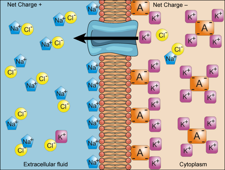
To move substances against a concentration or electrochemical gradient, the cell must use energy. This energy comes from ATP generated through the cell’s metabolism. Active transport mechanisms, or pumps, work against electrochemical gradients. Small substances constantly pass through plasma membranes. Active transport maintains concentrations of ions and other substances that living cells require in the face of these passive movements.
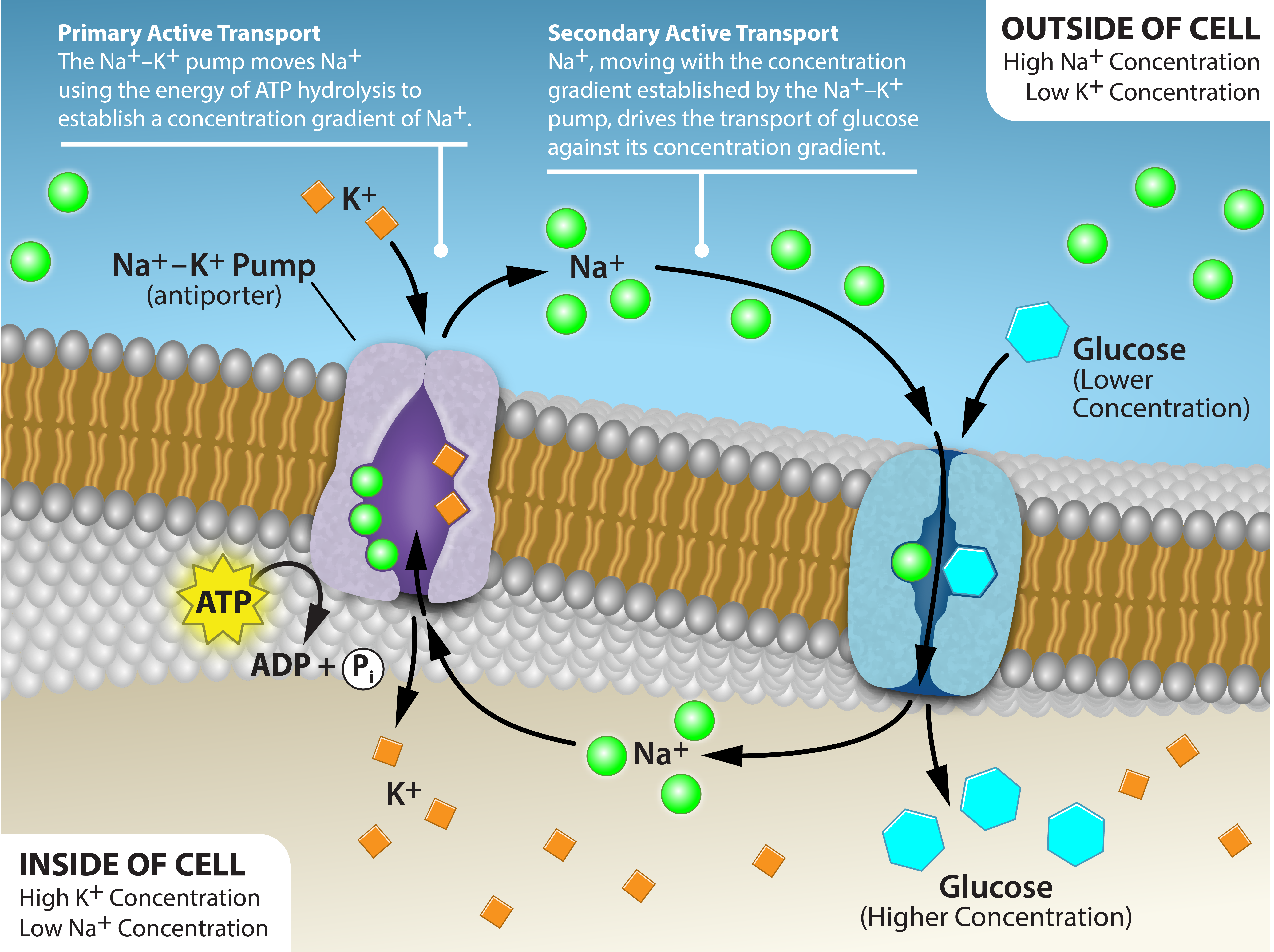
Two mechanisms exist for transporting small molecular weight material and small molecules. Primary active transport moves ions across a membrane and creates a difference in charge across that membrane, which is directly dependent on ATP. Secondary active transport does not directly require ATP: Instead, it is the movement of material due to the electrochemical gradient established by primary active transport.
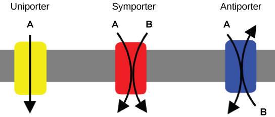
An important membrane adaptation for active transport is the presence of specific carrier proteins or pumps to facilitate movement: There are three protein types, or transporters. A uniporter carries one specific ion or molecule. A symporter carries two different ions or molecules, both in the same direction. An antiporter also carries two different ions or molecules, but in different directions. All of these transporters can also transport small, uncharged organic molecules like glucose. These three types of carrier proteins are also in facilitated diffusion, but they do not require ATP to work in that process.
One of the most important pumps in animal cells is the sodium–potassium pump, which maintains the electrochemical gradient (and the correct concentrations of Na⁺ and K⁺) in living cells and represents an example of primary active transport.
The sodium–potassium pump moves K⁺ into the cell while moving Na⁺ out at the same time, at a ratio of three Na⁺ for every two K⁺ ions moved in. The figure below shows how this occurs.
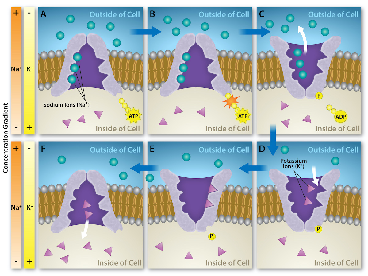
Several things have happened as a result of this process. At this point, there are more sodium ions outside the cell than inside and more potassium ions inside than out. For every three sodium ions that move out, two potassium ions move in. This results in the interior being slightly more negative relative to the exterior. This difference in charge is important in creating the conditions necessary for the secondary process. The sodium–potassium pump is, therefore, an electrogenic pump (a pump that creates a charge imbalance), creating an electrical imbalance across the membrane and contributing to the membrane potential.
The primary active transport that functions with the active transport of sodium and potassium allows secondary active transport to occur, which is described below.
Using the example above of the sodium-potassium pump, secondary active transport uses the kinetic energy (energy that results from motion) of the sodium ions to bring other compounds against their concentration gradient into the cell. As sodium ion concentrations build outside of the plasma membrane because of the primary active transport process, this creates an electrochemical gradient.
EXAMPLE
If a channel protein exists and is open, the sodium ions will move down its concentration gradient across the membrane. This movement transports other substances that must be attached to the same transport protein in order for the sodium ions to move across the membrane. Many amino acids, as well as glucose, enter a cell this way.This secondary process also stores high-energy hydrogen ions in the mitochondria of plant and animal cells in order to produce ATP. The potential energy that accumulates in the stored hydrogen ions translates into kinetic energy as the ions surge through the channel protein ATP synthase, and that energy then converts ADP into ATP.
In addition to moving small ions and molecules through the membrane, cells also need to remove and take in larger molecules and particles. Some cells are even capable of engulfing entire unicellular microorganisms. You might have correctly hypothesized that when a cell uptakes and releases large particles, it requires energy. A large particle, however, cannot pass through the membrane, even with energy that the cell supplies.
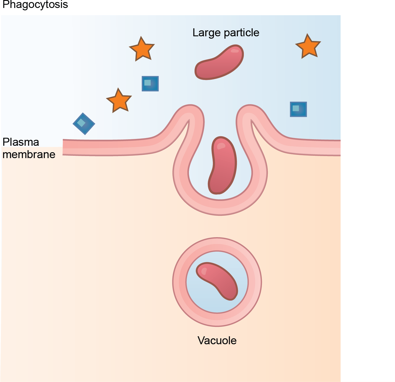
Endocytosis is a type of active transport that moves particles, such as large molecules, parts of cells, and even whole cells, into a cell. There are different endocytosis variations, but all share a common characteristic: The cell's plasma membrane invaginates, forming a pocket around the target particle. The pocket pinches off, resulting in the particle containing itself in a newly created intracellular vesicle formed from the plasma membrane.
Phagocytosis
Phagocytosis (the condition of “cell eating”) is the process by which a cell takes in large particles, such as other cells or relatively large particles. For example, when microorganisms invade the human body, a type of white blood cell, a neutrophil, will remove the invaders through this process, surrounding and engulfing the microorganism, which the neutrophil then destroys.
Pinocytosis
A variation of endocytosis is pinocytosis. This literally means “cell drinking.” Discovered by Warren Lewis in 1929, this American embryologist and cell biologist described a process whereby he assumed that the cell was purposefully taking in extracellular fluid. In reality, this is a process that takes in molecules, including water, which the cell needs from the extracellular fluid. Pinocytosis results in a much smaller vesicle than does phagocytosis, and the vesicle does not need to merge with a lysosome.
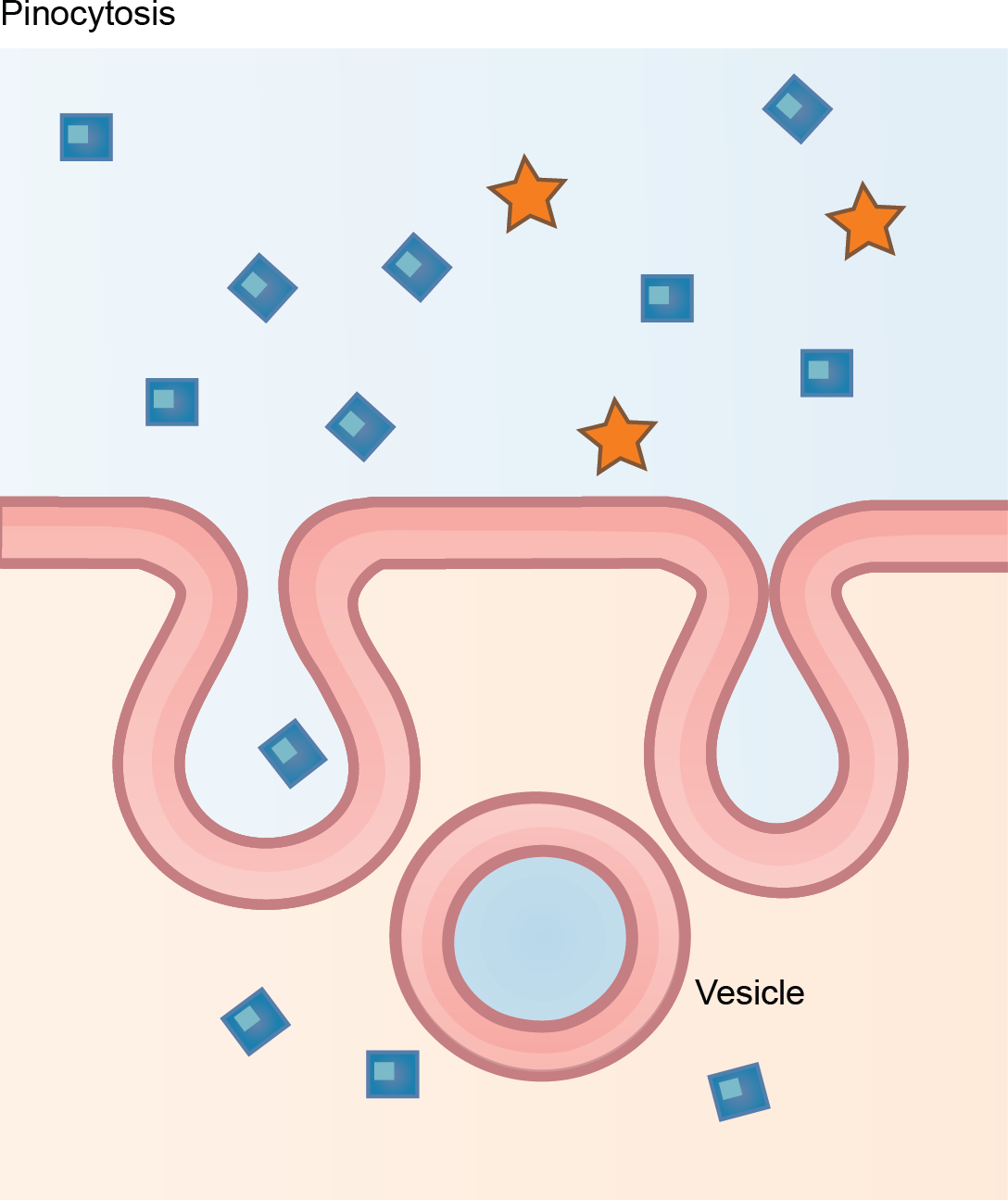
A variation of pinocytosis is potocytosis. This process uses a coating protein, caveolin, on the plasma membrane's cytoplasmic side. The cavities in the plasma membrane that form the vacuoles have membrane receptors and lipid rafts in addition to caveolin. The vacuoles or vesicles formed in caveolae (singular, caveola; a plasma membrane invagination that resembles a small cave) are smaller than those in pinocytosis. Potocytosis brings small molecules into the cell and transports them through the cell for their release on the other side, a process we call transcytosis. In some cases, the caveolae deliver their cargo to membranous organelles like the ER.
Receptor-Mediated Endocytosis
A targeted variation of endocytosis employs receptor proteins in the plasma membrane that have a specific binding affinity for certain substances. This is referred to as receptor-mediated endocytosis.
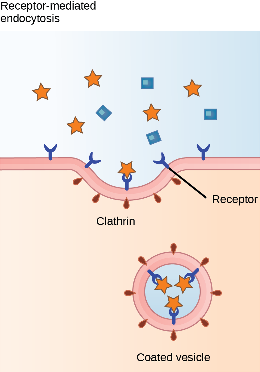
If a compound's uptake is dependent on receptor-mediated endocytosis and the process is ineffective, the material will not be removed from the tissue fluids or blood. Instead, it will stay in those fluids and increase in concentration. The failure of receptor-mediated endocytosis causes some human diseases. For example, receptor-mediated endocytosis removes low density lipoprotein, or LDL (or "bad" cholesterol), from the blood. In the human genetic disease familial hypercholesterolemia, the LDL receptors are defective or missing entirely. People with this condition have life-threatening levels of cholesterol in their blood because their cells cannot clear LDL particles.
Although receptor-mediated endocytosis is designed to bring specific substances that are normally in the extracellular fluid into the cell, other substances may also gain entry into the cell at the same site. Flu viruses, diphtheria, and cholera toxin all have sites that cross-react with normal receptor-binding sites and gain entry into cells.
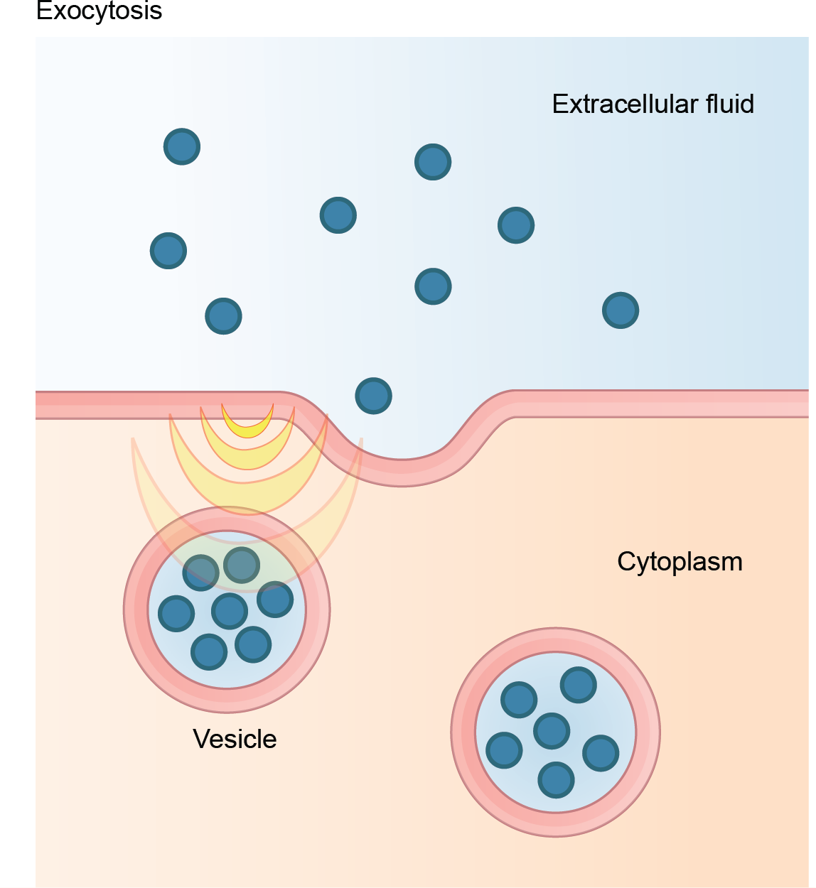
The reverse process of moving material into a cell is the process of exocytosis. Exocytosis is the opposite of the processes we discussed above in that its purpose is to expel material from the cell into the extracellular fluid. Waste material is enveloped in a membrane and fuses with the plasma membrane's interior. This fusion opens the membranous envelope on the cell's exterior, and the waste material expels into the extracellular space.
The table below summarizes the methods of transport that you have reviewed over the past two lessons.
| Transport Method | Active/Passive | Material Transported |
|---|---|---|
| Diffusion | Passive | Small molecular weight material |
| Osmosis | Passive | Water |
| Facilitated transport/diffusion | Passive | Sodium, potassium, calcium, glucose |
| Primary active transport | Active | Sodium, potassium, calcium |
| Secondary active transport | Active | Amino acids, lactose |
| Phagocytosis | Active | Large macromolecules, whole cells, or cellular structures |
| Pinocytosis and potocytosis | Active | Small molecules (liquids/water) |
| Receptor-mediated endocytosis | Active | Large quantities of macromolecules |
SOURCE: THIS TUTORIAL HAS BEEN ADAPTED FROM (1) OPENSTAX “BIOLOGY 2E”. ACCESS FOR FREE AT OPENSTAX.ORG/BOOKS/BIOLOGY-2E/PAGES/1-INTRODUCTION (2) OPENSTAX “ANATOMY AND PHYSIOLOGY 2E”. ACCESS FOR FREE AT OPENSTAX.ORG/BOOKS/ANATOMY-AND-PHYSIOLOGY-2E/PAGES/1-INTRODUCTION. LICENSING (1 & 2): CREATIVE COMMONS ATTRIBUTION 4.0 INTERNATIONAL.